






















This lecture presents research demonstrating that dysfunction in amygdala inhibitory circuits, particularly involving neurexin-1 alpha and ErbB4 signaling pathways, contributes to amygdala disinhibition as a common endophenotype in schizophrenia spectrum disorders. The research uses multiple mouse models to show that both neurexin-1 alpha knockout mice and ErbB4 intercalated cell knockout mice exhibit impaired fear learning, social interaction deficits, and disrupted synaptic plasticity in amygdala circuits, suggesting that targeting amygdala intercalated cells may offer therapeutic potential for treating schizophrenia.
Amygdala Inhibitory Neuron Dysfunction in Schizophrenia | Neuroscience Seminar
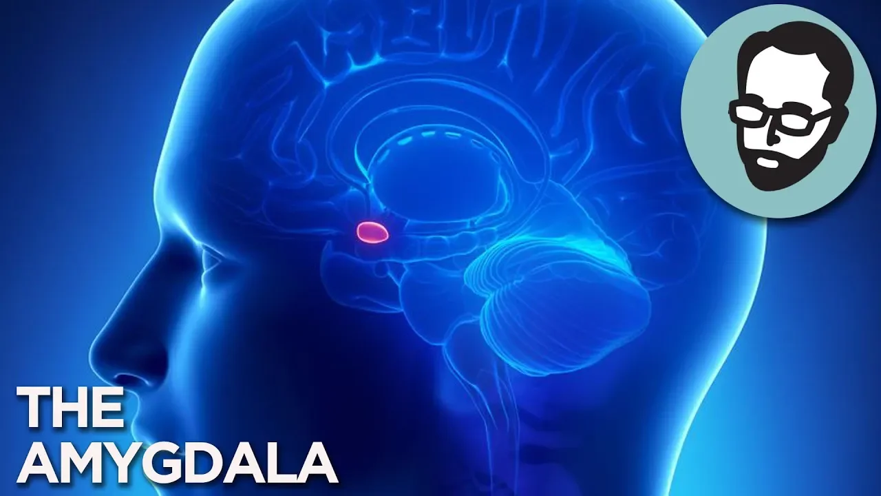
Added:Anatomy and functional role of the amygdala in emotional processing, threat detection, and social behavior.
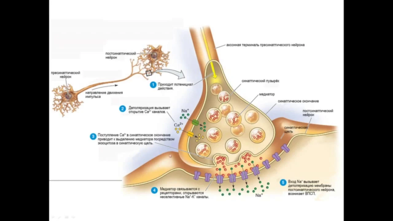
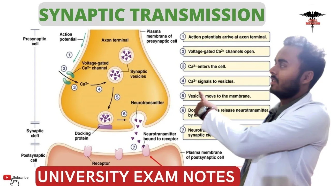
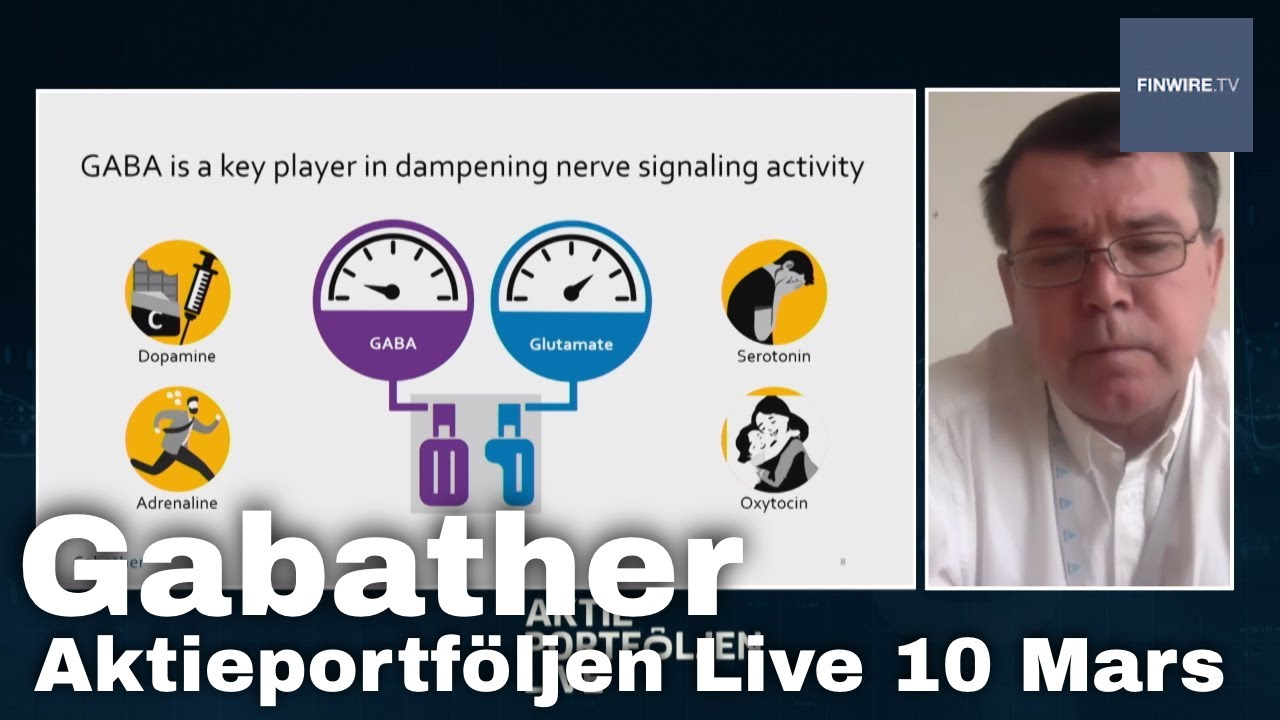
Fundamentals of synaptic transmission, specifically the distinction between excitatory (glutamatergic) and inhibitory (GABAergic) signaling.
The clinical presentation and neurobiological hypotheses of schizophrenia, including positive, negative, and cognitive symptoms.
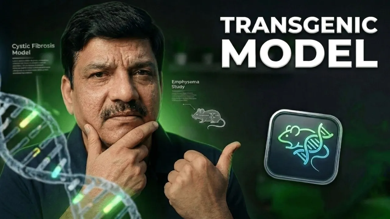
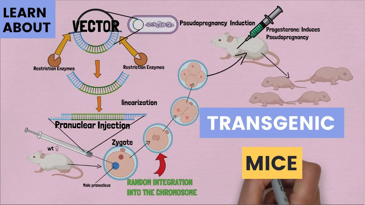
The methodology of using transgenic and knockout genetic mouse models to mimic human psychiatric disorders in laboratory settings.
Targeted pharmacological interventions, such as specific GABA receptor modulators, designed to restore inhibitory control in the amygdala.
The Excitation/Inhibition (E/I) imbalance theory of schizophrenia and its manifestations in other key brain regions like the prefrontal cortex.
Advanced neuromodulation techniques (e.g., optogenetics and DREADDs) used to selectively control and study interneuron excitability in vivo.
Translational biomarkers (such as EEG gamma oscillations or functional MRI connectivity) that bridge rodent model findings with clinical observations in human patients.
377 views6likes58:50Original Release: 2020-10-07
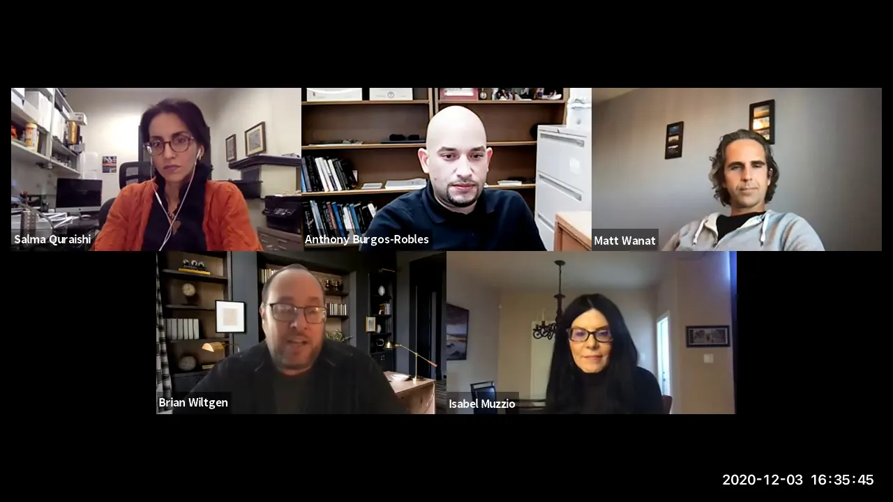
today we have a guest speaker from moxplock florida institute just part of our campus up in jupiter dr ken dawson scully is hosting so i'm going to throw it to ken thanks kate it's a pleasure to introduce dr mclean bolton dr bolton received her phd from duke in 2000 in neurobiology she worked as a research associate with the postdoctoral nrsa in dr mike ehler's lab where then she moved through a number of startup companies as a senior scientist dr bolton then became a research assistant professor in the department of pediatrics neurology at duke and then in 2011 she then transitioned to the max plank florida in super neuroscience where she became a research group leader and she focuses on the understanding alterations in neural circuits that underlie neuropsychiatric disorders such as schizophrenia and autism she's a very active in administration as the chair of academic programs and she's also a co-speaker for the international max planck research school shared between max playing florida institute and florida atlantic university she also has a very active laboratory with several graduate students and technicians where she's well funded by the max blank society she publishes regularly in high impact journals where the most recent papers were in 2020 in translational psychiatry and brain structure function take it away mclean so good afternoon first i wanted to thank randy for inviting me and ken for hosting me and it's really great to have the opportunity to share my research with my neighbors even if it is virtual so my research is motivated by the need to find treatments to psychiatric disorders such as schizophrenia so i want to first introduce schizophrenia so that we realize that i'm disease focused it's not just basic research and then i want to give talk about a trio of projects that explore how schizophrenia-rich genes may put the amygdala on overdrive okay so schizophrenia is a complex chronic and severe mental illness that affects how a person thinks feels and acts and the symptoms are divided into three categories the positive symptoms or psychotic behaviors cognitive symptoms and negative symptoms such as reduced emotions so some examples of positive symptoms are hallucinations which are perceived sensory experiences that occur in the absence of a sensory stimulus it's like hearing voices that aren't there or seeing people that aren't there and it can happen to any in any senses but it's most common in auditory um delusions are another positive symptoms they're strongly held false beliefs that persist even when there's evidence to the contrary so thinking someone that's placing thoughts in your mind or can control your thoughts or paranoid thoughts and some thought disorders are considered positive symptoms like having great difficulty in organizing your thoughts or speaking in garbled words that have no meaning but sound like a language there are also some movement disorders and agitated repetitive body movements or catatonia in some patients as far as negative symptoms um flat affect is one of the major ones so reduce expression of emotions and be a tone of voice and facial expression or gestures anhedonia or reduce feelings of pleasure in everyday life or abolition a lack of motivation or elogia or which is reduced speaking or less complicated patterns of speech patients have a lot of trouble with executive function or the poor ability to understand information and to be able to use it to make it make decisions and they have trouble with attention focusing keeping on tasks they also have difficulty with working memory you know remembering information for immediate use um so here are some statistics that's good training so one percent of the people in the united states and worldwide are affected by schizophrenia and this is across the board it doesn't vary by region um males are 1.4 times more likely to be affected than females and the average age of onset is 24 males and 27 females and the life expectancy is reduced by 10 to 20 years in patients and the annual cost of schizophrenia in the u.s alone is 90 billion dollars so clearly we need to understand what happens in the brain circuits and develop treatments that help people with the disorder initiation so how do we treat schizophrenia so medication is of course the first line of defense counseling and social support groups can make a world of difference and kind of training your brain with cognitive behavioral therapy to keep some of the symptoms in check can really minimize the need for a lot of medication the primary medications of course are antipsychotics the first generation antipsychotics were very strong dopamine receptor type 2 blockers and the second generations have had a milder effect on d2 and have serotonin blocking as well as well as many other things but they're not perfect and they do not affect the negative or the cognitive symptoms so what causes schizophrenia to develop well it's very strongly genetic with environmental influences so this graph helps me to think about that this shows the concordance rate between different types of relatives for schizophrenia so if you have a twin if you're monogamous zygotic twin and one of the twins has schizophrenia there's a 45 chance the other will have it if you have two schizophrenic parents then the chance that the child will have it is over 35 percent it goes down to seven percent with siblings and spouses which would be like the general population is one percent so clearly there's a genetic influence but clearly that's not all so there's environmental modifiers with genetic background so many genetic loci and candidate genes have been identified for schizophrenia there are examples of rare variants in genes where a single gene can make a major contribution but by and large the disorder is very polygenetic so mutations in many genes that have small effects combined to create the conditions for developing schizophrenia so this study here shows uh the top candidate genes that were identified across many guest studies we there are some rare variants that have strong effects so disc one which is a scaffolding protein regulan one which we'll talk about later and the choline elemental transferase gene they have a metabolic glutamate receptor an ampa receptor an nmda receptor the dopamine receptor a calcium channel that's norexin one and the receptor for an irregular one so these are genes that have been identified across the board as being rare mutations that can contribute a lot in certain cases um recently there was a study that identified 108 schizophrenia candidate loci and one interesting thing that they found was that the major histocompatibility locus was way above everything else and this was a bit surprising in the study and subsequent work has identified the c4 complement protein as in this area and that that's particularly interesting with respect to synaptic pruning okay so there are several hypotheses about what is imbalanced at a network level in schizophrenia and potential molecular mechanisms that undergo this imbalance so they're not mutually exclusive and we hope someday they'll merge into one complete picture of the etiology of schizophrenia but the main ideas are excessive mesolimbic dopamine excessive spine pruning particularly in the prefrontal areas nmda hyper function and oscillatory rhythm imbalances and oxidative stress so synaptic connections continue to form at a high rate throughout child childhood and then there's a period of synapse elimination and adolescence and then even it's kind of levels out into normal adulthood but with they found that in schizophrenia the period of adolescent spine pruning is much more profound than in normal individuals so you can see this on the bottom graphs here where there's a this shows dendritic splines in the prefrontal cortex on layer three and you see more spines in the control than the schizophrenic patients and you can see this summarized in the middle here the the yellow is the controls and the red is the schizophrenic group it's interesting in light of the fact that the c4 complement protein was identified by the schizophrenia consortium and that's involved in microglial assisted synaptic engulfment so the c4 protein binds to a synapse and tags it for microglia to come and eat the synapse so with respect to the hypothesis of increased mesolimbic dopamine so this circuit is important for reward and motivation and salience so here the here are the neurons in the vta and their dopaminergic neurons and they project to the nucleus accumbens the hippocampus the prefrontal cortex and the amygdala and they receive inhibitory projections from the ventral palatium and the nucleus accumbens and somehow in this loop the hypothesis in schizophrenia is that there there's too much dopamine released in the nucleus accumbens and not enough in the prefrontal cortex why this evolves is unknown but that's the the theory and a lot of the antipsychotics are based on the principle of blocking this excess mesolimbic dopamine another theory about what goes wrong with brain circuits is nmda receptor hyperfunction so glutamate as you know is the main excitatory neurotransmitter in the brain when glutamate vesicles are released glutamate goes across the synaptic cleft and binds to anti-receptors and nmda receptors and nmda receptors are particularly important for brain rhythms because they have us they are often expressed and drive the fast carbohydrate neurons in the brain and one of the original reasons why people thought that nmda receptor hyperfunction could be involved in schizophrenia was that drugs such as pcp and ketamine block the md receptors and they cause behaviors that are similar to the positive symptoms of schizophrenia in normal subjects and make the symptoms of schizophrenia patients worse like hallucinations delusions and hyperactivity there's also variation in the nmda receptor gene associated with schizophrenia and if you make a mouse that not you knock down the md receptor you also may have some of the positive symptoms so brain rhythms are caused by synchronized firing of many neurons the brain has oscillations at many frequencies from the low delta waves to the high gamma range which is 30 to 100 hertz or 400 actually the the power of these rhythms changes with the intentional state and during sleep synchronization of these rhythms between different brain regions is important for integrated processing of information for memory consolidation and gamma power is impaired during cognitive tests and schizophrenia so these are pictures of the eeg recordings um when schizophrenia and control patients are performing some kind of working memory task and you can see that there's reduced activation or power of the gamma rhythms in the schizophrenia patients shown in the darker blue colors and this is these are the difference plots but what about the amygdalin schizophrenia in in schizophrenic nations particularly those with paranoid schizophrenia fear and anxiety is absolutely debilitating but there's really little work that's been done on how the amygdala is affected in schizophrenia um there are studies that hint that this could be an issue but just if you observe the behavior you you would realize that this is something that should be studied so this study shows that there's increased resting state blood flow to the left amygdala and schizophrenic patients so the orange or schizophrenic patients the blue are controls and the green are actually non-paranoid schizophrenic patients and so there's an increase in the basal activation of the amygdala in these patients so it's like they're slightly on alert for for fearful stimuli but there's a decreased activation regulator during a task where you're trying to judge trustworthiness from facial expressions so here again the orange patients are the schizophrenia patients and you saw show less activation than the controls and this may be because they're already the basal activity is already so high that they don't have much room left to activate it further when something relevant it comes into play there's also reduced functional connectivity between the amygdala and the medial prefrontal cortex in patients so in this task the subjects were told they were shown a picture of a face and then they were shown two other patients and had the faces and they had to pick which one was the same face as the one they were shown before and they had two blocks one block was emotional faces and one were neutral faces and interestingly on the bottom here that the schizophrenic patients had difficulty with the accuracy of the test only in the emotional block not in the neutral block so it was the emotions of the faces were bothering them to the extent they were having difficulty performing the task and they when they did this they were measuring also the functional connectivity between the amygdala and the prefrontal cortex and they found um reduced connectivity in the dorsaline the dorsal um what is it i'm trying to say the singlet cortex and the dorsal medial prefrontal cortex and the rostromedial prefrontal cortex so and you can see that here with the um patients versus the controls so so given the evidence that the amygdala isn't functioning properly in schizophrenia i wanted to look at the amygdala function in a couple of knockout models of top schizophrenia candidate genes so the first project i want to talk about is the investigation of amygdala circuit function and behavior in the norexin one alpha knockout mouse model and this is the work from douglas sacedy who's a postdoctoral fellow in the lab and national joseph a postbackfell in the lab who is also a former fau student so norexin is a presynaptic protein that is on in the synaptic cleft it's important for the synapse formation and stabilization and function it's present at both excitatory and inhibitory synapses and it binds trans-synaptically to a number of post-synaptic partner proteins the neuroligans the lrtms the cerebellums lush villains dystroglycan it has six different alternate splicing sites so it can be put together in many different ways and with that kind of specificity you can imagine how different synapses in different brain regions could use the directions differently differentially so they have found copy number variations associated with various um psychiatric disorders with the norexin gene so in point one eight percent of schizophrenic patients and point five of autism patients those are actually high numbers for a rare gene so a knockout model of norexin 1 has some of the behaviors that you might expect for a mouse or a rat who was behaving with some of the symptoms of schizophrenia although it's hard because their mouths so they had the direction knockouts has difficulty with sensor sensory motor integration nest building self-grooming motor learning they have increased aggression they have increased startle decreased instrumental and spatial learning and decreased social fear learning so douglas and azela examined classical cued fear conditioning and then erection 1 often knockout mice so how the test works is the first day you put the mice in the cage just to get used to it and the second day you pair a tone with a shock several times and you appear there's another tone that you don't pair so it's a so it's not used to shock so they learn to freeze when they're they learn to associate the tone with the shock then the next day you test them in a different context and you just play the tone and not the shock and you see if they still remember the tone was was fearful by freezing so in the norexins um knockout mice when it on during the fear learning phase there was a trend towards decreased learning but it wasn't significant but on the next day after they had a chance to calm down and recollect their experience the control mice remembered that the tone was a pair with the talk because they shocked because they froze but the neuroxin wanting knockouts they did not learn as well to associate the tone with the shock so fear learning um is associated with the amygdala amygdala circuitry so this is a simplified diagram of the amygdala circuit so in fear learning you have information about the cs coming in from the auditory system through the solenoids in the cortex and entering the lateral amygdala the unconditioned stimulus or pain comes in to the amygdala through the thalamus and through the la and during fear learning the synapses here among other places are potentiated to learn to associate the tone with the shock there are other areas in the in within the circuit that are also altered in fear learning but that's the classic one from the la the it's a kind of a linear circuit where information flows from the la to the ba then to the central amygdala and out to the hypothalamus and brain stem to affect the fear of behavior there is um top down emotional control from the medial prefrontal cortex and contextual information and and kind of salience information from the nucleus accumbens and that's the fear of circuit okay so in these norixin one knockout mice they had the fear deficit so what we wanted to do was to investigate what in the amygdala was changing to underlie this deficit they had in fear learning so we wanted to look at this because the medial prefrontal cortex to amygdala circuitry is weakened or could be weakened in schizophrenia from top down control we looked at the synapse between the medial prefrontal cortex and the basal amygdala so how we did this is we expressed channel rhodopsin which is a protein that when you shine a light on this channel it opens and conducts cations and it can be used to polarize neurons so it's a very useful tool for us so we injected this channel rhodopsin into the prefrontal cortex and then the terminals from the channel orgasm um went on down and synapsed on the ba and the purple here is a cell that we were patch clamping in the ba so when we patch clamp the neuron in the va and shine the light on to activate the channel rhodopsin these synapses are activated and you see a postsynaptic current response in the neuron and these are excitatory responses so one way to determine how strong a presynaptic part of the synapse is is to look at what we call pair pulses so if you stimulate a neuron to make release glutamate and you have a postsynaptic response and then you do it again 50 milliseconds later if the synonyms has a high probability of release synapse then all the vesicles or most of the vessels can be released the first time and there aren't that many left over for the second guy so it's a measure so that the paired pulse response is a measure of pre-synaptic function so with what we found was that in the norrex and knockouts this second pulse was much smaller than the first pulse so and this could mean either that they were it's a high probability release synapse or that they just didn't have enough vesicles to follow along in the beginning so at any rate he has reduced presynaptic ability to follow a train of synaptic stimuli and that's summarized here and in this bar graph so we looked at the same neurons but we stimulated in the the lateral amygdala to see what the synapses from the lateral major lesion the basal lateral amygdala we're doing and here when we stimulate these synaptic responses you could see that the second pulse is almost as large as the first and it wasn't changing the knockouts and that's summarized here so at two different synapses on the same neurons norexin is having a very different effect in terms of the presynaptic function but what about on the postsynaptic side so the the postsynapse has different types of ampa receptors of glutamate receptors there are nmda receptors and their ampa receptors to name a few the nmda receptors have this really unusual feature in that they are blocked by magnesium at minus 70. when you depolarize the neuron it pops the magnesium out and then and the calcium and sodium can come into the nmda receptors so how we measure how many what percentage of the synaptic current is carried by amp and nmda is we hold the cell at different voltages so here we're holding the cell at minus 70 and we're looking at the ampa current going down and at plus 40 it's the ampa current plus the nmd current but we the amp current is very fast so if you measure out here it's just the nmd current so what you see here in the knockouts in the synapse from the mpfc to ba is that there is a major decrease in the ampa to nmda ratio and we don't know from this information whether this is due to the ampa receptors being smaller or the nmda receptors being bigger because it's just a ratio when we looked at the la to ba synapses and did the same thing looking at ampa receptor current here and nmd up here they were exactly the same so again the synapses from the medial prefrontal to the ba are behaving very differently than the lidba when you knock out an erection one so to try to figure out the question about whether the ampa receptors are smaller or the nmda receptors are bigger we did what we call a light response curve so here we have our optogenetic stimulation of the medial prefrontal cortex fibers and we just turn the light intensity up and we crank it up and then you can see that all across the board the knockout ampa receptor currents are slot smaller than the control so this indicates that we have decreased amp receptors in the knockout so the other thing that we wanted to check was to look at inhibition onto these same neurons so um what we did here is we we can hold the cells in such a way that we can put the reversal potentials at to hold through minus 70 is the reversal potential for chloride so when you're going down it will be only amp receptor currents and if you're holding it zero the excitatory currents are at reversal potential so you can only see the inhibitory so those are details but the short of it is that excitatory transmission is down and inhibitory transmission is up and what you can see in this case is that the in the knockouts inhibition is just decimated the those are tiny little yellow curves in in the knockout and they're tiny both at the mpsc to va synapse and the la to basin apps so here we have the the epscs are the same and the ipscs are decreased in amplitude in the knockout so from that we don't know if inhibition is tiny because the excitatory drive to this inner interneurons are smaller or because the interneuron synapsing on the next neuron are smaller so what we wanted to do is to take the excitatory drive out of the equation so here what we did was we expressed channelrhodopsin in the cell bodies of the neurons in ba and we shined the the light on and measured the inhibitory responses and and what we found was that the probability that one um inhibitory neuron was connected to an excitatory neuron was reduced three-fold in the knockout and then when you integrate the currents that reflects the three-fold reduction in in the amplitude of inhibition so to summarize this project the synapse from the medial prefrontal cortex to the basal amygdala it provides executive control of fear emotions the synapse is unusually dependent on schizophrenia susceptibility gene norexin-1 alpha it has a reduced pre-synaptic capacity to release glutamate at high frequency it has weaker ampa receptor-mediated postsynaptic responses um the inhibition in this in the amygdala is basically decimated by the loss of erection and there are fewer inhibitory connections onto the ba principle [Music] neurons so i wanted to switch gears and talk about uh the next project that i wanted to present and this is a project is called apical paracapsular intercalated cell cluster a novel sensory regulator in the amygdala this is a pretty esoteric project i have to admit so this project was led by douglas acety again a postdoctoral fellow and predominantly the work was done by division fau medical student who also was an undergraduate at fau um abigail chavis who was a post was a fau honors graduate and hopefully will come back and work in my lab in the future she did all the morphological analysis carolyn von walter who is in my lab now and hopefully will stay forever um as a graduate student and she did a lot of the surgeries and some of the imaging and morphological analysis so the itcs are clusters of inhibitory neurons that gate information flow into and within the amygdala itcs are potential therapeutic targets for schizophrenia at least i think so i don't think anyone else has ever said it but i think they would be a good target so um anyway so the inhibition is very important part of mental functions and the neurons within the immediate fire at low basal rates and under normal conditions and then when something that's relevant for emotional response the brakes or inhibitory brakes are removed and the synapses can undergo plasticity and they could have responses to to fear so the itc's are interesting because they receive dense dopaminergic input that hyperpolarizes them through d1 receptors so when the vta synapses on the itc's the inward rectifier potassium channel opens and it hyperpolarizes cell making them less excitable so each cluster has a lot of specific connections within the amygdala and receives specific information from outside for example the medial pair capsule itcs receive information from the thalamus the medial prefrontal cortex and several other cortical areas and the la and they project to the central lateral amygdala and the ba and the medial cluster the lateral itc's receive sensor information at from the sensory cortex and the entronal cortex and they project to la and ba the main itc cluster receives information from the medial prefrontal medial itcs and the central medial nucleus and and also the hippocampus and insular cortex so but what about the apical itc so there are tiny little cluster up at the top on top of la and it's missing in most of the diagrams that you see of immediate circuitry and it's missing because we don't know anything about it so we wanted to study the apitcs and and try to figure out um who it's connected to and what it's doing so in order to do this we needed to find out who the presynaptic partners of the apit sports these were so one way to do this is to use a monosynaptic rabies chasing um so monosynaptic radiation is nice because it takes advantage of a natural property of this rabies virus to jump across the synapse and infect the presynaptic terminal and so then you could identify everybody that was presynaptic to that cell but you have to stop it from reproducing and going back further until the whole brain is full of virus so the way this has been done is that they remove one of the necessary proteins the glycoprotein g protein and they also pseudotype the virus so it has an avian coke protein envelope a so that it will only affect things that have the receptor to the envelope protein and that's here so if you use your your first you inject a helper virus that give that adds in the delta g and then and has the the tva receptor for the envelope protein then if you wait and let that express for two weeks and then deliver a rabies virus that has this envelope a coating it can internalize into the cell it can go about its life cycle and then hop a synapse and then transfect the synaptic partner with whatever you have cloned into it so we used this approach to try to figure out who was who were the partners to the apitcs um so we injected the apitcs with their helper virus and then two weeks later with our rabies virus and here you can see the starter cells in blue and then the presynaptic partners are in green so what we found um was some things were expected and some things were not so we found the um the pin and mgm nucleus which conveys is expected because it would convey auditory information um that was necessary for the fear learning we found some midline nuclei that would be are useful because they they're they convey limbic information to the amygdala and to and from the medial prefrontal cortex so we also found um so then we went through and checked to see if these things were actually connected so and how we did this was we injected channelrhodopsin into the presynaptic partner which in this case was in the thalamus in the mgm pen and then the channelrhodopsin expressed we made slices and we patch clamped the apitcs and we shined the light on the terminals to see if we got a synaptic response so here so here you see the injection site here you see the channelrhodopsin expressing axons going over near the apidcs and if you and here's a blow up showing them right next door and here are the synaptic responses and we found that 100 percent of the ap itcs receive connections from the pin nucleus we also found a number of limbic relevant association areas we found insular cortex piriform cortex endoper claustrum auditory cortex temporal association cortex the amygdala hippocampal area ventral hippocampus and into rhino cortex so now we're in the process of going in checking and seeing which of those are actually connected and which are just artifacts of our rabies process so um so here we injected our channel rhodopsin in the temporal association cortex and again let it express for three or four weeks and you can see the again the axons coming through near the apatc and the patch clamped excitatory responses and in this case we found that 58 of the itc's received synaptic connections from the temporal cortex we also found synaptic connections from the insular cortex 60 of the neurons that we checked were responsive so sixty percent of apitcs receive synaptic inputs um for for insular cortex and and in the intra rhino cortex we found that a hundred percent of the api disease received connection from the intra-rhino cortex so now we know some of the areas that project to the apitcs now we wanted to find out where they project to so how these experiments were done was we patch clamped the apic neurons and we filled the electrode with biocytin and then reacted it with streptavidin and that was conjugated to a fluorescent dye and then we imaged with the confocal and then traced the axon in a program called neurolucida and what we found um was that 33 of the apitc is projected within the cluster here 35 of the apic is projected to la and 23 of them projected striatum and 10 projected stratum nla so now we wanted to just look at how they were functioning within the amygdala circuit so um ap igc we found form inhibitory synapses on la principle neurons so here if you put an ops in a fast option in this case chronos in the apitcs and you shine a light on them and you record from a principal neuron you see an inhibitory response so we find that they what they do is they form inhibitory connections on la principle neurons then we showed that if you can drive so since we think that the thalamus projects to the apitcs as a major input we wanted to know whether those afferents from the thalamus can drive the abc itcs to fire and activate the inhibitory synapses onto the principal neurons so here we shine the light on the channel docs and terminals and recorded from the principal neuron and we found a dissynaptic inhibitory current so this kind of verifies the circuit that the thalamus drives the apitcs to inhibit the la principle neurons so then we wanted to see well what happens how do they participate in fear learning so we know that they're in a position to integrate information from a number of higher order cortical association areas and receive direct sensory input relevant for fear from the thalamus so how do they work in the fear circuit so we did um fear conditioning on mice and then we met and they learned that your conditioning as normal here's the they freeze when they learn to associate the tone with the shock the next day they remember it but what we did was in addition to training them then we sacrificed the mice at different time points and looked at how this the synapses onto the apitcs from the thalamus were um changed by the experience of learning the fear so here is our so the so again these traces are amp currents going down and nmda receptor currents going up this is like the just the controlled home cage control animals and um we find that they have this is their their amp occurrence and this is the paired pulse ratio between the amp currents and here's your enemy currents so in the fear condition mice though if you you look here their the paired pulse ratio is is different so they increa increase the probability of release because the pair pulse ratio decreased so that's shown here in this bar graph um and if you in another control where you don't shock them with at this frequency of tone there was no change in their um paired pulse ratio and but but by the time the next day the fear memory this change in the paired pulse ratio was back to normal so that was a transient synaptic change during associated with the acquisition of fear but it was not present in part of the fear memory however if you look at the nmda currents in the same situation so here's the control and here's the fear condition the nmd currents um got smaller and in in there they remain smaller in the memory case so this is a permanent change in the amp to nmda ratio in these neurons with fear conditioning so in summary um the apitcs are strongly driven by sensory information related by the thalamus they receive information from a diverse range of higher order cortical association areas the apitcs project to la and they gain information entering this emotional processing hub and they also project to the stride i'm gaining motivational drive with emotional salience and it's regulated by dopamine so i have one final project to quickly go through if we have enough time this project was done again by douglas escadi and three students contributed saba ali james oko and dps and the it's about the functional consequences of herb before deletion in amygdala intercalated cells and the implications for schizophrenia yeah so noregulin is a member of the egf super family of growth factors it's expressed in the brain during development in adulthood it's the dominant receptor for an irregular in the brain and upon binding to its ligand the regulin the receptor dimerizes and autophosphorylates and activates the pi3 kinase and the rathmat kinase pathways which are involved in cell growth and cell proliferation and differentiation the herb receptor also is involved in keeping nmda receptors at the synapse by binding to a scaffolding protein that holds them under the synapse as we mentioned before neregulin and herb4 are highly top candidate genes for schizophrenia um in schizophrenia patients or regulating one expression is increased and rb4 is hyper phosphorylated and there are a lot of her before splice pattern alterations in patients um removal in a verb before in pv neurons leads to a schizophrenia mouse phenotype um in a number of tests and herb is expressed in gabaergic interneurons um tv neurons cck neurons and vip neurons and in gabaergic projection neurons in itc's and and also in dopaminergic neurons and noradrenergic neurons so it's involved in the migration differentiation and function of gaba neurons and it sets the window for visual system critical period plasticity it's involved in gamma rhythms and theta rhythms and synchrony between the ventral hippocampus and the prefrontal cortex so one of the striking features about this herb protein is it's expressed at very high levels in the intercalated nucleus of the amygdala so we really wanted to look at knocking out the herb before gene specifically in this population of neurons and see what it affected so in this case we're looking at the mpitcs and they receive strong projections from the thalamus and the medial prefrontal cortex they project to ba in the central lateral um in nucleus so how we did this is we cross the herb for knockout phlox mice with a dr r1 creline that is expressed specifically in the intercalated cells and is the main cluster and in also some scattered populations in the nucleus accumbens and the colostrum [Music] so when we looked at the behavior of these knockouts they had deficits in sociability so they in the control mice they prefer to spend time with another mouse compared to the empty cage if you put them in in the center here they'll go visit the mouse more than they would visit the empty cage and the and knockouts didn't seem to care whether it was an empty cage or a mouse and we also checked social novelty and so if you have a mouse that he knows and a new mouse normal mice generally like to go explore the new mouse so they spend more time with the unfamiliar mouse in the controls the norex and knockouts did didn't care if he was old or new it's all the same to him so they had some social deficits they were also hyperactive in the open field test so when we looked at fear conditioning in these guys um we found that they they had they were okay in terms of the training but they couldn't remember the fear of learning the next day they didn't they couldn't remember to that the tone was scary and that they needed to freeze they also um didn't forget right so so if you normally if you play a tone that you have previously shocked a mouse with over and over again and you don't shock them they say they learn this is not dangerous anymore so they stop freezing but these guys um the norex and knockouts kept on freezing they didn't learn to it was okay and this you might expect a schizophrenia so what we we wanted to look at what was going on in the mpitcs in these mice and we found when we looked at spontaneous synaptic transmission we found that excitatory transmission seemed to be okay in terms of ample receptor minis but the knockouts had larger individual responses to gaba released spontaneously so they had larger inhibitory quantum amplitude so we looked um at this process to see if it was when you evoke the stimulation so if you stimulate the thalamic afference to the mpitcs and you patch plant them and you do a light response curve you look at excitatory transmission on the bottom you turn the light up and the responses get bigger and they look about the same in the mutants in orange compared to the the wild type in black but as you increase the light intensity and look at the inhibitory currents it's clear that the inhibitory currents are much larger all across the board and the knockouts and that's shown here in the summary so so they're getting too much inhibition onto these neurons we also wanted to look at the amp at nmda ratio in in the mutants in the same way and and the paired pulse ratio in the same way that we did in the other projects and what we found was that there was no difference in the paired pulse ratio indicating there wasn't an issue with the pre-synaptic functioning but there was an increase in the ampa to nmda ratio in the mutants so here we wanted to also find out whether it was due to um a change in the amp current or the change in the nmda current so when we did the light response curve for just amp eps it was the same in the control and knockouts and but the nmda receptor current was decreased so we wanted to find out ultimately what do these synaptic pathologies have to do with how this synapse is going to behave in a fear situation so these neurons undergo ltp during fear learning and so we did an ltp protocol on while from the lamb gafferins to the mpitcs that you have a high frequency stimulus that would normally give ltp and you can see that in wild type mice they get ltp the open circles are normal ltp but in the mutants they didn't it's and it's in fact ltd if you block inhibition which helps with the depolarization of the neurons and allows an mda currents to open then the wild type mice had larger ltp and the mutants had some level of ltp and this is summarized here showing that the wild type have ltep the knockouts have ltd you block innovation the wild type gets bigger and the knockout is slightly rescued so in summary they they do not have normal learning plasticity at this synapse that would and this would have an effect on fear so to summarize this project the deletion of our before and itcs results in hyperactivity and fear learning and social interaction deficits and increased quantal amplitude of inhibitory synapses onto the mpitcs and increased strength of feed-forward inhibition of mpit species by thalamic afference and decreased nmda receptor mediated synaptic currents in the thalamus and synapses onto mpitcs and impaired ltp of thalamic synapses onto mphtcs due to enhanced inhibitory gaiting and the overall summary is that disinhibition of the base lateral amygdala is a common endophenotype among two mouse models of schizophrenia in norexin one a knockout mice feed forward inhibition from the mtsc and la to ba is profoundly compromised and in the herb before itc knockout mice aberrant enhancement of inhibition and a lack of ltp of excitatory thalamic afference to the npitcs leads to disinhibition of the ba and itcs may benefit be a beneficial target for developing therapies for schizophrenia so and i just want to thank the members of my lab for their dedication and support and team spirit it's a it's a really great place to be because they're all so hard working and great so thanks and thanks for listening so if anybody would like to ask any questions of dr bolton we invite you to use the hand raising mechanism in zoom on your screens mclean i'd like to ask you a question um you mentioned a couple of times the um contributions of dopamine signaling to the circuits that you mentioned and i wondered if in any of the projects that you described you've tried to manipulate dopamine signaling either in the animal or in the slice to take a look at differences in say dopaminergic modulation of synaptic function in the mutants that's a really great idea so we've we've verified that in the apitcs they those neurons are inhibited by dopamine as expected but we haven't tried to um see if we can enhance any of the pathologies or conduct them by dopamine that's a really that's a really cool thought um it's something that i'm going to write down right and you know maybe you know validity more validity to the model and with antagonists that might be therapeutic and just sort of to see about parallels i think that'd be really really cool and one one other little quick question have you looked at all at sex effects that might um distinguish um their responses at all so i didn't show some of the data in the herb mice there were some difference in the social phenotypes with the females versus the males and i think they were much slightly more sensitive to some of the the social behaviors but i haven't um spent a lot of time classifying we do in general do the behaviors on the males great thanks beautiful work hi yeah i don't know can i just jump in here it's claire um yeah i'm just wondering it's kind of actually a follow-up question to randy's question and it's looking at the opposite side have you looked at the glutamate uh effect uh and uh dis uh disinhibiting the or yes disinhibiting the gaba uh response so have we systematically manipulated the glutamate levels yeah with your with with some of these knockout mice i think it might be interesting to see you know what would happen so if you're if your nmda receptor is dysfunctioning and it's causing an inhibition it's not releasing glutamate into the gala or gabaergic system yeah so i think pushing the system with by doing uh perhaps you know high frequency stimulation at different places to to get at that question would be a good idea so yeah i just thought it might be interesting with some of those knockout mice yeah and i think it would have effects because you you when you have things that are changing especially the presynaptic function if you manipulate with a high frequency you can get at that question but in terms of globally putting on glutamate antagonists or agonists that haven't gone that way thank you well i'll ask another question because i just don't see anybody there and uh why not so the itc's are just fascinating i really didn't know anything about them and i'm wondering if in schizophrenia or autism or other things that are genetically connected they've been looked at in postmortem studies is there is there any morphological data cellular data at all no isn't it amazing that i mean it doesn't strike you is that this is something that should be played with right sure now i think so yeah yeah and and it just hasn't been especially given that the weird d1 receptor manipulation and um i i haven't found anyone that has looked at it i should look more carefully but i just haven't found it in the clinic but they're there in humans as well yeah yeah they're yeah they're they're kind of analogous to their reticular neurons and violence in some in some ways you know they just haven't been well studied right thanks um this is a real talk um i asked a question about the relationship because i'm really interested by the amygdala involved the schizophrenia disease how can you integrate the amygdala to the um the vitamin d to like your striatum because the main hypothesis the old days classic way is that too much dopamine in the striatum and the treated tobacco in the front of the cortex so yeah the uh yogurt you know i think you know it's first of all they're integrated right in the mesolimbic system you know it's all part of that integrated circuit and you know it's understudied how you know it's that how the amygdala is affected so that you don't maybe in five years you'll that that line will be there's too much mesolimbic dopamine in the nucleus that come in there's not enough in the prefrontal there's too much in the amygdala right we might hear that so because they're not you know it's you know it just where it's a work in progress but i think it would be integrated in that the same way that you subject and they may have features like these little odd shells that make it more um make a difference in the in the way it's gated right maybe there's another question while we wait for krista to connect that way um yeah actually i had a question can you guys hear me um so i just wanted to know if you could talk a little bit more about the role that the gamma and data frequencies play that you're observing in in the knockout mice so the gamma differences that i quoted i haven't measured in the mice it's just being fur that they would be important because they're important in the human and other people who have studied you know different models and different brain regions have seen decreased gamma rhythms in these mice and so i would only have to answer it with respect to the general understanding of the functions of the gamma rhythms which are partially in memory consolidation in terms of bi and binding different uh thoughts in the brain together and having different brain regions be on the same and neuron classes be on the same wavelength per se by having the same time links so just coordinating things and that's what people kind of think rhythms are important for thank you that's helpful sounds like it well thank you very much very much yay thank you thanks for having me randy
Up Next
51:08
Memory Consolidation & Retrieval Mechanisms | Neuroscientists Talk Shop
@utsaneuro
153 views•2020-12-21

21:55
Deciphering Neural Circuits Controlling Anorexia in Mice | Richard Palmiter
@AllenInstitute
2.3K views•2011-10-10

4:10
Vagus Nerve (CN X): Anatomy, Nuclei & Functions Explained
@Alilamedicalmedia
305.2K views•2022-10-31

13:03
How Exercise Benefits Your Brain: Science Explained
@TED
11.4M views•2018-03-21
Related Study Plans & Knowledge Roadmaps
Structured learning paths in Neuroscience
neuroscience
Appetite Regulation: Brain Circuits & Hormones
01Foundations of Endocrinology and Brain Anatomy
02Introduction to Energy Homeostasis
03Peripheral Signals: Leptin, Ghrelin, and Insulin
+ 2 more learning modules
14h5 modules
Explore Roadmap
neuroscience
Neuroeconomics: Value, Risk & Brain Mechanisms
01Foundations of Neuroscience and Brain Anatomy
02Introduction to Neuroeconomics and Subjective Value
03Dopaminergic Prediction Errors & Reinforcement Learning
+ 3 more learning modules
18h6 modules
Explore Roadmap
neuroscience
Spatial Navigation: Cells, Grids & Brain Maps
01Foundations of Brain Anatomy and Memory
02Place Cells and Head-Direction Cells
03Grid Cells: The Brain's Coordinate System
+ 2 more learning modules
12h5 modules
Explore Roadmap
neuroscience
BCIs: Neural Decoding & Prosthetic Control
01Neuroscience Foundations: How the Brain Signals Intent
02Signal Acquisition: Recording the Brain's Activity
03Neural Decoding: Translating Brain Waves into Data
+ 2 more learning modules
25h5 modules
Explore Roadmap