




![ASMR | Doctor Roleplay FULL BODY MEDICAL EXAM 👩🏻⚕️✨ Collab w/ 10 UNDERRATED ASMRTISTS [for Sleep]](https://i.ytimg.com/vi/gDPo64q-Q7c/maxresdefault.jpg)













This lecture teaches how to localize neurological lesions by understanding brain organization principles: the homunculus shows motor areas anterior to sensory areas, with the leg dangling between hemispheres, hand draping over the parietal lobe, and face at the bottom; dominant hemisphere (left in 95% of right-handed people) controls language and motor functions, while non-dominant controls spatial and creative abilities; Broca's area (frontal lobe) produces speech while Wernicke's area (temporal lobe) understands language; brainstem nuclei follow a midbrain (III-IV), pons (V-VIII), and medulla pattern; spinal cord dermatomes follow a 'six-shooter' pattern (C5-C8-T1) and a 'Christmas tree' for thoracic levels; and clinical vignettes demonstrate how these principles localize lesions to specific brain regions based on symptoms like gaze preference, facial weakness, and motor deficits.
Name That Lesion: Neuroanatomy Clinical Vignettes
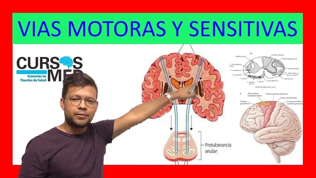
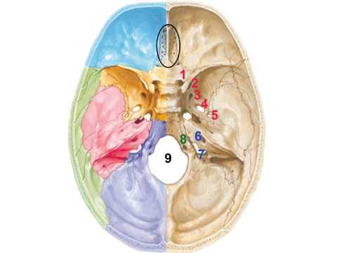
Added:[Music] Thank You cavae he and to the organizers including KJ for asking me to come and share some of my passion for neuroscience with you today so we're gonna go through name the lesion and KJ knows that I love to kind of torture the nurses and the residents and trying to figure out where the lesion is and my day when we learn neurology you weren't allowed to look at the imaging until you could figure it out so it's started it's looking at the brain and part of the interesting thing about being in neuroscience is that you can figure out because of the way that brains organized with the roadmaps this is a road map of Hawaii and around here usually it's red but this is green but there's roadmaps that help us follow what's going on with the patient so some of this is going to be obviously a lot of review for you experienced nurses but I thought that it's always kind of fun to bring it back to that baseline so going back to the homunculus of how our brain is organized we know that that there's the area that's the sensory which is in the posterior parietal and then the motor and I always think of it motor forward so before the pre Sylvian fissure here the motor area the motor strip runs in front of the sensory so the motor and thinking about how we organize this helps you decide where the lesion is so we know that the leg the homunculus again is dangling down between the two hemispheres so if you have say hydrocephalus that's stretching those fibers that you'll have patients that have leg wobbliness and then the trunk is a smaller area both our motor and sensory and then of course our hand is very large and drapes over the part of the monk is so lying on this part here and in the motor areas in the frontal region and then our thumb is obviously a very large motor area our forefinger is a very large sensory area and then the face dangles down in the lower part so the face and of course the tongue and the ability to move our tongue is going to be very important and then and here's the tongue here so and then the sensory again follows similarly so it's really helpful to know when we talk about different areas of stroke for example where you have maybe the anterior cerebral artery and we'll talk about that where the leg weakness comes because the anterior cerebral artery is lost and the anterior cerebral that comes up and feeds this part of the brain and the middle cerebral artery there's an interesting phenomena where if you have watershed like loss of blood flow where it's decreased so meaning where the ACA is coming up and the MCA is coming up there's a watershed so your watershed is gonna be losing this area here so you get what we call a man in the barrel syndrome where it's like a man who's in a barrel who they can kick their legs because this homunculus part is working and they can move their fingers because this part of the homunculus is working in their face but they can't move their trunk so just knowing these kind of things it makes it very interesting and this is again showing again in a cartoon cartoon fashion our motor function and again our hand skills the neck the arms the fingers the face down here on the sensory that's just behind there and then when we're describing strokes and injuries to patients tumors we can tell them that their speech areas that we all know the speech of the Broca's and then the Wernicke's area of language and then hearing that in the air will get back into this and a little bit our frontal lobes when we have patients with traumatic brain injury we know that they strike the front of their head so the thing that we know that's going to be affected is their personality their mood initiative so the ability to be interested in doing things so if any of you have seen the One Flew Over the Cuckoo's Nest where Jack Nicholson had by frontal lobotomies and that's how they used to treat very an recalcitrant psychiatric patients he just sat there he wasn't interested in anything though he had all those motor movements and a vision his initiative and interest was decreased by that and obviously our concentration our behavioral skills come from there as well and then we know that the temporal lobe is our memories our motion the limbic system that runs through there and the vision though coming through the eyes we know those motor tracks and we'll get to that get to the posterior occipital region and then the cerebellum as we know is our posture our balancing and coordination that we can do very well without but don't do well with the brain stem compression that might happen from a cerebellar stroke so again localizing lesions gonna it's going to be important that you know which side of the brain does different things so our dominant hemisphere for those of us who are right-handed about ninety ninety-five percent of us are going to be left hemisphere dominant that's going to be our spoken language our ability obviously move that right side on both arms and legs and hands are so I'd like to think of it the arse the reading writing arithmetic so numbers and the the ability to do math is going to come from that as well as your reasoning so written language scientific all comes from that dominant hemisphere and then we know that that's the apart that's what it's going to be affected if someone has a hemispheric dominant hemispheric lesion and when you have the non dominant dominant hemisphere you can tell patients or families that probably they're going to lose some artistic ability their ability to do spatial things so be able to find their way to the auditorium or to the bathroom once you've been there you would lose your way because you can't find that spatial area that that spatial orientation and then our art and awareness and creativity and music comes from that side as well so I'm going to back up a little bit excuse me so we talked a little bit about Broca's the frontal area the speech production and again experience one's know that and again I think of it motor forwards the motor four is the frontal lobes and also being able to generate that speech the motor being able to say the words so it's producing the speech someone has an area where their frontal areas an injury where their frontal area is not working they're gonna have problems generating those words getting the words out but fully understand what you're saying because the ability to hear and understand is going to come from that temple region so think of it I'm very visual again so I think of it is your ears are where you're hearing something and being able to decode it so when you have a temporal lesion or pointing at the wrong hemisphere for me it would be the left hemisphere the left temporal lobe then I would have problems if there was injury there when you talk to me and how would I tell families is it's like you're speaking a different language if you're not Russian it'd be like speaking Russian to them they just find more Swahili it's just words it sounds like things the content of emotion is there so if you're crying or it sounds funny they might laugh but the actual comprehension of the words are there because it's going through that temple load the hearing area the Broca's area again is motoring out those words so if you have a frontal lesion you're not going to be able to see say the words but your temporal region if it's not affected you'll be able to at least understand so as you know we'll tell people when we're doing our exam I always tell the new residents our new nurses don't show fingers and we don't squeeze hands so squeezing hands can be of course a grass reflex what we do is say show two fingers and kind of sometimes when you're first learning kind of have to hold your own hands here because otherwise you might have that tendency to mimic or show fingers and they're mimicking you so it's really important that we teach people not to not to move their hands and be able to just ask and see if they can understand if they can understand then they're temple region Wernicke's working but focus is not I'm not going to go through the pituitary gland we all know it's there but and we know that for us at least in the neuro ICU were really very interested in whether the patient has if as they progress to brain death they may have decrease in their antidiuretic hormone and have loss of year and very dilute urine in their sodium may climb I'm not going to get too much we know that the green matter is the area that has the highest energy there to have the cell body so when we have patients who have an oxy brain injury the area that they're going to lose are is the cortex and and the gray matter that fuels us and has the ability to have the cognition the white matter the axons are crossing the axons these are where we have problems with XO no injury when there's injury from say traumatic brain injury and then multiple sclerosis is affected there now I mentioned a little bit about the cerebral vascular chairs so again reviewing for you folks and describing it to patients families I always remind them that there's four blood vessels we all know the two carotid that run up front but there's also the two in the vertebral areas and we'll talk about that we talked about injury and vertebral artery injury with the basilar artery that feeds that that brain stem but then again the carotid arteries that go up to the middle cerebral and the anterior serval so remembering our homunculus and the parts that are going to be affected with our middle cerebral cerebral artery and anterior cerebral and I'll show that in just a second here not going to go through all these details just yet but this is very helpful and the slides will be available to you on the website as well as on the app so this is showing that where the areas of injury depending on the blood vessel so the blue here you see the anterior cerebral artery these are the areas that if you had a stroke you're gonna lose again those motor that the leg functions some of the frontal areas so again you might expect that personality may change we look at that large grey area that's the middle cerebral artery so we lose a lot there and the deep structures that are lost with the lenticular strikes that come off the middle cerebral artery so we can I just by looking at this we could start guessing from knowing our neural Anatomy what lesions are we would expect if we include a certain blood vessel you can see that the the cerebral artery the cerebellar arteries here are going to be affected here with the pika and then the brain stem is going to be affected with Basler so again these are available for you to look at a little more closely so the brain stem is another when I talk to again families about what's going on we talk about especially if they're herniating that we eat the cortex is where the upper part of the brains where we do our thinking where we initiate the ability to move things but the brainstem is our way station so it has as we know three areas the midbrain the pons and the medulla and knowing where your nuclei run through again will help you know where the lesion is without even looking at your MRI so the midbrain is going to be the cranial nerves three through four and actually some of the fifth cranial nerve actually runs throughout the entire brainstem even though most of it is in the pons the pons goes from five to eight and the medulla is the decussation of the pyramids which remember the cortical spinal tract our motor strength and if you have a lesion above the medulla if you have a lesion there it's going to be the opposite side of the body you're going to lose but if it's a lesion below that it's going to be the UNEP the lateral side and then the reticular formation so if someone's not awake and you're evaluating your patient and they're sleepy we know that either they've got a by hemispheric lesion or something's going on with the reticular formation which is in the midbrain dorsal midbrain the central pons and the central and here in central medulla the ascending reticular activation is the wakefulness the descending is postural and balanced the cranial nerves and this is this is there for you just for reference because I don't expect and it's trying to get people not to necessarily memorize the exact names of it but thinking as you're moving down so we'll go through in the neuro ICU we don't check out I felt a factory olfactory but we do to do the optic and oculus motor and then the other cranial nerves we'll talk about in just a minute as we move down here and again this is all here and we'll go through these for a second because I'd like to think of it not so much memorizing it's just moving your way down again if you're thinking when you're doing your exam and you're not thinking can i memorize the cranial nerves but you're just moving your way physically down your your brainstem you can remember what you're going to do so the vision when we're doing this we're looking to see and we know that the optic tracts run so that they cross that say something falls in our visual field on the left side so this is in this side of the eye it's falling on the retina here and it moves to the optic radiations on this side woops sorry and then if it's falling on this side it's going to be falling on the up the retina on this side and then crossing across the optic chiasm so we know that if we have a lesion in the temporal lobe as you know you're going to lose the entire the visual field in both eyes anything that's pre chiasm so before that where the crossing is it's going to be a problem with the eye itself so if you're seeing that the problem is in one eye and it's not both eyes then it's going to be pre chiasm if it's a lesion so you're checking for visual field so that's why when we check visual fields you cover one eye and see if they can mimic or see these fingers cover the other and seeing in that side and that's how we're checking to see if it's both eyes or one eye and when it's a temporal lobe the true optic tracts will turn around and if it's temporal lobes so if you have a patient who's had a temporal lobectomy for epilepsy you're gonna lose what we call the pie-in-the-sky so you're gonna lose the things that are up in the opposite hemisphere in the temporal if it were a parietal loss you would lose things down in this area but the temple lobe think of pie-in-the-sky loss because of the way that these optic radiations move so we know that looking at pupils and we're taught this very early that we look for it to see if people's are very small and reactive those may be narcotics if they're very large they may be that they've gotten epinephrine an anticholinergic or been on a lot of barbs these techno pupils and people talk about HIPAA Sande HIPAA is not always pathologic doing general Medicine Clinic and you're looking at young adults sometimes you'll see hippest so it doesn't necessarily mean that there's tectal injury it can be normal but it's important to watch to see if there's been a change we think of these pontine pupils so the difference between small reactive and pontine is that it's though you take a straight pin and poke it right through a little piece of paper and two neurologists when you say pinpoint pupils I automatically think pontine if you say small reactive people's I'm thinking of other things either there's got a scene on meiosis or they have they've had narcotics onboard but pontine pupils are pinpoint when you see unilateral small pupils this is a something like this where this pupil isn't necessarily dilate or fixed remember to think of whether the the pupil that may be abnormal is actually maybe the small people if the patient has ptosis not in this particular picture but because this one's unreactive but if they're both reactive think about Horner's as well so that's sympathetic loss the midbrain peoples are there's mid-position fix people's and we just talked the uncle herniation we all know that when the third cranial nerve is compressed and then the Dian's of phallic people's so moving down so we're taught we've done cranial nerve two so we've looked at the visual fields next we're going to be seeing how they move their eyes we know that the third cranial nerve and if you've read neural Anatomy made ridiculously simple the pillars of three hold that eye open so if you just remember three pillars will hold the eye open and the hook of seven will close the eyes you'll remember that seven will close and then if you think of that everything in the eye movement is three accept the fact that lateral so looking toward your ear and if you were again visual you think of the six here it looks like an ear so looking towards your ear is six and then your nose is like four looks like a four so that would be looking to your your nose other all the rest is cranial nerve three the other thing that's really important we'll get into this when we do those clinical on those clinical vignettes is eye deviation so when you look at eye deviation you should know exactly where you think is what's going on and but this is how it works is that your frontal I gaze center so in this little cartoon I want to drive the eyes to the left so that means that the right frontal I gaze Center is fire firing off that goes down crosses over to the para pontine so pontine it's in the pons reticular formation and that goes up to the six cranial nerve to move the left eye laterally this also goes up the MLF to the third cranial nerve to move this line medially so you can imagine then if you're having a problem with either if the eyes deviate so when you come to the patient's bedside and you're looking at them and their eyes are deviated there's three choices that are happening if they're having conjugate deviation one they're seizing on that right side driving the eyes to the contralateral side so looking to the last seizing here or a stroke on the left side so they can't look to the right so their eyes are deviated or a stroke down in the para ponting or an injury down to the pontine reticular formation on the opposite side so the best way to think about it again either seizing on the right to look to the left so contour outside or a stroke on the other side or a stroke in the brainstem or stroke or injury to the brain stem in the pons on that opposite side as well and that's what's causing your eye deviation so when we're looking at the patient and we'll get into that vignette we can figure out what's going on with their patient again without calling the doc without scanning patient you can figure it out also what's happening so the seventh cranial nerve as we work down their way and actually boy I did I miss five so I guess I sew five there's so five is obviously sensory so we do corneal response for that and then I don't know where sorry about that so five and seven did I have it in here anyhow I know I had three four and six in here okay so anyhow so it's so five is going to be sensory and then obviously you're gonna look on the face for sensory and also the corneal response will be the afferent of touching the eye very lightly with a cotton swab or with a drip on the seventh cranial nerve what we're looking for it's there's a little bit of sensory to that to the tongue which we don't usually test and the external ear canal but the most obviously is we know that seven remember we close the iowa seven the most important for you is its when you're doing seventh cranial nerve exam you have to also check whether they when they lift their eyebrows whether both eyebrows go up the reason why in this patient you can see that there's no furling of the eyebrow of the forehead here this makes it a peripheral seventh or it's either the seventh nerve from a Bell's palsy or the seventh nuclei because there's there's bilateral innervation of the seventh nerve of the seventh cranial nerve so if you have a patient who has just and you don't see it so well here but there's flattening of the nasolabial fold with nice furling of the eye the the forehead here then it's going to be a central seventh so again you can have the patient show the teeth close their eyes but you have to also have them raise their eyebrows some use the puffing the cheeks as well and then the the eleventh is going to be the shoulder shrug which we don't do a lot in the ICU but the tongue as well as the hypoglossal is that remember that the tongue if it's weak it's not going to protrude the tongue so the tongue will deviate to a weak side now if you have a patient who has a facial droop it looks like their tongue deviates so it's important when you have a patient with a pretty profound facial droop you have to lip lift their face up and have them have them protrude their tongue especially in the Bell's palsy often times that the patient's facial droop the ER doctor will call us and say well they also have a tongue deviation and said well did you lift your faith the face up and once they do that the tongue looks midline so again if you have a significant facial weakness you need to look and see if that problem is that there's a facial droop that looks like they're weak on one side but again remember that the tongue when it's weak the strong side is going to push the tongue out the weak sides not moving so the tongue will deviate to the opposite side so again so the very fast and in the neuro ICU I the nurses it's a very fast comb exam we do the blink to threat we look at the fundus copic well and and we'll do to see if they have people in reaction we use the light obviously and look for eye deviation will do oculus of phallic so doll's eyes and to see whether the patient is moving their eyes dolls eyes or when you move the head the eyes should stay looking to one side or another if you're all of you that are awake will not have dolls eyes response so it's only when you're comatose that it's present and your and if you're fully awake you're able to overcome that so a lot a lot of times we're not particularly using that in our usual exam for our nurses of the neuro ICU and then we look for nystagmus we'll do ice water calorics where we put the head of the bed when we're doing brain death exam the head of the bed and when you're watching your physicians or if you're doing as a nurse practitioner or a brain death examination had of bed is thirty degrees you do is 60 cc's of ice wandering in the ears when they're competitive panic membranes are intact and you're looking to see whether the eyes deviate slowly to the cold ear and then fast cortex movement back and that would be an intact oculocephalic ice water caloric response then corneal response we talked about and then grimace and then coughing to suction and gagging so motor exam we're looking at bulk we're looking at some cog wheeling or parkinsonian we're looking in especially in the ICU for any kind of Maya clonus which tells us that if the patient is post on anoxic injury that that's usually a bad prognostic indicator Maya clonus as opposed to the tonic clonic movement we see myclone is is where it's a rhythmic jerking and you can think of it as the cortex isn't good enough to mount responses that is synchronized so it usually tells us again there's bad injury we're looking for asymmetry and triple flexion response so who knows what a triple flexion response is pretty much so the triple flexion response probably you're all shy but you know that painful stimulus to the toe has a very primitive reflex and the ankle flexes the hip flex knee flexes and hip flexes and it looks exactly like this and it's dairy type that's a that's a spinal cord reflex you could be brain dead and have a triple flexion response so it's important when you're getting when you're checking that to see that if the patient does that you might actually grade that is withdrawal what you need to do is pinch the inner aspect of the thigh and see if the leg moves away that would be make it a withdrawal down or pinched the outer aspect see if it moves in that would make it a withdrawal if it looks exactly the same and it's licking when you pinch that toe and you have that triple flexion again it's a spinal cord reflex so I warn patients families if the patient's looks like they're brain-dead that the patient may move but it's a reflex as otherwise they're all excited because they think the patient's withdrawing and they're awake so important to understand that and sensory in the coma exam we're doing painful stimulus looking for grimacing facial movement and motor response now for motor response we know that the decorticate posturing when there's injury that's above the red nucleus the patient will do this decorticate or flexor response and decerebrate is downward and that means the injury is between the midbrain of the red nucleus and above the vestibular nucleus it means that the patient is not brain dead but it does mean that there's a lot of injury to the cortical spinal and four reflexes and I know this is something that typically we have our nurses doing but hyper reflexive the patient's very hypertonic we'll know that's usually an upper motor neuron lesion and hypo reflexive usually is lower motor neuron paralytic medications also will make the patient a reflex ik so something that I think that you'll hear later from dr. miles a little bit about more of the peripheral nerves so respiratory patterns again something that you should be aware of when you're looking at patients the chain Stokes when you ask people what is - where does chain Stokes come from they almost automatically say it's brain stem but it's actually not it's by hemispheric lesion when you see that patient take those rapid breaths become APNIC rapid browse so there's usually by hemispheric injury in the pons there's this central neurogenic hyperventilation so those that work in the ICU will see patients that are hyperventilating no matter what you do with the ventilator you pco2 is very low not the normal 40 but almost as low as 15 sometimes and that's because there's a pond lesion this Eponine stick is the cramp pause fake breathing so they'll come in and very exaggerated and that's a lower pontine and then the ataxic or agonal breathing that unfortunately we're very familiar with let's lower down and then of course apnea the lower injury of the brains doesn't do a lot so again the quick cranial nerve exam just to go through quick we look at the visual fields we count fingers for us we sometimes have the mimic fingers in the different visual fields you know two and three we're looking at pupil reactivity with the light three four and six we're having their move their eyes we're also looking for ptosis for the cranial nerve three and also four for the Horner's syndrome corneal response when we're touching on the cornea lightly and the cornea is not the the sclera the cornea needs to be over the iris we don't go when you're checking it we usually don't go over the pupil because if you happen to especially if you're using a little bit of cotton swab and now we're using mostly saline drops so this doesn't happen we don't want to cause a corneal abrasion but it's over the iris not over the people and certainly not over the sclera which doesn't have corneal and then facial sensation touching and the three areas of the of the face of the patients fully awake the facial droop again we're looking for any symmetry when they're smiling any kind of flattening of the nasal labial fold we talked about shutting the eyes making sure again they raise their forehead to raise their eyebrows on the oral pharyngeal is nine and ten they're having they're listening to their voice how like they are like you're listening for that we usually have them make the guts out got a ha those are phonation sounds shoulder shrug and turning their heads sometimes also to resistance and then the tongue movement as we talked about before i'm just gonna make a quick mention about the peripheral autonomic i don't expect you to remember all this but just remember if you your patient has had a paralytic medication remember there's nicotinic receptors that are our nervous somatic nervous system so that's all our motor movement that's what we're blocking with the neuromuscular blockading agents the muscarinic are all the autonomic stuff that happens with their intestines moving and blood vessels in our heart so that's why the heart is working the intestines are working when we give the neuromuscular blockading but it's the nicotinic so the one thing that you can examine on your exam when they've gotten a neuromuscular blockading agent remember they're not going to be able to move at all the only thing is the people are response so the people are response is muscarinic otherwise everything else is gone this is important for you to know if they've had a paralytic on board the only thing you're going to be able to examine on your patient or pupils until the patient gets their reflexes back not going to go through all this this is going to do this is in your your handout but just um the spine as far as the localization then we'll go into the vignette so knowing that we have seven cervical vertebra but remember eight levels so eight there are neurologic levels but seven spine then the twelve thoracic vertebrae and the the five lumbar vertebrae and we try and remember that think of the spine as the bones when you talk to someone spine is bones spinal cord is what is inside that spine that's protecting it so the next is when we're talking about injury the most common the neurologic or single neurologic level of injury is c5 so if we're counting it this is the patient's side dens this is two three four and five and this is normally where or well not normally but where the most common level of injury is going to be this is again just showing some injury and we have to worry about the blood vessels remember the vertebral arteries run through there so we have to think about those and we know that there are blunt cerebral vascular injuries so when you have patients who come in with cervical spine injury think about if they look like they're having a stroke you have to think that they might have injured their vessels whether it's vertebral or maybe the carotid they're screening that we do and every Center is a little bit different of what they use whether there's a fracture through the bones themselves like we see here the Trent the foramen transverse areum Fassett dislocation the there's subluxation there's Basler fractures and if the patient has a stroke we either use anti platelets or anticoagulants and then I mentioned again the the nerves there's seven of the cervical spinal cord a spinal spine but there's eight cervical levels here and then twelve thoracic five lumbar and five sacral and then the sensory as well as the motor so one of the things hopefully you'll take away from this is your dermatomes so if you can remember and this is the fastest way instead of pulling out a card if you can remember this so in the old days the gun is a six-shooter so if you remember this is a six shooter that's crema that's sorry cervical level six so C six that means sevens below there eight below there and five is above there so five goes across here as the Cape of C five so there's a cape a c5 c6 7 is here eight and then t1 so when you have a heart attack remember that some thoracic innervation that's why you have pain that radiates down your arms as t1 so if you remember that you don't have to go back and look at your chart so a six-shooter the other thing that if you remember then is two nipples that you we have two nipples but if you remember four nipples instead of two so if you have four nipples it's t4 at the nipple line and it's like a tree so and it goes in and out so if you're talking so this would be t2 except for the Cape that sometimes run along there but at the notch here sternal not just t2 in out is the nipples that's for six at the xiphoid that's in eight at the bottom of the ribcage ten at the bellybutton and 12 so it's it's like an exercise 2 4 6 8 10 and 12 so again that would be an easy way to remember your thoracic levels so four nipples instead you start here two four in xiphoid eat at the bottom of the rib cage ten at the bellybutton at the humble Lycus and twelve at the hips and then if you remember a barber stripe and this is l2 your finger here is two and you put it across your leg so you can see here so if my hand were here it would be l2 l3 would be your hand going across here l4 and 5 is going to be the l5 is a lateral aspect of the foot here so so sorry so l5 is the l5 is the toe and s1 is the lateral aspect so again if you put your hand here put it across here here l5 both motor and sensory and then s1 runs on the side so if you remember the great toes l5 but remember the barber stripe so six-shooter okay seven eight and then the c5 here remember you're sort of in and out Christmas tree so two four nipples six eight ten twelve then put your hand on your leg and that's a barber stripe then you've got your dermatomes you don't need any cards then as far as the motor exam goes is that if you remember well three four five is the diaphragm but the things that we use in that on the scale the the international standards for classification of injury is if you remember your bicep and again this is going to be easy so you're gonna remember this exam without having to go to your cards so c5 just like we talked about the brachial plexus remember C 5 C fives you're both the Delta but we check the bicep so five six is the wrist back okay so you have patient hold their wrist seven is the tricep extending their arm eight is the claw so the digital funded the profundus of the finger here digiti minimi is t1 so again six seven eight is gonna be the arm extension I'm sorry seven is arm extension eight and t1 so the in the surgical ICU they were just talking about recently like it's a basketball throw so it's like you're bringing the ball up and then you're gonna do the wrist back so and then you can extend your arm to throw the basketball and then you do a yes and then a high-five so that's their basketball way of remembering it but again it's a very quick easy biceps okay so c6 c5 c6 c7 c8 t1 you're done that's it for your break for your brachial plexus and then for your leg it's going to be very simple also it's hard behind this podium but to bring this leg up this L to okay straighten the leg is L three that's the quad four is bringing the foot up five is the remember I told you the great toe is straight L fires for bringing the toe up and then this one's pointing so now you can do your spinal cord exam very quickly that way okay so we've done that's the summary and we're gonna go into in my last few minutes hopefully the the fun part of this but just remember the exam starting at the top you're starting with mental status you work your way down the cranial nerves we do motor we do coordination sensory and gait but in the IC we are we aren't walking the patients immediately then you think to with during examination you're thinking where is the lesion and then little tips to remember motor forward so you're gonna be the frontal cortex is motoring behind that the postcentral gyrus is going to be sensory the nicotinic receptors are going to be what's blocked with your neuromuscular blockade but the pupils work and then your dermatomes six-shooter that Christmas tree in and out and the barber pole in the leg so if you remember those then you can get through this now we're gonna get to the fun part so where's the lesion and what's the differential diagnosis you guys ready okay so our first is a 55 year old right-handed man so we're thinking left hemisphere dominant he can't follow commands but he's talking gibberish it doesn't make a lot of sense he's got a right hemiparesis right field cut and mild right Hemi hippest Asia so where is the lesion so we think that it's going to be in the left hemisphere correct and we know that he's he doesn't understand what we're saying but he's putting a lot of words out and I should have said the fluent word output probably has a lot of neologistic errors or whatnot so he can't understand this and it's probably going to be a left hemispheric something that's happening in the temporal lobe and that's the Wernicke's area and so remembering that their difference is diagnosed is something that happens acutely and I apologize if some of you work more in the outpatient and you're looking at not more neuromuscular things you'll get that from dr. miles but we're going to talk more about strokes the but the differential and an acute event like this is going to be either they've got an ischemic or hemorrhagic event that's happening in this temporal lobe here with or the difference it could be that they had a seizure and the seizures gone you never saw it but the energy the the cortex is not working in that area so it's acting like a stroke so it could be like a Todd's paralysis and an eighth delay Faiza and a patient so that's in the differential as well so the next is a 78 year old right-handed woman I made it easy again dominant hemisphere no word output now I'm not saying anything can understand everything you're saying follows all verbal commands and we're not cueing her we have our hands at her side and we're asking her to show two fingers or to stick out her tongue she has a right Hemi Paris's right hip Nastasia you guys all have this one right it's gonna be a left hemisphere frontal lobe Broca's area so that can be this area here what it looks like so again in your differential when you're thinking you know as what you're going to do for imaging you're probably going to need to find out whether there's a stroke going on again same thing it could be a Tut nut with the image that looks like this but before you've gotten you're imaging it could again be another Todd's paralysis that's happening in that hemisphere okay the case 372 we have eight cases to do 72 year old man who's fallen he has a left gaze preference okay so here we go left gaze preference so either he's seizing in the right hemisphere or had an event in the left hemisphere or event in the right brain stem in the pons okay so now let's go to more of the what we're gonna examine him now now we see his left side jerking and shaking so hey that looks like a seizure probably he's not falling verbal commands so he's probably ictal he's not blinking to spread again he's it dull he doesn't have any obvious facial weakness that doesn't help us a little bit moving his arm or leg sometimes if someone's having you know shivering or just sort of little chronic movements if you move their limbs they might it might stop but this doesn't so this is most likely a seizure and it's coming out of the right hemisphere right because their eyes are driven this way it's positive phenomena so it's not likely to be a stroke it's more likely to be something like a seizure and this patient ended up having actually a subdural that was causing seizures from that right hemisphere causing that so again you could have localized where the lesion is and what you have to think about what was going on here so a 70 year old man who has decreased mental status now we have a left gaze preference again okay so now we're thinking left gaze preference they read the seizing on that right hemisphere got a stroke on that left or stroke and the right brain stem so now we see they with the gaze preference now they have a facial weakness okay so facial weakness that involves the forehead oh boy okay so that's either they've got a peripheral seventh the Bell's palsy which wouldn't make sense or they've got something in the nuclei on that left side and they have right arm numbness and weakness so now what's happening probably is this patient has a pair of pontine or lesion so something in the pons on this left side does that make sense so so they're decreased Mental Status the left gaze preference the right the left facial weakness and the right arm and leg weakness so left brainstem likely the pons involving that reticular formation so they're a little sleepy the left 7th nerve the left cordial cortical spinal tract causing the right side weakness so that's before the decussation the decussation happens in the medulla so we could localize that to the brainstem so the patient had a left brainstem lesion here okay so the 21 year old man fell off a balcony he's awake alert fallen commands he's got good strength in his bilateral deltoids that's as that's actually this should be also bicep so so deltoids and biceps so those are c5 the wrist extensors working also so c5 c6 bilaterally but they're under he's unable to extend his elbows so that's seven and he can't move his fingers or legs so his sensories decreased below two so going back to motor then we could probably localize it to the fact that he's the last good motor areas c6 okay so wrist extensors and then the sensory is decreased on the thumb on the second and third fingers and both sides and below both shoulders so actually I think I meant to say it was present on decreased below I'm sorry it's supposed to be decreased below and this the thumb the second third fingers both sides are okay but there's no rectal tone so what that was is the motor level like we said was c6 the sensory level so they had he had two and three present and the thumb present everything below that was down so again if you go six shooter and seven and below that there's nothing then the sensory levels c7 so the way that we grade this by using the single neurologic level is the last normal level should be the motor level as actually a c6 and I did a typo in there sorry KJ I'll correct that so this is an Asia AE meaning that the patient is completely absent with no rectal tone they're totally out below that c7 but the last normal for us was c6 so the highest area of the most north of the last normal area which is c6 so c7 sensory and c6 essentially but the last motor c6 so that would make them a c6 level okay so this patient she's jumped off the wall Waikiki headfirst we see this off and when the jumping in the water so four out of five strength moving in the biceps so c5 is good but Flickr movements in the wrist extensor so six the seven is out the finger intrinsics including that digit II the flexor profundis and the digiti minimi are out and moving the lower extremities four out of five so she's weak and and everything below the c5 she's got good rectal tones so the motor level is gonna be c5 because that's the last normal sensory level I'm gonna put that there a sensory level c5 but she has a central cord syndrome so this is someone who has a c5 injury and she does have rectal tones so this puts their in what we call an Asia D so where she has greater than 50% of the muscles below than the single neurologic level of c5 is strong because she's moving her legs strongly but again the level is going to be the last normal level that everything's normal both sensory and motor are both normal k7 is a 65 year old with diabetes hypertension sudden onset of being lethargic he's got a right gaze preference so again either seizing in the left hemisphere a stroke in the right side or a stroke or an injury and in the brain stem on the left side in the pons got a left facial droop and then so that's putting it at the left facial droop so it's putting up in the right hemisphere and then the pupils are equal is able to look bilaterally and a left hemiparesis so now we've localized this that it's going to be in the right hemisphere that this patient has probably had an event because again the left side is weak and there's the gaze preference is going to be to that right side so unable to look to the left so the right side so this patient had a right hemispheric lesion again differentials either stroke until you at the image or a hemorrhage in this patient and then the last case is a 66 year old man who's diabetic hypertensive who's unresponsive he's intubated in the field because he's found at home in bed no medications that you hear he's got no bling to threat his pupils are small they're pinpoints so these are really tiny and no corneal is no coffee and motor no movement at all and reflexes are present so they're not there's no paralytics on board because they've got reflex we don't we could think that the pupils are small due to medication but they said that there's no medications no narcotics so we talked about the pupils being very small and the problem is is as we're doing our exam there's one thing in this that I've missed so and that is the eye movements okay so I didn't say it here I didn't report it but if you don't look at eye movements you may miss this this patient has slight eye movements to command just a tiny bit of vertical gaze and what this poor patient has is locked-in syndrome but they've had a big pontine injury pupils are small and the only hint that you have that this patient's fully awakened aware and living a nightmare because you can't move can't do anything but fully awake is that his eyes move so very important and your examination to look at eye movements in these patients and that concludes my part of the program I appreciate you bearing with me with my little typos and errors thank you you [Applause]
Up Next
Motor Neuron Lesions: UMN vs LMN - Neurology Guide
@NinjaNerdOfficial
880.5K views•2020-11-01

Deciphering Neural Circuits Controlling Anorexia in Mice | Richard Palmiter
@AllenInstitute
2.3K views•2011-10-10
Neuroanatomy Basics: A Simplified Guide for Medical Students
@worldfederationofneuroscie7473
1.2M views•2015-12-30

How Exercise Benefits Your Brain: Science Explained
@TED
11.4M views•2018-03-21
Related Study Plans & Knowledge Roadmaps
Structured learning paths in Neuroscience