











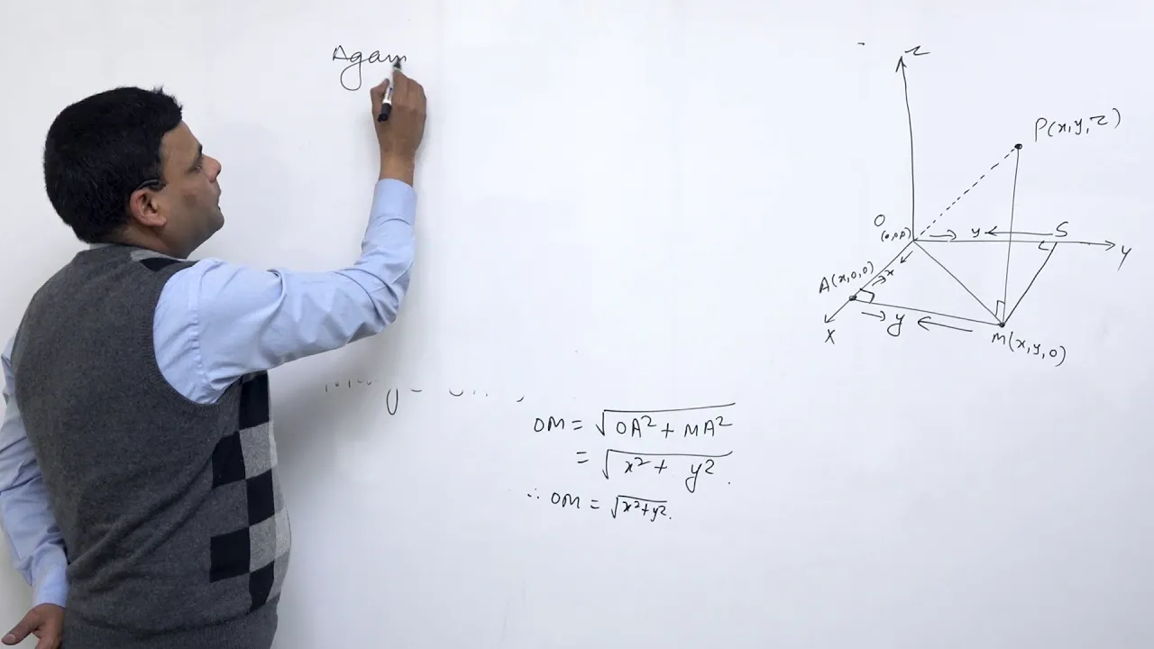

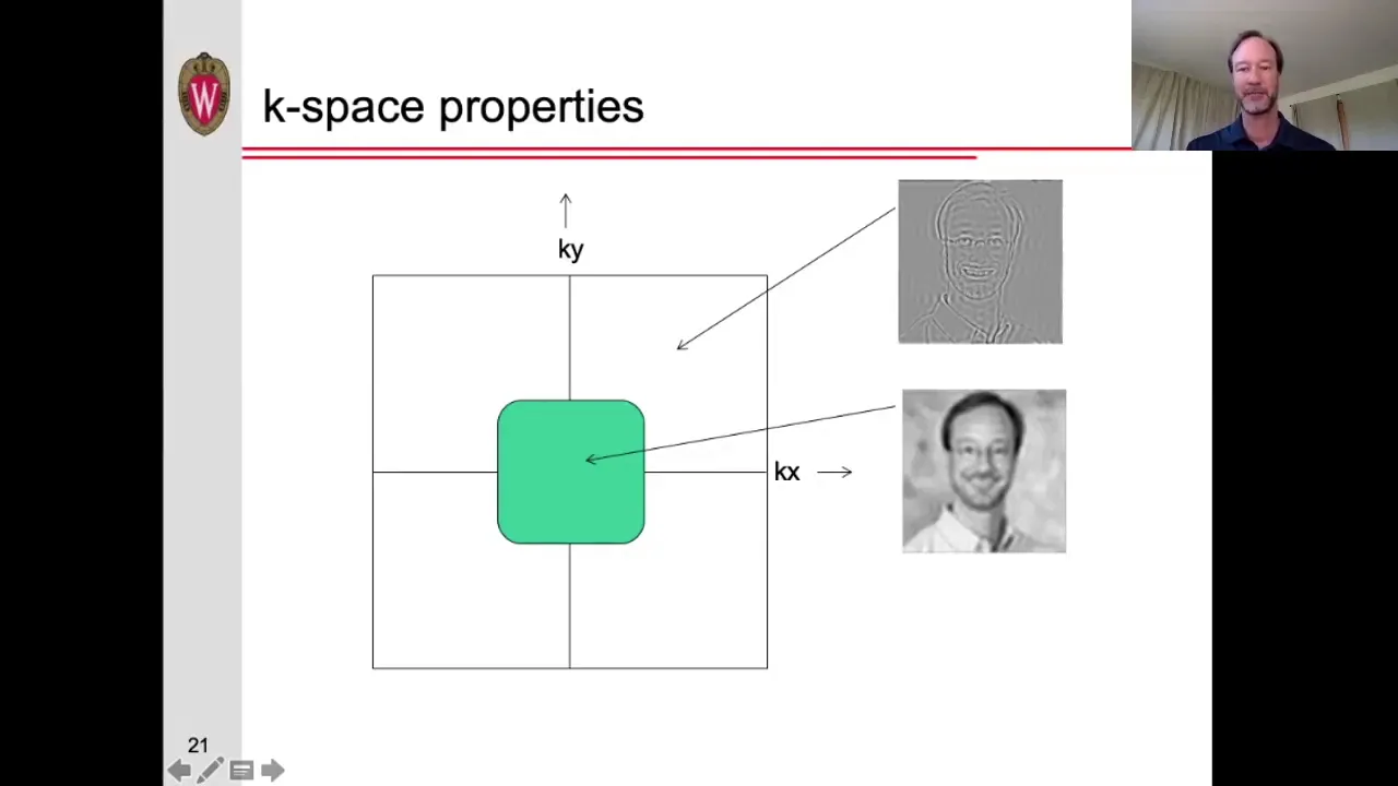

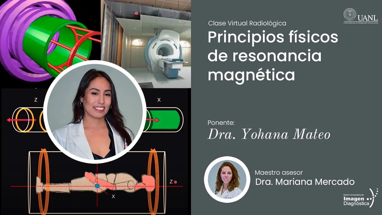











Magnetic Resonance Imaging (MRI) operates on the physics of Nuclear Magnetic Resonance (NMR), where hydrogen protons in the body act as tiny magnets that align with or against a strong external magnetic field; when exposed to radiofrequency pulses at their resonant frequency (Larmor frequency, approximately 42.5 MHz per Tesla), these protons absorb energy and flip from low-energy to high-energy states, creating transverse magnetization that generates detectable signals; the subsequent relaxation processes—T2 relaxation (spin-spin relaxation where protons lose phase coherence) and T1 relaxation (spin-lattice relaxation where energy dissipates as heat)—differ between tissues based on their molecular environments, enabling contrast generation through pulse sequences with specific repetition time (TR) and echo time (TE) parameters to produce T1-weighted, T2-weighted, or proton density-weighted images; spatial localization is achieved using magnetic field gradients along the x, y, and z axes combined with phase and frequency encoding to map signals to their precise anatomical locations within a matrix of voxels.
MRI Physics and a Brief History of Magnetic Resonance Imaging
Added:[Music] magnetic resonance imaging is based on the physics principle of nuclear magnetic resonance or NMR first described in the 1930s and 40s the basic principle of NMR deals with the interaction of certain Atomic nuclei radio frequency energy and a strong magnetic field this is a tough concept to grasp so for comprehension purposes we'll start with the basics first a few definitions radio frequency energy is part of the electromagnetic spectrum that includes visible light and x-rays as you can see the radio frequencies are at the far left of the spectrum visible light in the middle and x-rays to the right all of these waves are defined by a wavelength and frequency the wavelength is the distance between peaks of the wave and the frequency is how many cycles the wave completes every second the amplitude is the height or power of the wave when talking about two or more waves we can also describe a phase the blue and orange waves on the screen have the the same frequency and line up completely so they are also in Phase if we shift the orange wave to the right the frequency Remains the Same but the waves are now out of phase or have been phase shifted on the other hand these two waves have different frequencies but start at the same time so they are in phase for the purpose of NMR and MRI radio frequency signals will be described by their unique phase and frequency a point on this computer screen is called a pixel a concatenation of the words picture and element Mr images are made up of a series of voxal or volume elements each Square on the picture corresponds to a volume of tissue on the body the Mr machine is designed to measure the NMR signal from each of these small volumes localize them in 3D space and plot them on a 2 56x 256 or 512x 512 Matrix to make a visible picture with this in mind we'll first describe the principles of NMR in a single small square or voxal of [Music] tissue I suspect most of us have some recollection of constructing an electromagnet as a kid utilizing a common battery a switch an iron nail and a coil of wire throw the switch pick up some small steel balls and consider yourself a mad scientist great stuff what we unknowingly demonstrated with that little project was the basic principle of electromagnetism where a flowing current of electrons has an Associated magnetic field oriented 90° from the direction of current flow the battery pushes the electrons around around the wire we coil the wire to add up all those little magnetic moments and add the steel nail to conduct the magnetic field to the object we want to pick up the direction of the magnetic field associated with the coil is defined by the right hand rule curl the fingers of your right hand in the direction of the current flow through the coil your outstretched thumb will point in the direction of the associated magnetic field with that in mind let's look at the simplest of all elements hydrogen the hydrogen atom consists of a single proton in the nucleus with a single circulating electron orbiting around that proton for the principle of NMR we don't care much about the electron we're interested only in that single Central proton or the nucleus hence the descriptor nuclear in the acronym NMR that unopposed proton in the nucleus just doesn't sit statically in the center of the atom it actually rotates on its axis similar to the planets and [Music] stars because the proton is positively charged if we curl our fingers in the direction of rotation our thumb will point in the direction of the small magnetic field associated with each and every one of the hydrogen protons or spins in our body and fortunately for Imaging purposes we have a lot of hydrogen atoms in our bodies an adult is about 60% water which contains two hydrogen atoms for every oxygen the energy storing molecules of fat and carbohydrates also contain an abundance of hydrogen with glucose supporting 12 hydrogens on a carbon oxygen hexagonal [Music] backbone and the free fatty acid containing two to three hydrogens attached to every atom of the linear or Branch chain carbon backbone now while we're walking around the Earth all of these little proton magnets or spins are randomly oriented canceling each other out and therefore we're usually not magnetic however when we put any of us in a strong magnetic field such as the one found in a standard MRI machine all of these little spins or proton magnets line up most of them line up with the main magnetic field while a few line up directly opposite the main magnetic field and nothing in between what determines the orientation is the amount of energy associated with each of the individual atoms or protons the ones with a little extra energy possibly from some local increased heat will line up against the main magnetic field and therefore considered to be in a high energy State the ones lining up with the main magnetic field are in a low energy State these protons don't simply point with or against the main magnetic field they actually precess much like a spinning top as it falls to the solid surface the rate of precession can be termined exactly by the lore frequency equation which states that the rate of rotation is directly proportional to the strength of the local magnetic field at one Tesla the lore frequency of a hydrogen proton or spin is 42.5 MHz at two Tesla 8516 MHz and at three Tesla 127.71 MHz or basically 42.5 MHz per Tesla this will be important when we're talking about making an image from NMR data later on for demonstration purposes let's move all of our precessing protons to a common origin on a 3D graph as before most of the protons are in a low energy State pointing in the direction of the main magnetic field a few energetic protons are oriented against the main magnetic field in the high energy State even though each proton is precessing in space if you cancel all the opposing vectors you end up with a net magnetization pointing with the large main external magnetic field as demonstrated in the simplified diagram on the right this is called the longitudinal magnetization because it is in the same direction as the large main external magnetic field it cannot be measured or detected directly and is therefore inferred but we can change all that let's see what happens when we put energy into these spins or protons assuming our protons are sitting in a homogeneous one Tesla field the lore equation states that the precession rate of these spins is 42.5 MHz if we transmit a radio frequency pulse of exactly 42.5 MHz in the vicinity of the protons two things happen to make this a little easier to see we're going to stop the procession of the protons for a moment first the prot will absorb that energy and flip some of the spins into the higher energy state if we put in enough energy to push 50% of the proton population into the high state in our case four up and four down you can see our longitudinal magnetization reduces to zero as the opposing magnetic forces cancel each other out in addition the sinusoidal radio frequency pushes the protons to synchronize and spin together this is the resonance portion of NMR if we add up all the magnetic moments you can see that we now have a net magnetic force oriented horizontally or 90° to the longitudinal magnetization this is called the transverse magnetization and this magnetization can be detected with a coil or [Music] antenna just as a current can create a magnet a magnet can create a current if we have a coil of wire connected to an AM meter or current meter and we place a magnet through the coil we will generate an electrical current through the wire when we pull the magnet back the current flows in the opposite direction if we spin the magnet we generate a sinos soidal or alternating electrical current the basis of a generator similarly the transverse magnetization rotates around as the protons press and generates a small but measurable current in a regional coil of wire this is the result we're looking for in nuclear magnetic resonance but it's still not the whole story after we remove the radio frequency signal the protons will relax back into their Baseline position again for demonstration purposes will temporarily stop the procession the first thing that happens is the protons or spins being all positively charged will repel each other and move apart as they spread apart we lose that transverse magnetization this process is called the t2 or spin spin relaxation because it has to do with the interactions of the protons or spins themselves no net energy transfer occurs with this relaxation the other relaxation occurs as the high energy protons fall back into the low energy State as this happens the energy that was previously absorbed by the protons is dissipated into the surrounding tissues in the form of heat and thus involves an actual transfer of energy as these protons fall back down to Baseline we Reg grow the longitudinal magnetization this is referred to as the T1 or spin lattice relaxation because it involves the transfer of energy from the spins to the surrounding tissues or [Music] lattice putting this all together a sufficient radio frequency pulse tuned to the Natural precession frequency of the pressing protons is put into the tissues to flip 50% of the spins into the high energy State and cause the prot to synchronize in Phase or spin together thus moving the longitudinal magnetization 90° into the transverse plane the transverse magnetization pressing at the resonant frequency of the local protons produces a radio signal of the same frequency that can be detected by a coil of wires as the energy is removed the protons first move apart in a T2 or spin spin relaxation destroying the transverse magnetization and then through T1 one or spin lattice relaxation fall back into the lower energy State dissipating the previously absorbed energy into the surrounding tissues in the form of heat while regrowing or restoring the original longitudinal magnetization because these protons in our bodies have different local environments some associated with the free flowing water molecules While others are fixed in position associated with the structural or energy storing molecules of protein and fat they have characteristic differences in their T1 and T2 relaxation we can accentuate and measure these differences by changing how quickly we put in the radio frequency energy or the repetition time designated TR and how quickly we choose to listen to the signal coming back from the transverse magnetization or Echo time designated te of the pressing protons this process is referred to as the pul sequence and we will use a sample of fat and water to demonstrate these differences as before to simplify and maximize comprehension we're going to stop the procession of the protons on the left is a group of spins or protons associated with fat and on the right water when we put in our resonant radio frequency pulse all the protons absorb that energy flip into the high energy State and spin together to produce our 90° pulse or transverse magnetization if we wait a sufficient amount of time the protons will move apart in a T2 or spin spin relaxation and the transverse magnetization will Decay the protons associated with the free fatty acids being relatively fixed in position Decay rapidly as the spins push away from one another they also give up their absorbed energy more rapidly as they fall back to Baseline in a T1 or spin lattice relaxation depositing heat energy into the surrounding tissues and regrowing the longitudinal magnetization on the other hand the protons in the freely flowing water can hold on to their energy and continue to spin together in Phase thus maintaining the transverse magnetization at this point when we turn on our receiving coil and measure the signal coming back from the proton the relatively large transverse magnetization in water will give a strong signal while the smaller or absent transverse magnetization in fat will give a weak signal by convention the strong water signal will be assigned a grayscale color of white and the weak signal of fat will be dark gray or [Music] black hence to accentuate the different T2 relaxations of the protons in our bodies we would wait a long period between radial pulses referred to as a long repetition time TR and wait a long time to listen for the return signal or Echo referred to as a long Echo time te and these differences can be measured and recorded using these same tissues in their Baseline state to accentuate the differences in T1 relaxations we again put in our 90° resonant radio frequency pulse which flips the protons into the high energy State and pushes them in phase to produce our transverse [Music] magnetization T2 relaxation occurs as the protons move apart faster in fat than in water and then the protons fall back into the low energy State dissipating the absorbed energy as heat into the surrounding tissues and regrowing the longitudinal magnetization again because the protons in water are fluid and move freely they tend to hold on to that energy longer and the protons stay in the higher energy state with little regrowth of the longitudinal magnetization whereas the tightly packed fat protons more rapidly give up that energy and return to the low energy State thus rapidly regrowing the longitudinal magnetization if we then quickly put in another resonant 90° radio frequency pulse the fully recovered fat protons will produce a large transverse magnetization and a strong measured signal that we can Rec record if we listen to the return signal or Echo shortly after the second radio frequency pulse however the water protons are still in a high energy state with little regrowth of the longitudinal magnetization therefore the new radio frequency pulse flips more of the low energy protons into the high energy State and can only produce a small transverse magnetization as well as a net longitudinal magnetization oriented 180° or directly opposite the main magnetic field this configuration in the water protons will give a low amplitude or low energy wave when we listen for the echo or return signal in other words the protons in the water are saturated with energy and can no longer produce a strong transverse magnetization therefore to accentuate the differences in the T1 relaxations between the protons in fat and water we want to rapidly put in our resonant radio frequency pulses or a short repetition time TR and quickly listen for the return signal or short Echo time te now this is a difficult concept and you may have to listen to it a few times to get it to sink in but to summarize the T1 relaxation effects are accentuated by rapidly exposing the protons to radio frequency energy and keeping the spins in the high energy State and thus reducing the effect Ive longitudinal magnetization tissues that recover quickly will have a large signal while those slow to recover will have a low signal T2 effects on the other hand are accentuated by a prolonged Echo time to allow the spins to move away from each other and accentuate the differences in T2 relaxation between Regional tissues or chemicals so a T2 weighted image is obtained with a pulse sequence of a long TR and a long te decreasing the T1 effect the long TR and enhancing the t2 effects with the long [Music] te a T1 weighted image on the other hand is obtained with a pulse sequence of a short TR and a short te enhancing the T1 relaxation and minimizing the t2 effects in between these two is the proton density pulse sequence which is obtained with a long TR and a short te effectively minimizing the T T1 and T2 effects and basically giving us an idea of the absolute number or density of protons in the region the first who proposed the use of NMR to diagnose disease in humans was Dr Raymond dadian who in the March 1971 edition of the journal science published a short article entitled tumor detection by nuclear magnetic resonance in which he suggested that the NMR signal from tumors in the body could be differentiated from normal tissues and therefore could be detected his original concept was not to make an anatomic picture but to build a detector large enough to accommodate a human perform a whole body NMR and look for a characteristic T1 and T2 tumor signal that would suggest you have a malignancy somewhere in your body sort of a quick screening tool for malignancy [Music] while Dr dadian tumor detector never came to fruition we do use Mr spectroscopy in conjunction with a standard Mr image to help distinguish benign from malignant processes in the body to make an NMR picture or MRI as we know it today we have to be able to localize the signals coming from the sample or tissues in 3D space the first to propose a technique to do just that was Dr Paul Laur in the March 19 1973 edition of nature Laur published a brief article entitled image formation by induced local interactions examples employing nuclear magnetic resonance in the article he describes a technique using magnetic gradients to identify the location of two 1 mm capillary tubes filled with water both of which were submerged in a larger 4.2 mm tube filled with heavy water heavy water has the symbol d2o because the hydrogen atoms in heavy water are isotopes of the standard prodium configuration with one proton in the nucleus the dyum atom on the other hand contains a neutron as well as the proton which limits the spin of the nucleus and therefore does not give a good NMR signal lur placed his apparatus in the Highfield NMR magnet as shown and developed a technique to identify the actual origin of the NMR signals from the standard water this is the image produced very primitive by today's standards but clearly shows the location of the two capillary tubes of standard water Laur called his technique zgat ography from the Greek zukma which means that which is used for joining to show you how we make an actual Mr picture we're going to move our perspective of our spins from the side to the top and represent each grouping simply by the net magnetic field rotating around the [Music] axis remember that the lore or resonant frequency of our spins is determined by the strength of the local magnetic field when we first get into the MRI machine a superconducting magnet creates a near homogeneous magnetic field from one side to the other that determines the strength of the MRI machine common systems are 1 1.5 and 3 Tesla in strength there are three sets of gradient magnets in the MRI used to localize locations in 3D space the [Music] z-axis x-axis and Y AIS to select a particular slice of tissue in the body we can turn on the set of electromagnets along the z-axis that create a magnetic gradient from head to toe we now put in a radio pulse with a frequency that will cause the desired area to resonate as described earlier we have now selected our slice through the body because the local magnetic gradient is homogeneous all of these net magnetic moments in the slice are in Phase spinning together in sync and can't be distinguished from one another to further localize these magnetic moments and their signal strength we have two more gradients that we can use to isolate the source of the signals the first gradient is called the phas and c and gradient to demonstrate the effect we're going to slow down our net magnetic moments the phas and coding gradient is briefly turned on creating a gradient along the Y AIS in this particular case resulting in the magnetic moments at the bottom of the gradient to slow down and the ones at the top where the magnetic field is higher to speed up the gradient is quickly turned off and the spinning magnets return to the base frequency spinning at the same rate but they have now experienced a phase shift in the Y AIS which we can use to localize the spins in the y direction we then tune our system to focus on a particular phase in The Matrix and use our third gradient in the X direction to definitively localize each of these signals in the selected row this gradient again causes the spins to the right to slow down and the ones on the left to speed up this frequency encoding gradient remains on while the signals are recorded now each of the signals has a unique phase and frequency which can be localized in 3D space the whole process is repeated for each row localizing in this example in the Y Direction with the phase encoding gradient and in the X Direction with the frequency encoding gradient until the entire Matrix is complete [Music] [Music] each of these squares or voxal are assigned a grayscale value corresponding to the strength of the local signal by convention white being a strong signal and black being no signal at all in this simplified example our 4x4 Matrix doesn't look like much but a standard MRI with a 256x 256 or 512x 512 Matrix will provide Exquisite anatomic details of the body [Music]
Up Next
How MRI Works: Magnetic Resonance Imaging Explained Simply
@dr.paulinemoyaert
283.8K views•2022-09-28

21cm Hyperfine Transition in Neutral Hydrogen: Radio Astronomy Basics
@AaronRobertParsons
12.4K views•2011-10-13

NMR Spin Physics I: Zeeman Effect, Resonance Condition & Larmor Frequency
@nptel-indianinstituteofsci8064
2.3K views•2024-01-17

Entropy and the Second Law of Thermodynamics Explained
@veritasium
27.5M views•2023-07-01
Related Study Plans & Knowledge Roadmaps
Structured learning paths in Physics