
![Anatomical Position and Directional Terms [Anatomy MADE EASY]](https://i.ytimg.com/vi/t6-ueqFK1IE/maxresdefault.jpg)


![Localisation of Function in the Brain - Biological Psychology [AQA ALevel]](https://i.ytimg.com/vi_webp/Nu3fFB9rxKE/maxresdefault.webp)

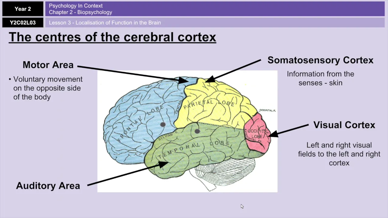













The human brain consists of the cerebrum (with two hemispheres containing the cerebral cortex divided into frontal, parietal, occipital, and temporal lobes), the cerebellum (responsible for motor control and coordination), and the brainstem (including midbrain, pons, and medulla oblongata). Key structures include the hippocampus (memory formation), amygdala (emotional processing), basal ganglia (movement regulation), thalamus (sensory relay), hypothalamus (autonomic control), and white matter tracts connecting different brain regions. The ventricular system (lateral, third, and fourth ventricles) circulates cerebrospinal fluid throughout the brain.
Human Brain Anatomy: 3D Animation of Structure & Function
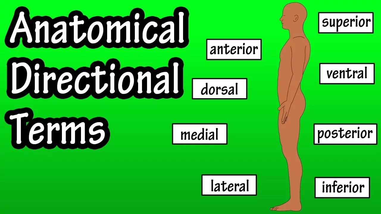
Added:Basic anatomical terminology for directional orientation (such as anterior/posterior, dorsal/ventral, medial/lateral) to navigate 3D space.
The fundamental division of the human nervous system into the Central Nervous System (CNS) and the Peripheral Nervous System (PNS).
The basic cellular units of the brain, specifically the general structure and function of neurons and glial cells.
The concept of biological localization of function, which posits that different physical regions of the brain control distinct bodily and cognitive functions.
The pathophysiology of neurological disorders, such as understanding how damage to the basal ganglia leads to Parkinson's disease, or how ventricular enlargement relates to hydrocephalus.
The study of neuroimaging modalities (like fMRI, PET, and CT scans) to see how structural anatomy correlates with real-time brain activity.
The biochemical pathways and major neurotransmitter systems (like dopamine and serotonin) that flow through and regulate these anatomical structures.
The principles of neuroplasticity, exploring how these anatomical regions adapt, reorganize, and form new connections in response to learning or injury.
21.4K views352likes35:00Original Release: 2021-01-27
welcome to the 3d biology youtube channel visit the3dbiology.com site linked in the description for articles animations and more so today we will be looking at detailed human brain anatomy in 3d the cerebral cortex is the outer gray matter of the syrup room and is made up of two hemispheres the two cerebral hemispheres consist of depressions or sulci and elevations or gyri it is partially separated by a deep longitudinal fissure the medial longitudinal fissure separates it into two hemispheres the cortex can be divided into the frontal parietal occipital and temporal lobe the cortex includes primary sensory areas which receive sensory information and association areas the frontal lobe includes bronchus area responsible for production of language and the temporal lobe includes vernicus area responsible for speech comprehension the frontal cortex as the name suggests can be found in the anterior of each cerebral hemisphere it is separated from the parietal lobe by the central surface and from the temporal lobe by the lateral surface the pre-motor as well as the primary motor cortex are found in the frontal cortex the prefrontal cortex can be found in the interior frontal lobe it is where executive function is carried out and high level filtering meaning acceptable behavior predicting outcomes determination of good and bad working toward a goal it receives connections from the brainstem arousal systems the middle frontal gyrus is found between the superior and the inferior frontal sulci it has a role in the reorienting of attention the inferior frontal gyrus a part of the prefrontal cortex is the location of rocus area responsible for speech creation and language processing the orbital gyrus can be found on the inferior surface of the frontal lobe this area of the frontal lobe rests on the orbital plate of the frontal bone there are four alveolar gyro the posterior the lateral the medial and the anterior the precentral gyrus is brodmann area 4 it is located at the rear of the frontal lobe just before the central surface it has a diagonal orientation and is continuous with the post-central gyrus behind it is the post-central gyrus it is the location of the primary motor cortex involved in the execution of voluntary motor movements and skeletal muscles the postcentral gyrus is located in the anterior parietal lobe it includes broadman areas 1 2 and 3 it is where the primary somatosensory cortex can be found it is the location of urines that integrate sensory information from distinct parts of the body in front of it is the central sulcus in the back of it is the post-central sulcus the supramarginal gyrus is located anterior to the angular gyrus it is part of the somatosensory association cortex it is involved in the interpretation of tactile sensory data the angular gyrus is in product area 39 it is posterior to the supermarginal tyrus in the parietal lobe and close to the superior edge of the temporal lobe it is involved in complex language functions such as reading writing and their interpretation it sends visual information to vernix area its other functions involve attention memory retrieval and spatial cognition including distinguishing between left and right the superior parietal lobule includes broadman area 5 and 7.
its functions include spatial orientation it receives visual input and sensory input the occipital lobe of the cervical cortex is named after the occipital bone and is the smallest lobe of the cortex it is located above the temporal and below the parietal lobe the occipital lobe includes the visual cortex mainly broadman area 17 or v1 it also includes v2 or the ventral stream which is a secondary visual cortex underneath the occipital lobe is the tentorium cerebelli which divides the cerebellum from the cerebrum the lateral occipital sulcus separates occipital gyri the superior temporal gyrus is located in the temporal lobe below the lateral surface it includes broadman area 41 and 42 and wernicke's area it is the location of the auditory cortex processing of sounds the middle temporal gyrus is located in the middle of the temporal lobe it has been linked to accessing word meaning while reading figuring out distance and recognition of known faces the inferior temporal gyrus is the lowest of the gyri of the temporal lobe it is separated from the middle temporal gyrus by the inferior temporal sulcus the occipital temporal sulcus separates it from the fusiform gyrus it is involved in visual stimulus processing and the level of object recognition based on form and color the white matter is found deep in the brain including the sedum and superficially in the spinal cord white matter is made up of axon bundles that connect gray matter areas within the white matter i can find gray matter nuclei like brainstem nuclei and the basal ganglia the name comes from the color of fatty myelinated axons the cingulate cortex includes the whole singular gyrus and can be found immediately in the cerebral hemispheres above the corpus callosum it is part of the limbic system it functions both in respiratory control executive function as well as learning memory and emotions the ceredrum or tonsefon is the largest part of the brain it contains not only the two cerebral hemispheres made up of an outer cortex of gray matter and an inner white matter but also subcortical structures such as the basal ganglia the hippocampus and the olfactory model the cerebrum is involved in voluntary motor actions sensory perception memory and thoughts a cerebrum develops from the present cephalon the third bone is located in the posterior underneath the cervical hemispheres the pons and the medulla are anterior to it the actual name means little brain it is made up of a gray matter cortex deeper white matter with myelinated fibers deep gray matter cerebellar nuclei inside the white matter and a fluid-filled ventricle the cerebellum is made up of two hemispheres and a protruding midline thermos or worm in latin the white matter is called the arbor vitae or tree of life due to its appearance one section the cortex is made up of a continuous layer of tissue folded repeatedly although it appears as parallel grooves on the surface the gyri of the cerebellum are called folia the lateral cerebellum is called the cerebral cerebellum and the medial cerebellum is called the spinosaur bone the cerebellum is divided into three lobes the anterior above the primary fissure the posterior below the primary fissure and the floccular nodular below the posterior fissure a cerebellum is associated with movement including motor learning timing coordination and precision as well as language and attention the cerebellum receives input from the sensory systems of the spinal cord neurons most commonly found in cerebellum include the purkinje and granule cells there are three important action types in the cerebellum mostly fibers farming fibers and parallel fibers three pairs of cerebellar peduncles connect the cerebellum to the nervous system the superior cerebellar peduncle connects to the cerebral cortex the middle cerebellar peduncle connects to the bones the inferior cerebellar peduncle has output to the reticular formation and vestibular nuclei the medulla oblongata is part of the brain stem medullary pyramids contain the corticospinal and cortical bulbar tracts the swellings or olives are due to the inferior alveolar nuclei the medulla connects higher brain levels to the spinal cord the medulla functions in involuntary actions its autonomic control includes heart rate respiration and sleep the word palms comes from the latin for bridge it is in the brain stem between the midbrain and the medulla within it are tracts that carry sensory signals up and other signals down in the rear it is made up of two pairs of cerebellar peduncles the middle cerebellar peduncle connects the pons to the cerebellum it is located in front of the cerebellum it has two major divisions the ventral pons and the momentum the pons contain several cranial nerve nuclei including the vestibular cochlear nucleus the facial nerve nucleus the nucleus of duties and the motor nucleus of the trigeminal nerve it is part of various autonomic functions such as arousal sleep regulation equilibrium and muscle tone the hippocampus or seahorse in latin is part of the limbic system and can be found as a pair one on each side by the floor of the lateral ventricle the para hippocampal gyrus conceals the hippocampus it is made up of the hippocampus itself and the dentist gyrus which is surrounded by hippocampal gray matter the name corner armonis or ram's horn is given to it because of its shape as shown in cross-section the abbreviation ca given to its parts including ca1 ca2 ca3 and ca4 come from this name the hippocampus takes part in storing of explicit memory such as information about objects places and people this is done through long-term potentiation hippocampal damage is seen in dementia such as alzheimer's disease it is also affected in schizophrenia and ptsd the three major pathways in the hippocampus include the muscle fiber pathway the schaefer collateral pathway and the perforin fiber pathway input comes from the internal cortex outputs include the internal cortex and the prefrontal cortex it is modulated by dopamine serotonin and norepinephrine the hippocampus is one of the few brain regions where new nerve cells are born the putamen is a round structure that is part of the basal ganglia together with the code nucleus it forms the dorsal striatum to the medial of it lies the globus pallidus it functions in regulation of movement and is connected to the globus pallidus and substantia the putamen is affected in parkinson's disease where involuntary muscle movements occur together with the globus pallidus it is known as the lentiform nucleus as they appear like a lens like shape the global spiders which is part of the basal ganglia translates into pale globe there are two parts the parse externa and the parse internet it projects mainly to the substantia and thalamus and functions in regulation of voluntary movement through inhibitory action whereas the cerebellum provides excitatory action it receives inputs from the caudate and the putamen the v-shaped internal capsule is a white matter structure that separates the cardiac nucleus and putamen and also the caudate and thalamus from the putamen and globus powders it contains ascending and descending tracts connecting the cortex the band in the v of the internal capsule is called the genome it has an anterior limb and a posterior limb much of the internal capsule is the corticospinal tract which carries information from the primary motor cortex to motor neurons in the spinal cord the caudate nucleus along with the putamen form the dorsal striatum which is divided by the internal capsule in each hemisphere it forms a thick anterior part called the head which tapers to a body and a narrow tail giving it a c-shape the tail curves toward the front it is located on both sides of the thalamus it functions in associative and procedural learning inhibitory control of action and movement there is a link between the caudate and sleep patterns movement functions include posture as well as speed of directed movements neuronal connections from the caudate reach the substantia on globus paridius and it is innervated by neurons from the substantia the caudate has projections to the hippocampus and receives projections from the amygdala there are data showing effects on emotion hyperactivity and drive [Music] the pair of amygdala deep in the temporal lobes are part of the limbic system their almond shape the amygdala are made up of the basolateral complex the medial nucleus the cortical nucleus the central nucleus and the incarcerated cell clusters the amygdala is larger in males the amygdala play a role in emotional responses decision making and processing of memory this involves memories associated with emotional events each amygdala has an independent memory system the right amygdala is concerned with fear conditioning and declarative memory as well as in associating time and place with emotions the amygdala are involved in memory consolidation they are the main structure involved in the fight or flight response projections of the amygdala include the locus cerebellus the hypothalamus the nucleus accumbens and the thalamus there is data showing that the left amygdala has a role in the reward system it has also been linked to obsessive and compulsive behavior anxiety disorders and ptsd the left amygdala develops first while the right amygdala grows for a longer span of time the thalamus is a great matter structure in the forebrain it is found medially the thalamus is a hub for information relay it relays sensory signals regulation alertness arousal sleep and consciousness every sensory system utilizes the thalamus except for the olfactory system it is connected to the cortex by many thalamocortical radiations the hypothalamus is part of the limbic system and is found under the thalamus the name hypothalamus means under chamber the hypothalamus links the nervous system and the endocrine system via the pituitary therefore it has a neuroendocrine function it works to control sleep hunger body temperature and activities of the autonomic nervous system the hypothalamus synthesizes and secretes hormones which control the pituitary gland its connections include the reticular formation the brain stem the amygdala and the septum other functions of the hypothalamus include controlling defensive behavior the midbrain is the anterior part of the brainstem the dorsal side of the midbrain is the tacum which means roof the tactum has four colliculi or bumps on its surface the tegmentum is the floor and is eventual to the circle aqueduct it is involved in homeostasis superior colliculi process visual information and inferior follicular process auditory information it functions alertness sleep temperature regulation hearing and vision the superior colliculus can be found on top of the midbrain and is a paired structure it is a multi-sensory structure some of its functions include directing eye movement head turns and shifts and attention the inferior colliculus is located over the trochlear nerve and below the superior colliculus it is a large main nucleus of the auditory pathway in the midbrain where auditory pathways converge it also functions in spatial localization through hearing as well as an integration station and switchboard its inputs include the auditory cortex and brainstem nuclei the name inferior colliculus means lower hill inputs include several brainstem nuclei the lateral geniculate is a small thalamic nucleus located at the end of the optic tract it is a visual pathway relay center information from the eye and optic tract goes through the lateral geniculate on its way to the primary visual cortex the lateral geniculate has six layers of neurons the medial geniculate body is made up of several nuclei it is part of the auditory system the medial geniculate is a relay between the inferior colliculus and the auditory cortex the pineal is a small endocrine gland which is not paired the name comes from the shape of the gland which looks like a small pine cone it produces melatonin a serotonin-derived hormone which modulates sleep cycles the pineal can be found where two of the halves of the thalamus meet behind the third ventricle and is part of the epithalamus the pituitary is an endocrine gland located at the bottom of the hypothalamus hormones secreted here control blood pressure temperature regulation sex organ function thyroid function pain relief and water regulation the posterior pituitary is connected to the hypothalamus by the pituitary star the pituitary has an anterior intermediate and posterior lobe the posterior or epitheliamic commissure is a white matter tract which connects the two hemispheres of the cerebrum it crosses the midline dorsally of the cerebral aqueduct it forms one of the stalks that attach the pineal gland to the wall of the third ventricle the posterior commissure connects language processing centers of the two hemispheres the anterior commissure is a white matter tract much smaller than the corpus callosum it connects the two temporal lobes of the cerebrum in front of the fornix it is important in pain sensation and olfaction as it includes fibers from the neo-spinal thalamic tract and olfactory tracts there are two c-shaped cerebral spinal fluid-filled lateral ventricles they stretch from the inferior horn of the temporal lobe all the way down to the third ventricle they can be divided into three horns and a body the frontal horn can be found in the frontal lobe of the brain while the occipital horn enters the occipital lobe the lateral ventricles are the largest of all the ventricles the third ventricle is filled with cervical spinal fluid and is found between the two halves of the thalamus it is narrow the epithalamus is behind it the cerebral aqueduct is found central to the cerebellum and dorsal to the pons also called the sylvian aqueduct it contains csf and connects the third and fourth ventricles the gray matter around the aqueduct is called the periaqueductal gray the fourth ventricle carries cerebral spinal fluid and can be found extending from the aqueduct of silvius to the albex in the coral medulla it is at the level of the pons the cerebral peduncles form the sides of the fourth ventricle the rhomboid fossa is the floor of this ventricle the fourth ventricle has a diamond shape and cross-section csf flows into the fourth ventricle through the cerebral aqueduct the central canal runs through the spinal cord it is filled with cerebrospinal fluid and it transports nutrients to the spinal cord in the upper regions of the spinal cord it can be found in the anterior portion later becoming more central and ultimately posterior the widest part of the central canal is the terminal or fifth ventricle the corpus callosum which translates into tough body is a thick nerve tract that connects the two cervical hemispheres it can be found below the cervical cortex crossing the midline the corpus callosum is the largest white matter body with several hundred million axons it can be divided into the restroom the general the body and the splenium the corpus callosum allows for communication between the two hemispheres of the cerebrum the septum pellucidum is a membrane between the two cerebral hemispheres it separates lateral ventricles and encloses the fifth ventricle it stretches from the corpus callosum to the fornix the fornix is a part of the limbic system and is an output of the hippocampus it starts on each side of the hippocampus as fimbriae hippocampi and continues as the acrora or posterior pillars at the commissure of fornix the fibers meet forming the body around the anterior commissure the body divides again into the anterior pillars or columns which continue onto the mammillary bodies the fornix is important in establishing episodic memories and fornix damage leads to problems with spatial memory thanks for watching for licensing inquiries or custom work email contact 3dbiology.com don't forget to subscribe and turn on notifications to see new videos as they are released disclaimer the information on this channel is not intended or implied to be a substitute for professional medical advice diagnosis or treatment all content including text graphics images and information is for information purposes only you
Up Next
5:35
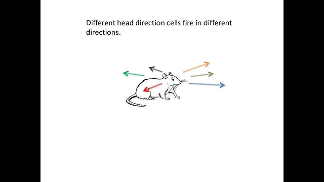
Head Direction Cells: Neural Compass in Mammalian Brains
@spatialcognition7071
3.9K views•2016-01-27

21:55
Deciphering Neural Circuits Controlling Anorexia in Mice | Richard Palmiter
@AllenInstitute
2.3K views•2011-10-10

4:10
Vagus Nerve (CN X): Anatomy, Nuclei & Functions Explained
@Alilamedicalmedia
305.2K views•2022-10-31

13:03
How Exercise Benefits Your Brain: Science Explained
@TED
11.4M views•2018-03-21
Related Study Plans & Knowledge Roadmaps
Structured learning paths in Neuroscience
neuroscience
Appetite Regulation: Brain Circuits & Hormones
01Foundations of Endocrinology and Brain Anatomy
02Introduction to Energy Homeostasis
03Peripheral Signals: Leptin, Ghrelin, and Insulin
+ 2 more learning modules
14h5 modules
Explore Roadmap
neuroscience
Neuroeconomics: Value, Risk & Brain Mechanisms
01Foundations of Neuroscience and Brain Anatomy
02Introduction to Neuroeconomics and Subjective Value
03Dopaminergic Prediction Errors & Reinforcement Learning
+ 3 more learning modules
18h6 modules
Explore Roadmap
neuroscience
Spatial Navigation: Cells, Grids & Brain Maps
01Foundations of Brain Anatomy and Memory
02Place Cells and Head-Direction Cells
03Grid Cells: The Brain's Coordinate System
+ 2 more learning modules
12h5 modules
Explore Roadmap
neuroscience
BCIs: Neural Decoding & Prosthetic Control
01Neuroscience Foundations: How the Brain Signals Intent
02Signal Acquisition: Recording the Brain's Activity
03Neural Decoding: Translating Brain Waves into Data
+ 2 more learning modules
25h5 modules
Explore Roadmap