The MTT assay is a colorimetric method for measuring cell viability, where metabolically active cells reduce MTT reagent into a purple formazan precipitate; the protocol involves vacuuming media, adding MTT reagent, incubating for 1-2 hours, solubilizing the precipitate with DMSO, mixing on a plate shaker for 10-20 minutes, and measuring absorbance with a plate reader to quantify cell survival.
MTT Cell Viability Assay Protocol: Day 3 Cell Block Analysis
Added:Deep Dive
Understanding the biological principle of the MTT assay, specifically how mitochondrial dehydrogenases in living cells reduce the yellow MTT tetrazolium salt into purple formazan crystals.

Mtt Assay
151.7K
The MTT assay is a colorimetric method to assess cell viability by measuring the reduction of the yellow tetrazolium dye (MTT) to purple formazan by NADPH-dependent oxidoreductase enzymes in viable cells' mitochondria; the intensity of the purple color indicates the number of living cells, enabling both qualitative and quantitative analysis of cellular health.
Click to watch →

In vitro Anti cancer Assay: Dr. Bhushan P Pimple
4.3K
The MTT assay measures cell viability through reduction of yellow tetrazolium salt (MTT) to purple formazan by mitochondrial enzymes (NADH/NADPH). Only viable cells with active mitochondria can convert the yellow MTT to purple formazan. Dead cells maintain yellow color because their mitochondrial enzymes are inactive. After incubation, detergent lysates cells, and intracellular formazan is solubilized and measured spectrophotometrically at 550-600 nm. Purple color indicates low cytotoxicity (many living cells), while yellow color indicates high cytotoxicity (cells killed by the test compound).
Click to watch →

Cómo estudiar la toxicidad celular | MEDNIGHT 2020 | Ana Juan García (COAL)
6.7K
The MTT assay measures cell viability by detecting the reduction of yellow tetrazolium salt (MTT) to purple formazan by mitochondrial succinate dehydrogenase in living cells; lower color intensity indicates higher cytotoxicity, enabling determination of the inhibitory concentration 50 (IC50) through spectrophotometric analysis at 570 nm.
Click to watch →

MTT Assay Made Simple: How to Measure Cell Viability?
373
The MTT assay is a widely-used calorimetric method that measures cell metabolic activity by detecting the conversion of yellow MTT tetrazolium salt into purple formazan crystals by living cells; this color change occurs through mitochondrial enzymes and indicates cell viability, making it valuable for drug screening, toxicology studies, and cell biology research despite its limitations including interference from colored compounds and cell-type variability.
Click to watch →

MTT assay | Principle | How does it work | Rapid fire Q.24
1.7K
The MTT assay is a colorimetric technique used to assess cell viability by measuring metabolic activity; healthy cells generate NADH through glycolysis, Krebs cycle, and oxidative phosphorylation, which reduces yellow MTT dye to purple formazan crystals via oxidoreductase enzymes, and the intensity of purple color (measured at 540nm absorbance) indicates cell health, with more intense color reflecting higher metabolic activity and healthier cells.
Click to watch →
Deep Dive
Familiarity with basic cell culture techniques, including cell seeding, cell density optimization, and aseptic techniques used on Days 1 and 2 of the protocol.
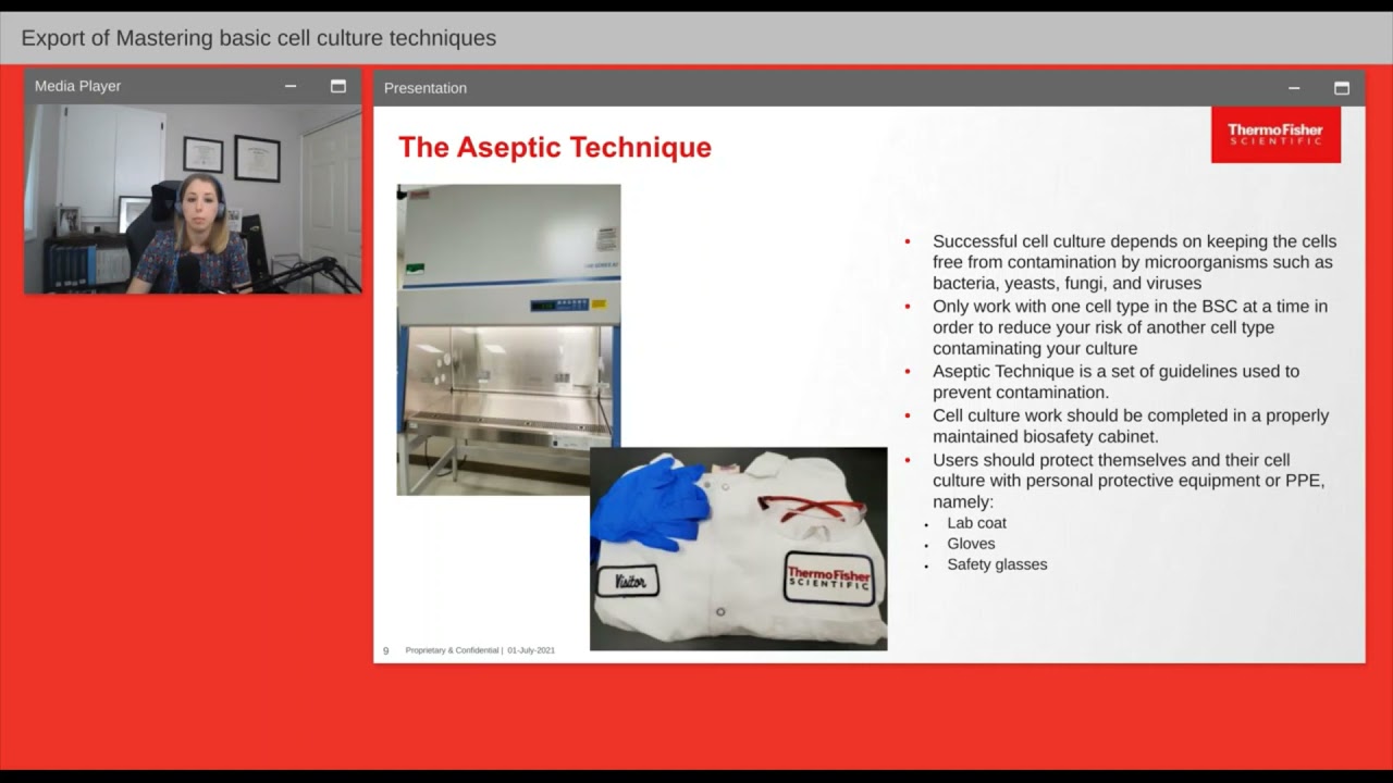
Mastering basic cell culture techniques
21.6K
Cell culture is the process of maintaining plant and animal cells alive and dividing in vitro, requiring specific tools including complete cell culture media (basal media plus supplements like serum and glutamine), appropriate culture vessels, and an incubator. Key techniques include subculturing (passaging) cells at optimal confluency (70-80% for adherent cells), practicing strict aseptic technique to prevent contamination, and regularly monitoring cell health through microscopy and media observation. Different cell types (adherent vs. suspension) require different handling protocols, and maintaining genetic stability requires cryopreserving original stocks to prevent drift over multiple passages.
Click to watch →
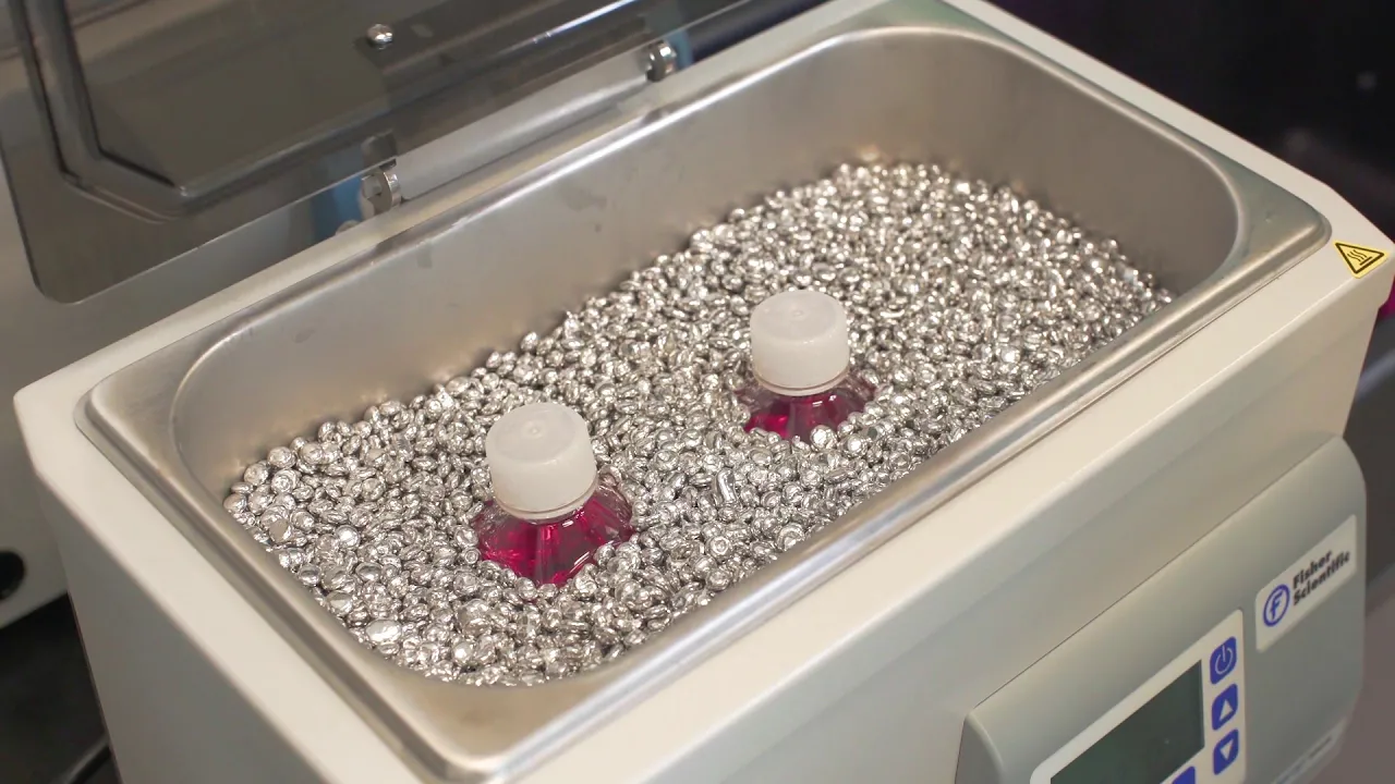
Aseptic Technique Training – Essential Cell Culture Video
457.2K
Successful cell culture requires proper laboratory setup with essential equipment including a cell culture hood, incubator, water bath, centrifuge, refrigerator/freezer, hemocytometer, and microscope; maintaining a sterile environment involves opening all materials within the hood, wiping surfaces with 70% ethanol, organizing items systematically (pipettes in front, reagents at back, waste containers in corners), and avoiding breathing or talking into the hood; mammalian cells should be cultured at 37°C with 5-7% CO2 and high humidity, with flasks evenly spaced for proper gas exchange; reagents should be warmed to 37°C for 10-20 minutes but not left excessive time to prevent degradation; daily microscopic examination is essential for monitoring cell health and detecting contamination.
Click to watch →

Cell Tissue Culture: Biosafety & Risk Factors by Prof. Mohamed Labib Salem,, Tanta University, Egypt
139
Proper cell culture workstation organization is essential for maintaining sterility and efficiency: work systematically from left to right, keep materials organized within easy reach, never reach across the work area, and keep the hood door closed when not actively working. Cell seeding density is critical for successful culture: fast-growing cells require higher densities (10,000 cells per well) while slow-growing cells need lower densities (1,000 cells per well). Proper seeding density ensures adequate cell attachment and growth without overcrowding.
Click to watch →
![Cell & Tissue Culture [Part 1]: The Basics](https://i.ytimg.com/vi/6HfRlAmtc6I/maxresdefault.jpg)
Cell & Tissue Culture [Part 1]: The Basics
3.2K
Cell culture provides a reproducible method for testing drugs, vaccines, and treatments by isolating cells from organisms and creating controlled artificial conditions; successful cell culture requires strict contamination prevention through proper aseptic technique including 70% ethanol sterilization, safety hood usage, and regular UV irradiation, followed by careful cell handling processes such as resuscitating frozen cells by thawing in a 37°C water bath and washing with PBS, then subculturing cells when they reach approximately 80% confluency by detaching them with trypsin-EDTA and transferring them to fresh media at appropriate seeding densities to maintain healthy, proliferating cell populations for experimentation.
Click to watch →

SWdB trainings: 2D HepaRG cell culture basics (part 1)
430
Calculate seeding volume using the formula: desired cell count ÷ cell concentration = volume needed. For example, to seed 0.5 million cells from a 1.2 million cells/mL suspension, take 0.42 mL. Add sufficient fresh medium to reach the desired total volume (e.g., 5 mL total = 0.42 mL cells + 4.58 mL medium). Distribute cells evenly using a 'battering' motion: three times down, three times up, three times left-right, three times right-left. Incubate at 37°C with 5% CO2. Do not move cells for several hours post-seeding to allow proper attachment. Work quickly when opening the incubator as temperature and CO2 levels drop rapidly.
Click to watch →
Deep Dive
Knowledge of spectrophotometry and how a microplate reader functions to measure optical density (absorbance) at specific wavelengths (typically 570 nm).

Spectrophotometers, calibration curves and Beer's Law
113.4K
Spectrophotometers measure light absorption by passing a beam of incident light through a sample in a cuvette. The machine compares incoming light intensity (I₀) with transmitted light intensity (I). When molecules absorb light, less light reaches the detector. Absorbance (A) is calculated using the logarithmic formula A = log₁₀(I₀/I). For instance, if 100% of light enters and only 1% exits, absorbance equals 2 because 10² = 100. This logarithmic relationship means absorbance increases slowly with increasing light absorption. The machine performs these calculations automatically, displaying the absorbance value directly.
Click to watch →
![Microplate Reader for Absorbance Measurement [Surface and Colloid Science]](https://i.ytimg.com/vi/dkyKbRBi3dg/maxresdefault.jpg)
Microplate Reader for Absorbance Measurement [Surface and Colloid Science]
539
This video explains how microplate readers measure absorbance using the Beer-Lambert Law (A = εbc), where absorbance (A) equals the product of the extinction coefficient (ε), path length (b), and concentration (c); the practical protocol involves preparing 96-well plates with samples and blank controls, using reverse pipetting to avoid air bubbles, configuring Gen 5 software to set wavelengths and plate layouts, and generating blank-subtracted absorbance data for applications like determining critical micelle concentration (CMC) or measuring turbidity without requiring calibration curves.
Click to watch →

Azure Biosystems Webinar: ELISA on Ao Abosrbance Microplate Reader: Basics & Applications
851
Absorbance microplate readers measure optical density by passing light through samples and detecting absorbed wavelengths at specific positions. Key components include lamps, filters, and detectors enabling flexible kinetic assays. Standard wavelengths cover ELISA applications (405-630nm), protein assays (562-595nm), MTT/XTT cell viability (490-570nm), and nucleic acid quantification (260nm). Advanced features include dual-mode reading with reference subtraction, built-in shakers, single/dual-channel detection, and eight-position filter wheels. Data analysis encompasses pre-processing operations and kinetic parameters: average rate, maximum rate, time to peak, and absorbance kinetics. Practical applications demonstrate the technology's versatility: histamine detection in food samples uses competitive ELISA where HRP-labeled conjugates compete with sample histamine for antibody binding sites, measured at 450nm reference 630nm. In antibiotic resistance research, microdilution methods determine minimum inhibitory concentrations using MTT-based viability assays measured at 570nm. These examples illustrate how standardized ELISA protocols enable quantitative analysis across food safety, clinical microbiology, and environmental health research domains.
Click to watch →

spctrophotometry | optical density | sabar khan lectures
4.4K
Spectrophotometry is a technique that measures optical density (OD), defined as the ratio of incident light intensity to transmitted light intensity (OD = I_incident/I_transmitted), which indicates a substance's ability to absorb or resist light; according to Beer-Lambert's law, cell mass and solute concentration are directly proportional to optical density, meaning higher concentrations absorb more light and produce higher OD values, and the spectrophotometer instrument uses a light source, collimator, monochromator with prism, wavelength selector, sample compartment, detector, and display to measure these properties.
Click to watch →
MTT Assay Protocol: Cell Viability & Proliferation Guide
120.4K
Measure dissolved formazan using a microplate reader set to absorbance wavelengths of 550-600 nm. This wavelength range captures the purple color intensity accurately. Ensure the microplate reader is properly calibrated and configured for the specific plate type being used.
Click to watch →
Deep Dive
Awareness of safety protocols regarding handling chemical reagents like MTT and solubilization solutions (such as DMSO or acidified isopropanol).
MTT Assay Protocol: Cell Viability & Proliferation Guide
120.4K
The MTT assay measures cell viability and proliferation by detecting metabolic activity; viable cells reduce the yellow MTT reagent to purple formazan crystals, which are dissolved in DMSO and quantified by absorbance at 550-600 nm, with percent viability calculated by normalizing experimental values against positive and negative controls using the formula: [(Experimental - Background)/(Control - Background)] × 100%.
Click to watch →

SOSIALISASI PETUNJUK TEKNIS KEGIATAN PENDIDIKAN DAN PENELITIAN DI LAB TEKNOLOGI REKAYASA PANGAN
233
Laboratory rules include mandatory obligations (enter 5 minutes early, wear laboratory coat, follow instructions, use equipment properly, borrow tools from staff, report damage immediately, clean after use, dispose trash properly, check laboratory condition before leaving, notify in writing if unable to attend) and prohibited actions (no smoking/eating/drinking, no open shoes, no joking around, no jewelry, no touching equipment without permission). Safety protocols include studying hazard labels, washing hands before/after handling chemicals, wearing masks and gloves, tying back long hair, handling glass equipment carefully, using appropriate tools, rinsing with water if chemicals contact body, contacting staff for injuries. Chemical handling procedures include working in fume hoods for hazardous chemicals, not mixing strong acids/bases without knowing reactions, pouring only needed amounts, not returning chemicals to original bottles, diluting concentrated solutions before discarding, wafting chemicals instead of smelling directly, directing tube mouths away from face when heating, and adding chemicals drop by drop through tube sides.
Click to watch →

Forbidden Juice You Should Never Drink - Safety Third #23
260.4K
Chemical solvents like DMSO can facilitate the absorption of other substances through the skin, creating serious safety hazards. The hosts explain that DMSO is a polar solvent that can carry other chemicals through the skin into the bloodstream, potentially causing serious health problems. They share personal experiences of working with solvents and emphasizing the importance of proper protective equipment when handling chemical solvents. The hosts recommend that anyone working with chemical solvents should always wear appropriate protective equipment and understand the specific hazards associated with each solvent.
Click to watch →

مكتسبات قبلية 3 : زجاجيات وسائل المخبر ، إحتياطات أمنية ، بروتوكولات التمديد والتخفيف
310
This section covers essential laboratory safety protocols and solution preparation procedures. Three critical safety measures include wearing lab coats to protect clothing and skin from chemical spills and hot liquids, wearing gloves to protect hands from harmful chemicals and hot surfaces, and wearing safety goggles to protect eyes from chemical splashes and flying debris. For preparing solutions from solids: weigh the required mass using an electronic balance, transfer to a volumetric flask using a funnel, add distilled water to dissolve, fill to the calibration mark, stopper and invert to mix, then label with chemical formula and molar concentration. For dilution: use a volumetric pipette to measure the concentrated solution volume, transfer to a new volumetric flask, add distilled water to mix, fill to the calibration mark, stopper and invert to mix, then label with the diluted solution's formula and concentration.
Click to watch →

1. Introductory Lecture to 5.310
47.1K
Air-sensitive chemicals require immediate covering after use; leaving them exposed causes degradation (e.g., iron(II) chloride oxidizes to brown iron(III) chloride). Always use TA-prepared stock solutions in labeled beakers rather than direct pipetting from stock containers to prevent contamination. When adding chemicals, always add concentrated solutions to less concentrated ones—never add water to acid, which causes dangerous splashing.
Click to watch →
Prerequisite Knowledge
- Concept 01Understanding the biological principle of the MTT assay, specifically how mitochondrial dehydrogenases in living cells reduce the yellow MTT tetrazolium salt into purple formazan crystals.
- Concept 02Familiarity with basic cell culture techniques, including cell seeding, cell density optimization, and aseptic techniques used on Days 1 and 2 of the protocol.
- Concept 03Knowledge of spectrophotometry and how a microplate reader functions to measure optical density (absorbance) at specific wavelengths (typically 570 nm).
- Concept 04Awareness of safety protocols regarding handling chemical reagents like MTT and solubilization solutions (such as DMSO or acidified isopropanol).
Subsequent Learning
- Step 01Data analysis techniques, including how to calculate percentage cell viability, subtract background absorbance, and plot dose-response curves to determine IC50 values.
- Step 02Troubleshooting common issues in MTT assays, such as uneven seeding (edge effects), chemical interference from test compounds, and incomplete dissolution of formazan crystals.
- Step 03Comparison of the MTT assay with alternative cell viability and cytotoxicity assays, such as CCK-8, Alamar Blue, LDH release, and ATP luminescence assays, to understand when to use each.
- Step 04Application of cell viability data in preclinical drug discovery, toxicology screening, and evaluating the efficacy of cancer therapeutics.
Add MTT reagent
Solubilize formazan
Read and compare
Add MTT reagent
0:00Playing Section
- 1
Aspirate media from each well and add plain media with MTT reagent.
- 2
Use one pipette for all samples, dispensing carefully to avoid dislodging cells.
Limitations of Metabolic-Based Viability Assays and the Rise of Direct or Real-Time Alternatives
While the MTT assay is a classic, cost-effective standard for assessing cell viability, it has significant technical limitations. First, MTT measures mitochondrial metabolic activity rather than direct cell death or membrane integrity; treatments that alter cell metabolism without killing the cells can produce misleading results. Second, the assay is endpoint-only and destructive, as it requires dissolving the cells in DMSO to solubilize the formazan crystals, preventing any downstream analysis or real-time kinetic monitoring. Critics and modern researchers often advocate for alternative methods, such as Alamar Blue (resazurin-based) assays, which are non-destructive and allow for continuous monitoring, or direct cell-counting and membrane-integrity assays (like Trypan Blue or Flow Cytometry) to obtain a more accurate measure of true viability.
Deep Dive
Data analysis techniques, including how to calculate percentage cell viability, subtract background absorbance, and plot dose-response curves to determine IC50 values.
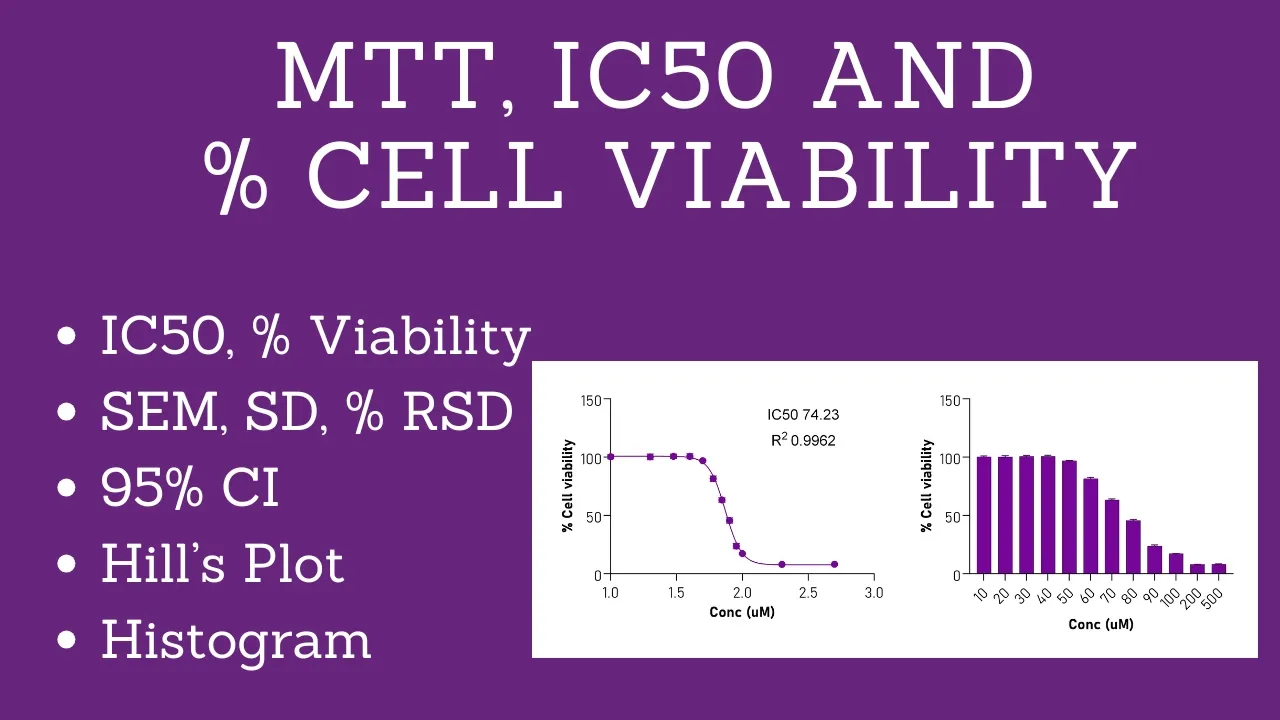
Complete MTT Assay Analysis | How to Calculate IC50, % Cell Viability, Dose-response curve | Prism
12.8K
This tutorial demonstrates the complete workflow for analyzing MTT assay data to calculate IC50 values and generate publication-quality dose-response curves. The process involves: (1) calculating corrected absorbance values by subtracting 630nm from 570nm readings, (2) computing percent cell viability relative to control, (3) determining statistical parameters including standard deviation, percent RSD, and standard error of mean, (4) plotting sigmoidal curves using nonlinear regression in GraphPad Prism with log-transformed X-axis, (5) generating bar graphs for visual comparison, and (6) exporting publication-ready figures. Key quality indicators include percent RSD below 10% for acceptable data and IC50 values obtained through dose-response inhibition curve fitting.
Click to watch →
MTT Assay Protocol: Cell Viability & Proliferation Guide
120.4K
Calculate percent viability using the formula: [(Experimental mean - Negative control mean) / (Positive control mean - Negative control mean)] × 100. Subtract background (negative control) from each experimental well, then divide by positive control to normalize to 100% viability. Multiply by 100 for percentage. Present results as line graphs with percent viability on y-axis and treatment conditions on x-axis. For drug treatments, use logarithmic scale (1, 2, 5, 10 μM) to properly display dose-response curves. Show positive control but typically omit negative control since it represents background subtraction. Include clear labels, error bars, and statistical significance indicators. IC50 represents the drug concentration causing 50% cell death. Different cell lines exhibit varying sensitivities reflected in different IC50 values. Higher IC50 indicates greater resistance; lower IC50 indicates higher sensitivity. Example: GBM 12 cells may have IC50 ~10 μM while other lines require >100 μM. This methodology enables comparison of drug effects across multiple cell lines and determination of appropriate experimental doses.
Click to watch →
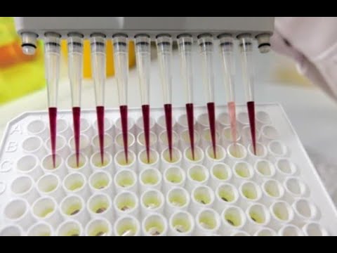
MTT Assay
1.2K
Post-assay analysis involves measuring absorbance at 570 nm (reference 630-690 nm) using microplate readers. Background correction uses blank wells with only culture medium. Cell viability percentage is calculated as (mean treated absorbance / mean control absorbance) × 100, where positive controls represent 100% viability and negative controls establish baseline. Normalization strategies include crystal violet staining for cell counting and protein quantification for metabolic normalization. Dose-response curves plot viability against compound concentration, fitted to sigmoidal curves to determine IC50—the concentration causing 50% cell death. Statistical analysis requires triplicate experiments with ANOVA/t-tests for significance determination.
Click to watch →
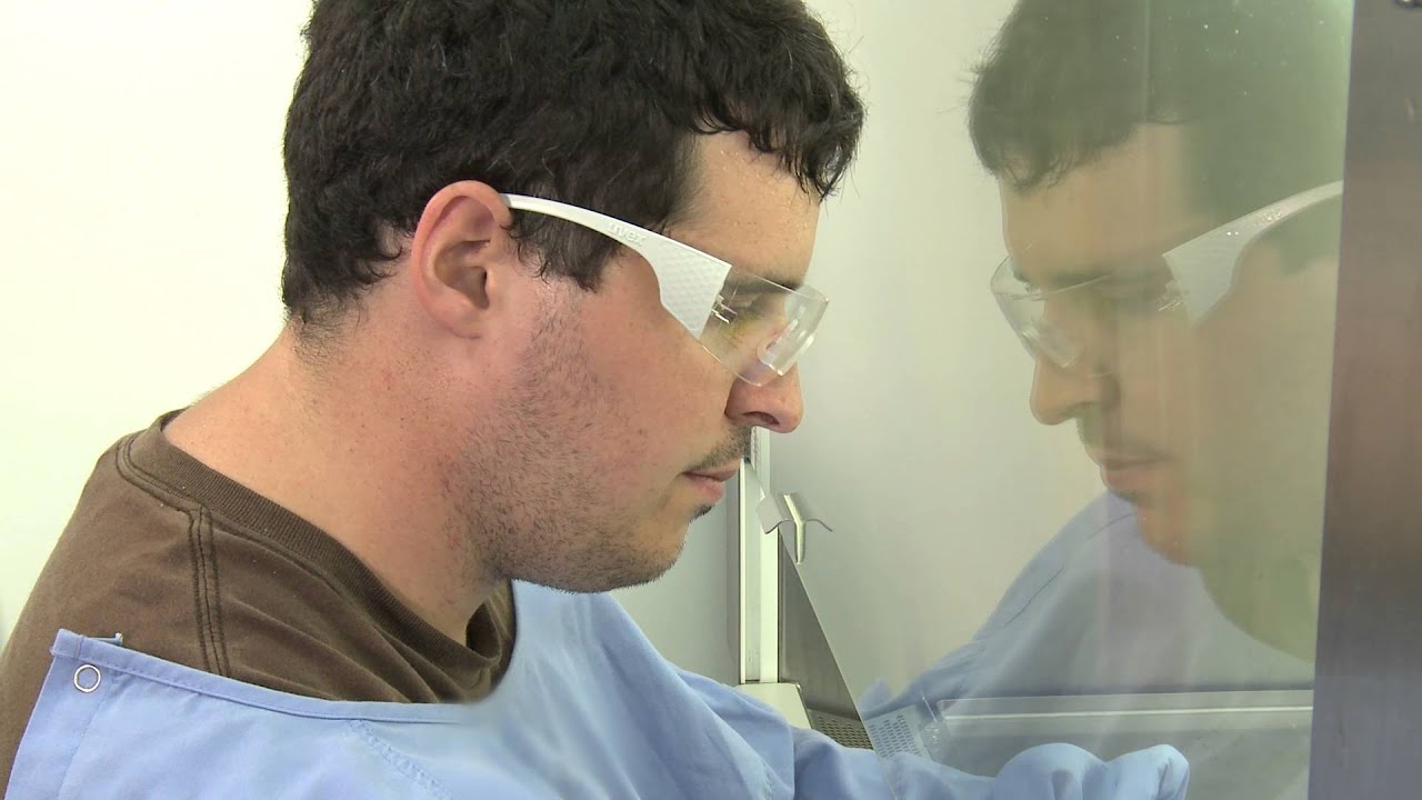
IC50 or cell viability experiment
52.7K
To determine the IC50 (concentration causing 50% cell death), seed cells in a 96-well plate, expose them to serially diluted treatments, stain with Alamar Blue, measure absorbance at 570nm and 600nm, calculate percent viability relative to untreated controls, plot log concentration versus viability on a sigmoidal curve, and identify the inflection point as the IC50 value.
Click to watch →

MTT assay and IC50 calculation
15.2K
The MTT assay is a colorimetric method that measures cell viability by detecting the metabolic activity of living cells, which convert the yellow tetrazolium salt MTT into purple formazan crystals; cell viability is calculated using the formula [(OD_sample - OD_blank)/(OD_control - OD_blank)] × 100, and the IC50 value (the concentration causing 50% cytotoxicity) is determined by plotting concentration-response curves and finding the point where the response equals 50% using linear regression analysis.
Click to watch →
Deep Dive
Troubleshooting common issues in MTT assays, such as uneven seeding (edge effects), chemical interference from test compounds, and incomplete dissolution of formazan crystals.
MTT Assay Protocol: Cell Viability & Proliferation Guide
120.4K
The MTT assay measures cell viability and proliferation through metabolic activity detection. MTT reagent is reduced by NADPH-dependent enzymes in viable cells to form insoluble purple formazan crystals. The assay requires metabolically active cells; cells with slow metabolism produce false negatives. Complete protocol: seed 3,000-5,000 cells per well in 96-well plates with identical cell counts across all wells. Allow 24-hour attachment before treatments. Prepare MTT stock at 5 mg/mL in sterile PBS, dilute 1:1000 to 5 μg/mL working solution. Add 110 μL per well and incubate 4-6 hours at 37°C in a sterile hood. Carefully remove media without disturbing crystals, add 100 μL DMSO per well, and pipette vigorously to dissolve formazan. Measure absorbance at 550-600 nm using a microplate reader. Effective experiments require careful plate organization with replicates down columns and conditions across rows. Use logarithmic spacing for dose-response curves (e.g., 10, 20, 50, 100, 500 μM) rather than linear increments. Include positive (0 nM drug, 100% viability) and negative (no cells, background subtraction) controls. Prepare master mixes for consistent cell seeding: calculate total cells needed plus total volume, mix thoroughly, and dispense equally using multi-channel pipettes.
Click to watch →

Cell Viability Assay Using MTT| Dr. Sitaram Harihar| Animal Biotechnology| Genetic Engineering | SRM
7K
The complete MTT assay procedure follows a systematic workflow: First, seed 10,000 cells per well in 200 microliters of media in a 96-well plate and incubate at 37°C for 24 hours for cell attachment. Next, remove media and add test compounds (e.g., quercetin at 0.1, 1, 10, and 100 micromolar) in triplicate, then incubate for another 24 hours to allow drug effects. After treatment, remove media and wash wells with PBS. Add 100 microliters of MTT reagent in the dark, incubate at 37°C for 3 hours for formazan crystal formation, then solubilize crystals with DMSO. The resulting purple-colored solution is ready for absorbance measurement at 570 nm. Blank wells (no cells) and control wells (vehicle-only) are included to account for background and ensure accurate readings.
Click to watch →

MTT assay - Protocol, principle and troubleshooting - DETAILED EXPLANATION
3.7K
The MTT assay is a colorimetric method used to measure cell viability and cytotoxicity by detecting mitochondrial metabolic activity; the yellow MTT reagent enters viable cells and is reduced by mitochondrial reductase enzymes to form purple formazan crystals, which are then solubilized with DMSO and quantified using a microplate reader, where higher absorbance indicates greater cell viability.
Click to watch →

Troubleshooting Cell based Assays Ask the Experts to Avoid Common Pitfalls from Cell Seeding to An
1.1K
Three common viability assays are based on reduction equivalence: MTT assay (CellTiter 96), MTS assay (CellTiter Aqueous MTS), and Resazurin-based assays (CellTiter Blue). MTT is a cell-permeable tetrazolium dye reduced intracellularly to an insoluble formazan product requiring solubilization (lytic endpoint assay). MTS has a negative charge preventing cellular entry, requiring an electron transfer reagent to shuttle electrons onto MTS, producing a soluble product that preserves cells for downstream applications. Resazurin is reduced intracellularly to fluorescent resorufin. All three require extensive incubation (1-4 hours) and share limitations including potential cytotoxicity from the assay reagents themselves.
Click to watch →
MTT Assay Made Simple: How to Measure Cell Viability?
373
The MTT assay is a widely-used calorimetric method that measures cell metabolic activity by detecting the conversion of yellow MTT tetrazolium salt into purple formazan crystals by living cells; this color change occurs through mitochondrial enzymes and indicates cell viability, making it valuable for drug screening, toxicology studies, and cell biology research despite its limitations including interference from colored compounds and cell-type variability.
Click to watch →
Deep Dive
Comparison of the MTT assay with alternative cell viability and cytotoxicity assays, such as CCK-8, Alamar Blue, LDH release, and ATP luminescence assays, to understand when to use each.

Lecture 7. Cytotoxicity evaluation
416
Metabolic assays measure mitochondrial dehydrogenase activity as proxies for cell viability. MTT assay converts yellow MTT to purple formazan crystals inside viable cells. XTT and other variants offer water solubility advantages. LDH release assay measures lactate dehydrogenase enzyme released from damaged cells into culture media. These are indirect methods providing only viability information without revealing underlying mechanisms. Decreased metabolic activity indicates reduced mitochondrial function but not the cause of damage. They cannot distinguish between different types of cell death or provide mechanistic insights.
Click to watch →
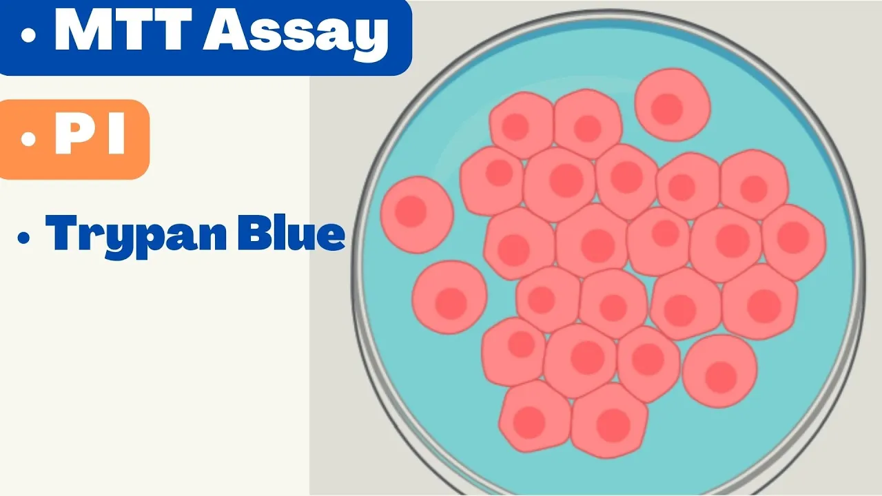
🛑 CELL VIABILITY ASSAYS | MTT Assay | Alamar Blue | Propidium Iodide | Cell TIter Glo
7.9K
Cell viability assays measure living cells through different mechanisms: dye exclusion tests (like Trypan blue) stain dead cells but not live ones; tetrazolium reduction assays (MTT, MTS, XTT, WST-1) rely on metabolically active cells reducing colorless compounds to colored formazan products; resazurin-based assays use fluorescent detection where viable cells convert dark blue resazurin to pink fluorescent resorufin; protease release assays detect dead-cell proteases released after membrane damage; and ATP-based luminescent assays measure ATP production, which only viable cells can synthesize, offering the highest sensitivity.
Click to watch →

The Seahorse Analyser, flow cytometry, and cell viability assays
599
Cell viability assays measure living cells through colorimetric conversion of tetrazolium salts to formazan by cellular dehydrogenases. Three common methods differ significantly: MTT uses NADH-dependent oxidoreductases producing insoluble formazan requiring DMSO dissolution (measured at 570 nm, less sensitive, cheaper); MTS produces water-soluble formazan (measured at 490 nm, faster, more sensitive); CCK8 targets dehydrogenases directly (measured at 450 nm, highly water-soluble, preferred for cancer cytotoxicity studies). All follow similar protocols: treat cells, add reagent, incubate, measure absorbance. These assays quantify cellular proliferation, assess drug cytotoxicity, and evaluate overall cell health in research applications.
Click to watch →
In vitro Anti cancer Assay: Dr. Bhushan P Pimple
4.3K
Two alternative cytotoxicity assays complement SRB methodology. The MTT assay measures cell viability through mitochondrial-dependent reduction of yellow tetrazolium salt to purple formazan. Viable cells with active mitochondria convert MTT to purple formazan; dead cells retain yellow color. The procedure includes 2-4 hour MTT incubation, detergent lysis, formazan solubilization, and spectrophotometric measurement at 550-600 nm. Purple color indicates low cytotoxicity; yellow color indicates high cytotoxicity. The Brine shrimp lethality assay uses Artemia nauplii larvae for in vivo screening. Artemia cysts hatch in artificial seawater (3.8% salinity) under light conditions. After ~36 hours, nauplii emerge and migrate toward light. Test compounds are dissolved in suitable solvents and added to tubes containing 10 nauplii each. After 24-hour exposure, survival rates are counted and percent cytotoxicity calculated against negative controls.
Click to watch →
MTT Assay Protocol: Cell Viability & Proliferation Guide
120.4K
The MTT assay measures cell viability and proliferation by detecting metabolic activity; viable cells reduce the yellow MTT reagent to purple formazan crystals, which are dissolved in DMSO and quantified by absorbance at 550-600 nm, with percent viability calculated by normalizing experimental values against positive and negative controls using the formula: [(Experimental - Background)/(Control - Background)] × 100%.
Click to watch →
Deep Dive
Application of cell viability data in preclinical drug discovery, toxicology screening, and evaluating the efficacy of cancer therapeutics.
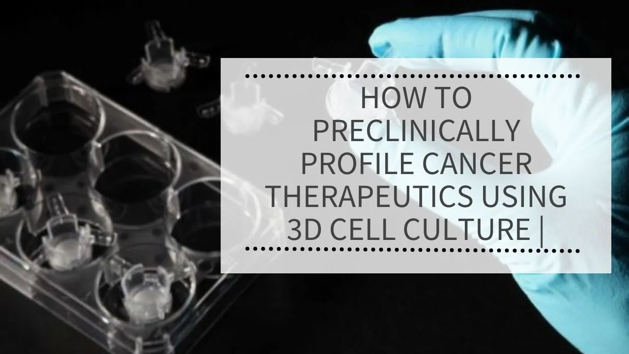
How to preclinically profile cancer therapeutics using 3D Cell Culture | REPROCELL (Alvevtex)
117
Drug development suffers from poor translation between early-stage screening and clinical outcomes, partly due to limitations of conventional 2D and soft agar assays. Alvetex technology addresses this gap by enabling post-treatment recovery for downstream molecular analysis including Western blotting and RNA extraction. Data correlates well with conventional soft agar assays while offering superior sensitivity (80+ fold signal-to-background vs. ~20 fold). Combination therapy studies reveal biological effects missed by viability assays alone, detecting phosphorylation changes at concentrations where viability remains unaffected. This comprehensive approach enhances understanding of drug mechanisms and resistance pathways.
Click to watch →
Cell Viability Assay Using MTT| Dr. Sitaram Harihar| Animal Biotechnology| Genetic Engineering | SRM
7K
Data analysis involves transferring absorbance readings to Excel, calculating average optical density values for duplicates, and subtracting blank values from all measurements. Percentage cell viability is calculated using the formula: (OD of test sample / OD of control) × 100. Vehicle control typically shows 100% viability, while increasing drug concentrations show progressively lower viability. Results are plotted with drug concentration on the x-axis and either absorbance or percentage viability on the y-axis. The IC50 value, representing the concentration that kills 50% of cells, is determined graphically from these plots. This quantitative approach enables researchers to assess compound toxicity, compare drug potencies, and establish appropriate concentrations for further biological studies.
Click to watch →
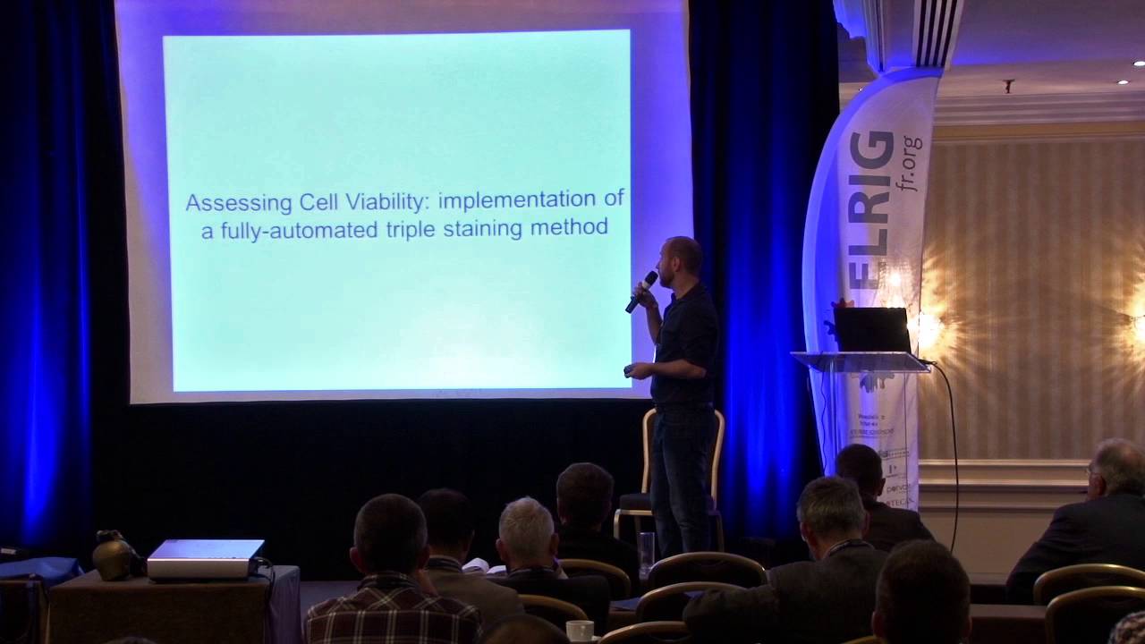
Quantification of cellular viability by automated microscopy (...). Allan Sauvat
189
Advanced data analysis transforms cell-by-cell data into meaningful biological conclusions through systematic filtering, clustering, and statistical analysis. The process involves excluding debris and poorly segmented cells, defining thresholds for fluorescent signals, and distinguishing normal cells from pyknotic cells and debris based on nuclear characteristics. Drug combination index (CI) calculation quantifies interactions between drugs in matrix treatment designs, revealing synergistic (CI < 1), additive (CI = 1), or antagonistic (CI > 1) effects. Time-course analysis extends endpoint assays by measuring viability at multiple time points, capturing dynamic changes and distinguishing cytostatic from cytotoxic effects. Absolute cell viability measurement distinguishes between reduced proliferation and actual cell death, providing deeper mechanistic insights than relative viability calculations.
Click to watch →

Maximize your cell health assays data acquisition with latest tools in cancer research
149
Presto blue is a resazurin-based assay where metabolically active cells convert the blue dye to fluorescent resorufin, providing stable signals for 4-6 hours. CellTiter-Glo measures ATP content through luciferase reaction, offering linear detection from 50-50,000 cells ideal for high-throughput screening. Combining these assays on the same plate provides comprehensive viability assessment. Microplate readers offer absorbance, fluorescence, and luminescence detection modes. Software enables pre-programmed protocols, blank subtraction, normalization, standard curve generation, and dose response curve analysis with automatic gain adjustment. The median effective dose (ED50) calculation demonstrates quantitative analysis capabilities.
Click to watch →
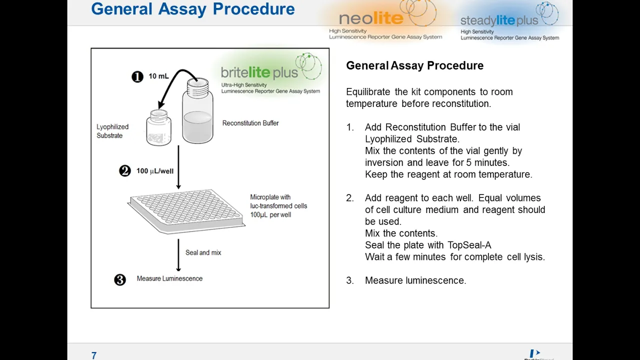
Answering the Challenges of Cell Viability and Reporter Gene Assays Screening Cost and Quality
537
Cell viability assessment is fundamental to drug development for eliminating toxic compounds and identifying effective therapeutics. Traditional methods including chromium release assays, DNA synthesis assays, and microscopy-based approaches are labor-intensive and time-consuming. ATP-based luminescent assays provide a sensitive, automated, and easily miniaturized alternative that correlates directly with metabolically active cell numbers. The ATP Light One-Step kit offers rapid results while two-step variants provide enhanced stability and flexibility for frozen sample storage. Key practical considerations include optimizing solvent concentrations (typically ≤0.5% DMSO), validating against literature values, and ensuring assays remain within linear detection ranges. Both single-step and two-step formats achieve detection limits below one cell per well, though practical screening typically uses hundreds of cells per well to minimize variability. This technology enables efficient cytotoxicity screening in high-throughput formats essential for modern drug discovery pipelines.
Click to watch →
Add MTT reagent
Solubilize formazan
Read and compare
Add MTT reagent
0:00- 1
Aspirate media from each well and add plain media with MTT reagent.
- 2
Use one pipette for all samples, dispensing carefully to avoid dislodging cells.
Limitations of Metabolic-Based Viability Assays and the Rise of Direct or Real-Time Alternatives
While the MTT assay is a classic, cost-effective standard for assessing cell viability, it has significant technical limitations. First, MTT measures mitochondrial metabolic activity rather than direct cell death or membrane integrity; treatments that alter cell metabolism without killing the cells can produce misleading results. Second, the assay is endpoint-only and destructive, as it requires dissolving the cells in DMSO to solubilize the formazan crystals, preventing any downstream analysis or real-time kinetic monitoring. Critics and modern researchers often advocate for alternative methods, such as Alamar Blue (resazurin-based) assays, which are non-destructive and allow for continuous monitoring, or direct cell-counting and membrane-integrity assays (like Trypan Blue or Flow Cytometry) to obtain a more accurate measure of true viability.
Understanding the biological principle of the MTT assay, specifically how mitochondrial dehydrogenases in living cells reduce the yellow MTT tetrazolium salt into purple formazan crystals.
Familiarity with basic cell culture techniques, including cell seeding, cell density optimization, and aseptic techniques used on Days 1 and 2 of the protocol.
Knowledge of spectrophotometry and how a microplate reader functions to measure optical density (absorbance) at specific wavelengths (typically 570 nm).
Awareness of safety protocols regarding handling chemical reagents like MTT and solubilization solutions (such as DMSO or acidified isopropanol).
Data analysis techniques, including how to calculate percentage cell viability, subtract background absorbance, and plot dose-response curves to determine IC50 values.
Troubleshooting common issues in MTT assays, such as uneven seeding (edge effects), chemical interference from test compounds, and incomplete dissolution of formazan crystals.
Comparison of the MTT assay with alternative cell viability and cytotoxicity assays, such as CCK-8, Alamar Blue, LDH release, and ATP luminescence assays, to understand when to use each.
Application of cell viability data in preclinical drug discovery, toxicology screening, and evaluating the efficacy of cancer therapeutics.
431 views3likes4:05@CellBiologyEducationConsortiumOriginal Release: 2019-09-18
okay so this is the last day of your MTT assay we're gonna actually see results today make sure again you look at the colors of the media it's hard to tell right now but these actually turn more yellow orange then the other ones which is what we would expect and make sure you look at them under the microscope to see how the cells look so all I'm gonna do in the first step is vacuum off each well and then I'm going to add my plain media plus M 2 T reagent and this will be provided for you okay so because these are all the same cell line I can use just one pipette to vacuum off all the media remember tilt it go down the side and just chop one little spot [Music] okay and then remember I can use just one pipette I'm going to pull up six mils and let out half a milliliter into each well and remember I don't wanna knock the cells off the bottom so I'm going to be careful and just lightly place the tip on the side and let our crack a little at a time [Music] all right so now you just replace the pepper and put it in the incubator for one to two hours [Applause] so which will then soluble eyes the precipitate [Music] and then for this we want to make sure you add DMSO to three extra Wells to use as bling I'm going to take up seven and a half milliliters and again empty half a mil into [Music] all right so then you put the lid back on and put it on the belly dancer for 10 to 20 minutes so set it in the center of the belly dancer and if there's someone else going at the same time you can have multiple plates on there and just turn it and these can go fairly fast set your timer 10 to 20 minutes okay so it's been going for 10 minutes I'm going to take it off the belly dancer and take it to the plate reader but before make sure you take a picture so you can compare these colors that you're seeing to the numbers that you get you
Up Next

10:44
Pregnancy & Development Explained: Crash Course Anatomy
@crashcourse
2M views•2015-11-23

50:12
Bacterial Communication: Quorum Sensing and Biofilm Formation Explained
@JHUAAP
12.6K views•2011-06-28

5:35
Enteric Nervous System Explained: The Gut's Brain | Neurobiology Lecture
@alumniu6029
438 views•2018-09-12

7:08
Bacteriophages: Earth's Deadliest Killers and Future Antibiotics
@kurzgesagt
34.6M views•2018-05-13
Related Study Plans & Knowledge Roadmaps
Structured learning paths in Biology
biology
RNAi: Biogenesis, RISC & Gene Silencing
01Molecular Biology Foundations: DNA to Protein
02Introduction to RNA Interference (RNAi)
03Molecular Biogenesis of miRNA and siRNA
+ 2 more learning modules
10h5 modules
Explore Roadmap
biology
Mycorrhizae: Signaling, Biophysics & Ecology
01Foundations of Mycorrhizal Symbiosis
02Molecular Signaling and Root Colonization
03Biophysics and Bioenergetics of Nutrient Exchange
+ 1 more learning modules
11h4 modules
Explore Roadmap
biology
Circadian Clocks: TTFL, SCN & Periphery
01Foundations of Molecular Biology & Gene Expression
02Introduction to Chronobiology and Actograms
03The Core TTFL: Molecular Architecture of the Clock
+ 3 more learning modules
14h6 modules
Explore Roadmap
biology
Population Genetics: Math, Drift & Selection
01Genetic & Probability Foundations
02The Hardy-Weinberg Principle and Baseline Models
03Mathematical Modeling of Natural Selection
+ 3 more learning modules
18h6 modules
Explore Roadmap