











![Tolerância Imunológica - Resumo [ Imunologia ]](https://i.ytimg.com/vi/ocvGryHpAg0/maxresdefault.jpg)



T cells are essential components of the adaptive immune system that recognize and respond to specific antigens through their unique T cell receptors (TCRs), which are generated through V(D)J rearrangement of gene segments; they are categorized into three main types: CD4+ helper T cells that coordinate immune responses by producing cytokines, CD8+ cytotoxic T cells that directly kill infected or cancerous cells, and regulatory T cells that suppress excessive immune responses to prevent autoimmunity; T cell activation requires three signals: peptide-MHC binding (signal 1), co-stimulatory molecule engagement (signal 2), and cytokine receipt (signal 3); TCR diversity is achieved through random recombination of V, D, and J gene segments, with allelic exclusion ensuring each T cell expresses only one receptor type; MHC class I molecules present intracellular peptides to CD8+ T cells, while MHC class II molecules present extracellular peptides to CD4+ T cells, with MHC restriction ensuring T cells only recognize antigens presented by their own MHC molecules.
T Cells Explained: Types, MHC, and Activation | Immunology Lecture 2
Added:Okay, then we are all silent.
Also the grass mower.
No, he doesn't listen.
[Music] Welcome to easy iminology. My name is Stefan and in our today's lecture we want to talk about TE-C cells. And in order for me to not do this completely alone, I have uh today again with me Alex. Hi Alex.
>> Hi Stefan.
>> Hi. Thank you very much for joining me.
And I have with me Hana. Hi Hannah.
>> Hi Stefan.
>> Thanks again also for joining me. The two will support me with questions. So they can ask the questions that you might have also during uh this this recording and then we can address them directly here in this video. And this as we learned is very helpful for everyone.
So, as I said, we're going to talk about TE- cells. We are starting with a teeny tiny bit of repetition at the beginning.
So, what we learned in the first video is that our immune system protects usually very efficiently from pathogens.
So these are microorganisms or viruses that want to make us sick because they want to grow inside or on our body on our costs and our immune system as I said protects us from these uh pathogens and it broadly speaking consists of four different components. We have learned the last time that they are the anatomical barriers that just protect us from these pathogens by preventing the pathogens from coming from the non-sterile outside into the sterile inside of our body. If the pathogens manage to come inside, they are attacked by the compliment system and the antimicrobial peptides. And if this also doesn't work or in parallel to this, they are also attacked by cells of the innate immune system. This is what we managed to go through in the first video and today we are starting starting at looking at the adaptive immune system.
So which are the T- cells and the B cells and the antibodies produced by B cells. And this is very very complicated. uh we have to say therefore we were able to look at the first three barriers in in the first video and for the the last barrier the adaptive immunity we will probably need probably three videos to go through all the the concepts but it's very important because the first three barriers are able to uh eliminate 95 to 98% of all pathogens but the last two to 5% for which we need the adaptive immune system for this we actually need it because there are patients who do not have an adaptive immune system and they are not able to live in a normal environment with all these pathogens because the adaptive immune system is so crucial and eliminating them. So what we also learned I think this is where we stopped the last time is that the inert immune system which consists of different cells that we looked at is available immediately after birth. It can react within minutes and hours and it does so by recognizing the pathogens as dangerous and foreign using a limited number of pattern recognition receptors.
We looked at the different classes and uh this is rather effective as I said eliminating 95 to 98% of all pathogens but uh broadly speaking uh it doesn't remember uh what it did yesterday and therefore if you are reinfected with the same pathogen you have to start the whole cascade again and this is not the most efficient way to do this and what we want to do over the next three videos is look at the adapter site and uh we are going to start with looking at the cells. As we learned in the last video, the inner immune system consists of different cells. We have looked at the dendritic cells and macrofasages as sensor cells that recognize pathogens.
We have looked at the aenophils, musels and basopils which are able to fight big extracellular pathogens like parasites, multisellular parasites that can be very very big up to 12 m in length. And we looked at the neutrfils which are very important the most frequent cells in the human blood because they are able to fight pathogens with many different effective mechanisms. We didn't look at the ENK cells and the NKT cells yet. We will do this at the end. uh of the the following video because we need to first understand the T- cells and then it's very easy to understand the NK cells and the NKT cells because if I spoil you these are like evolutionary precursor cells to the T- cells we are going to start looking at today on the adaptive side we have the T- cells uh they are there are different types we will look at today and together they are referred to as the cellular part of the adaptive immune system and we have the B cells and the antibodies produced by B cells and they are referred to as the hummeral part of the immune system. Uh the the the term cellular and hummeral are historically grown they are not totally correct because as you can see the the cells of the adaptive immune system are not concentrated in the cellular part. So the cellular part is just the T- cells but also the B cells are cells in the end. So they are also cellular. Uh but these are historical concepts because as we will see it is very hard to differentiate uh T and B cells with just a light microscope. So there's basically no difference unless they are activated. Therefore people couldn't couldn't keep them apart from each other. And what people were able to do is they were able to draw blood and analyze the blood and they found like the antibodies in there. And therefore they said okay the the antibodies are in the the humorus. This is like the bodily fluids and they protect us from many pathogens. So the the antibbody must have something to do with the hummeral uh fluids and therefore it was called the hummeral immune system. Okay. So today we are going to focus on te- cells and there are three different types of tea cells we going to look at in more detail in the next slides.
So first here is like like a lymphocy.
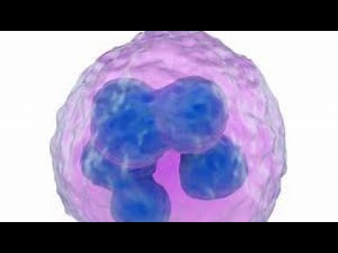
So T and B cells together are called lymphocytes. Among uh a lot of red blood cells. The red blood cells are the the pale ones in this picture. And you can see that they are like disc shaped and they do not have a a nucleus. They do not have a core inside the cell. And you have the single big lymphocy which consists of the nucleus. That's the pink part and the more pale part around is the cytoplasm. And you can see that you basically cannot see a lot. It is a round round cell without any characteristics and it has a very big nucleus and only very little cytoplasm.
So that's that's what you can see and that's why people were also not able to distinguish between T and B cells because at this stage a T cell just looks like AB cell and it's very hard to to distinguish if you do not have like for example genetical markers that you can look at on this level of course you can distinguish a T cell from AB cell there they are very different in terms of stages we can differentiate three three and a half stages which is for T and B cells. We have the uh precursor cell that is the gray cell that is like the lymphoid progenitor cell that develops into either a B cell that is the the left part the blue part or a T- cell that is the green part and we can differentiate between naive T- cells and B cells. These are the small round guys that I showed you and they are as I said very hard to distinguish because there's not much to see. Once TNB cells get activated, they differentiate. So they change. You can see actually they look very different.
They are much bigger and they proliferate. So they form a lot of identical daughter cells because these cells are needed during the infection to fight the the respective pathogen. So these are the activated TNB cells. And there you can already clearly see differences with a good microscope between a T and AB cell. And then once they find the pathogen and start to fight it, they fulfill their so-called defector function. For the B cell, it is producing the antibodies. You can see the the B cell is surrounded by a lot of antibodies in our case. And for the T- cell, it is at least for this type of T- cell, it is the production of cytoines.
So messenger molecules that direct the immune response. There you can also find differences. So, so these are the the three and a half stages for for the T and the B cells that we can differentiate. So, where do the cells of the immune system come from? Where do they develop? They develop in the so-called primary lymphoid organs. This is the bone marrow and the thymus. And in the bone marrow, we have so-called pur potent stem cells. So, these are stem cells are cells that can develop into many different cell types. They are not able to develop into every type of cell. It would be omnipotent like the fertilized oside is able to and has to develop into all cells in our body. This is this is less potent if you want to call it like this and it develops into first the common myoid progenitor and this then forms or is the the predecessor for all the or most of the cells in the inate immune system like dendotic cells microfers neutrfils and the granular sites and the common lymphoid progenitor develops in the bone marrow into the the B cell progenitor and then into the B cell and uh it can migrate the common lymphoid progentor gentor can migrate to the thymus and there uh become the T- cell progenitor and develop into the T- cells. And Alex has a question is the B cell called B cell because it comes out of the bone marrow like the T- cell comes out of the thymus.
>> Yeah. Uh one could could assume that and it's a nice nice uh way to remember it but actually not. Uh the the B cells are for some crazy reason uh called B cells because they develop in the BZA fabrii.
This is an organ in chicks uh where they were first developed because some some some guys did or some guys and girls did experiments on chicks. So that is not why they are called called B cells >> but you can remember them quite good with bone marrow is B cell and thymus is T cell.
>> Yeah but in the end if we are 100% correct it's it's not correct. Yeah.
Okay, so today we want to look at the T- cells and this is an overview how actually an adaptive immune response is formed and it starts with um a pathogen being recognized by an antigen presenting cell. We looked at this in in the last lecture already. And then it activates this this activated antigen presenting cell activates a naive T- cell that turns into aector T- cell. And this then this then activates B cells and they turn into antibbody producing plasma cells. And we're going to start with the first step the activation of the naive T- cell. And here we have our antigen presenting cell. Usually this is some kind of dendritic cell or also can be a macrofase but usually it's the denditic cells. They have so-called path recognition receptors on their surface or in the inside also. And these receptors are able to recognize certain structures. They are called pumps, pathogen associated molecular patterns.
And these are structures that only exist in in this shape or form in uh these microorganisms. And in contrast, they do not exist on our own cells and tissues.
So this is something that is definitely foreign. So if uh these receptors bind these path recognition receptors bind to these pumps this means that something foreign is around and this for this cell means just okay in this case here there's a there's a bacterium around which has a fleella and uh therefore an immune response needs to be made against this bacterium. So here we have the the fleella protein. It's called fleagellin.
It binds to the path recognition receptor and this triggers different things in our antigen presenting cell.
First is a change in gene transcription.
So there is through a very complex uh set of adapter molecules a activating signal transduced into the nucleus into the core of the cell and this results in many many different genes being turned on or turned off. So if you have a antigen presenting cell that is infected by a virus this you can do in an experimental settings as thousands of genes are up or down regulated. And if you remember that we only have between 19,500 and 23,000 different genes.
Thousands 56 7,000 genes that are up or down reggulated is quite a lot.
Among these genes that are upregulated are genes that code for so-called coimulatory molecules. These are molecules that are then expressed on the surface of the antigen presenting cell and they are only expressed there if the cell has recognized something dangerous, something foreign and dangerous via its pattern recognition receptors and therefore the presence of these molecules is just a way uh for the antigen presenting cell to show uh to the T- cell that it wants to activate that it has found something dangerous.
for the T- cell doesn't matter what the cell has found. Uh it just needs to know okay there's something dangerous around.
Also uh among the genes that are activated as genes that that encode for the production of cytoines small uh soluble messenger molecules that transmit information between immune cells and non-immune cells.
Additionally, the cell uh either triggered by the activation of the pathon recognition receptors or also generally takes up everything that floats around the cell. So these cells are like like PhD students. They are always always eating uh and these cells just just eat everything around them and uh including whole pathogens or parts of the pathogen and um these molecules are then inside the cell uh digested turned into peptides. So genes are long polyeptides and they are cut into short fragments and these fragments are called peptides and these peptides are presented on so-called MHC molecules major hisystocompatibility complex proteins. We will look at them in the second half of the lecture once we have understood what a T- cell is and what kinds of T- cells there are and then uh these the cell the antigen presenting cell shows on its surface what it has has taken up that's why it also called an antigen presenting cell because it presents peptides derived from the pathogen molecules usually proteins on its surface and then uh the cell that is activated It moves into the moves from the place where it's found the the pathogen. This can be everywhere in our body. It moves to a lymphoid organ. This is usually a lymph node where where the T- cells are also run wandering around. So the T- cells are wandering around in our body. It takes 24 hours approximately for a T- cell to reach every lymph node in our body and uh the the antigen presenting cell goes there and just waits for for the T- cell. So there are many T- cells coming and going and every T- cell checks what the the antigen presenting cell has to offer. So it has like this T- cell receptor going to highlight it uh in post. This is the the gray and green structure and this is able to recognize the peptides from from our blue bacterium together with the MHC molecules. And if this matches this means this is the T- cell that that was born. uh if you want to say it like this to fight this blue bacterium and this just results in the two cells sticking together it's not enough and it's not strong enough uh to to stick together long enough for the T- cell to become activated and this is where the coaster military molecules come into play because they result in the two cells sticking together even more strongly and even longer and both signals then transmit an activating signal into the nucleus of the T- cell and then it it can be activated. So it it knows via its T- cell receptor that uh the the T- cell receptor it has is useful for fighting this this blue pathogen and via the coastlatory molecules it gets the information that uh it is allowed to fight the pathogen because the antigen presenting cell has decided that the blue bacterium is dangerous and foreign but it doesn't know what to do and uh the information what to do and what kind of T- cell to become is is coded in in the cyto cytoines. So these cytoines bind to receptors on on on the T- cell surface and then they trigger the differentation or the professionalization if you want to call it like this of the T- cell into different uh types of T- cell because it could become for example antibacterial T- cell, antiviral T- cell, antiparasitic T- cell and antifungal T- cell. Um this is basically what the iminologist calls the three signal theory of T- cell activation. And the first signal is called is is the presentation of peptides in combination with MHC molecules. The second signal is the expression of co stimulatory molecules.
And the third signal is uh the production of cytoines.
And uh as I said the signal one is the binding of the T- cell receptor to the MHC complex with the peptide. The signal two is the expression of co stimulatory molecules and the signal three is the secretion of cytoines. And what these signals do is signal one ensures that only a te- cell specific for a certain pathogen gets activated. Signal two makes sure that this only happens once the antigen presenting cell has decided that this cell is dangerous because only then we have the expression of the coilatory molecules and signal 3 makes sure that the T- cell is is is aware and knows what to do and what kind of T- cell to become.
So unfortunately as as I tell my students all the time it is more complicated uh because there are different types of T- cells that can can develop. So we have uh three types of of T- cells. We have helper T- cells, we have regulatory T- cells and we have cytotoxic tea cells. And I want to just go through them uh one by one and explain you uh what they do and what their distinct functions in the immune system are. So let's start with the uh helper tea cells. They are CD4 positive.
This is something that we will look at in the later later half of the lecture.
But once they are activated uh by the the three signals that we just discussed, they have their main function in producing cytoines. They produce more cytoines and different cytoines than the antigen presenting cell did after activation. And these cytoines are then uh directing, modulating and maintaining the immune response. And they can activate for example macrofasages. These are cells that are able to fagocytose to eat up the pathogens and destroy them and induce inflammation. Also neutrils as we learned in the first lecture are very important cells that induce inflammation and they can also activate the other kinds of tea cells the cytotoxic tea cells. These are the red guys and they can activate these cells.
In a way these CD4 positive tea cells are the managers of the immune system.
So they tell all the other cells or most of the other cells what to do and then they can coordinate in this way the immune response.
Unfortunately it gets even more complicated because there is not just one uh CD4 positivity helper cell. There are different types and it is even more complicated than I have put it here on the slide. On this slide I've put five different types but there are even more as as Alex and Hannah know. um what kind of T-H helpper cell is generated depends on the signals that are transmitted by the antigen presenting cell during the activation of the naive T- cell. And if for example the antigen presenting cell produces the cytoines interfering gamma and 12 this results in the binding of these molecules to their receptors on the T- cell surface and this triggers the activation of molecules inside the T- cell. They are called stats signal transducers and activators of transcription. And there are many different stats and interferon gamma and 12 lead to the activation of start one and start 4 and this triggers a the activation of a transcription factor that we call T-bed and this is called the master transcription factor of TH1 cells. So if cells activate this T-B this then in turn activates many many different genes in this T- cell in this T- cell and turns into a so-called TH1 cell which in turn produces a lot of interferon gamma and this interfering gamma uh tells the immune system that there are intracellular bacteria that need to be fought and this is how they uh then fight these bacteria by activating all these other cells. In contrast, if the antigen presenting cell or some other cell provides interlucan 4, this triggers the activation of stat 6, which in turn activates the master transcription factor of TH2 cells, which is called gata 3. These TH2 cells then produce 4,5 and 13. And their main function is to coordinate the elimination of big multisellular parasites. And in turn, we have for example TH7 cells. They depend on raw gamma tea. They are responsible for coordinating immune responses against extracellular bacteria. We have follicular to helper cell. They are mainly in the in the lymph nodes and they help uh B cells to become activated. Uh they provide uh the perfect environment for for the cells in the lymph node to become activated to proliferate and to start immune responses. And we have regulatory T- cells which is like a a special kind we will look at also in more detail later because their function is not to induce immune responses but to shut them down because once you have started an immune response have eliminated a pathogen you also have to stop making the immune response at some point because this takes a lot of energy this takes a lot of resources and if the pathogen is no longer around because it has been eliminated you don't need this anymore and this is what the the regulatory tea cells are For we have also the the cytotoxic tea cells they are CD8 positive. They are induced if the antigen presenting cell produces interlucan 7. This triggers start stat one and start 4. You see this is very similar to the TH1 cell. They also like a TH1 like uh phenotype because they probably were the same cell during evolution and then this this diverged into the these two different cell types. They can produce also a lot of interferon gamma but also perfor and gran which they use to kill uh intracellular pathogens, virus infected cells and tumor cells but we will look at this in more detail in one of the next slides.
So these are the the helper cell their main function is to produce cytoines and these cytoines then coordinate the immune response in eliminating the pathogen.
Then we go through the CD8 positive cytotoxic tea cells. Their main function is to kill virus infected cells. So here we have a normal cell in our body for example a liver cell that is heavy uh producing liver enzymes and also these cells are able to show on their outside what they are working on in their inside. They also have these major hisystocompatibility complex proteins. You can see they look a bit different than the ones on the antigen presenting cells. Why this is the case we will look in in a few slides. But also these cells also always show on their surface what they are working on on the inside. And if they just are happy and work on liver enzymes, everything is fine. Then the T- cell will will not do anything. And what the T- cells are doing, they are circulating in our body and check uh what kind of peptides are presented on these MHC molecules. And if this is just a liver peptide from a liver enzyme, then everything is fine because the cell is doing what it's supposed to do. Yes, Hannah.
>> Does every TE-C cell only have one T- cell receptor for one specific peptide or do they can they recognize more peptides?
>> Yeah, there that's an excellent question. We would have gotten through this at at later stage, but we can also just quickly spoil it here. You can see on the surface of the T- cells there are 1 2 3 4 5 6 seven T- cell receptors.
They are all identical. So one T- cell only expresses one type of T- cell receptor. But we have millions millions of different T- cells and everybody has has a different T- cell receptor because this is generated randomly as we will learn in the second half of the lecture and it can only recognize I wouldn't say one specific peptide because uh peptides are in the end are just a certain amount of amino acids next to each other and there are different amino acids that have similar similar properties. So it can can be that one T- cell receptor can maybe recognize two three four different peptides which are not totally different but they are differing only at certain positions in in their in their sequence and there they only have very similar amino acids which have very similar biochemical properties. So but in theory a T- cell receptor is very limited in what it can can recognize only a very small subset of very similar peptides.
Okay.
Okay. So if our cell gets now infected by a virus, uh this the virus forces the cell to produce more virus proteins. And then we will have not only the pink liver peptides on on the liver cell, but we also have blue peptides generated from the blue virus proteins. And now the T- cell says, "No, no, no, no, no.
This is not correct. This doesn't belong here. You are very likely infected and I will kill you." uh and it does so by producing among many other molecules two molecules which are called perphrine and granzyme and the perphine just perforates the cell. This is as you can imagine not very healthy for the cell and what's also not very healthy is the granzyme because it induces apoptosis.
So this is directed cell death and this results in basically the virus infected cell getting a signal to kill itself and it does so because it is it is trained to to listen to the signal of of the T- cell and then this cell uh kills itself in a very controlled way without uh releasing all of its content and all the viruses inside the cell just in the surrounding. And this is a very elegant way to very specifically and very targeted uh killing of just this one virus infected cell in the middle and the one on top and the one on bottom are not affected by this. This is just the function of the uh CD8 positive tea cells and that's why they're also called cytotoxic. Toxic means something dies or something gets damaged and what gets damaged is the cyto. So the cells are killed by these by these cytotoxic tea cells. That's why they also called killer cells.
Here I looked a long time in the internet to find a nice copyright free picture. Here we have a cancer cell in the middle and we have three T- cells around it. And what was was stained here is in blue is the nucleus. So the the the genetic material which is in the nucleus and in in green we have the cytokeleton. So this is just the it's like kind of like the bones or the muscle of the cell what they the proteins they use to to keep their shape and to to migrate around. And in red cytotoxic granules were um were stained.
So these are small small uh vesicles small bags filled with toxic molecules and you can see that uh there are no toxic molecules in the cancer cell but they are in the T- cell and the T- cells surround the cancer cell and they concentrate these toxic granules at the contact area to the to the target cell to the cancer cell. Then they release these the content of these cytotoxic granules and this will lead to the death of the cell.
So this is what we call an imunological signapse because there is a a contact form between the the cytotoxic T- cell and the target cell and this results in a very specific and very targeted killing of this cancer cell in this case.
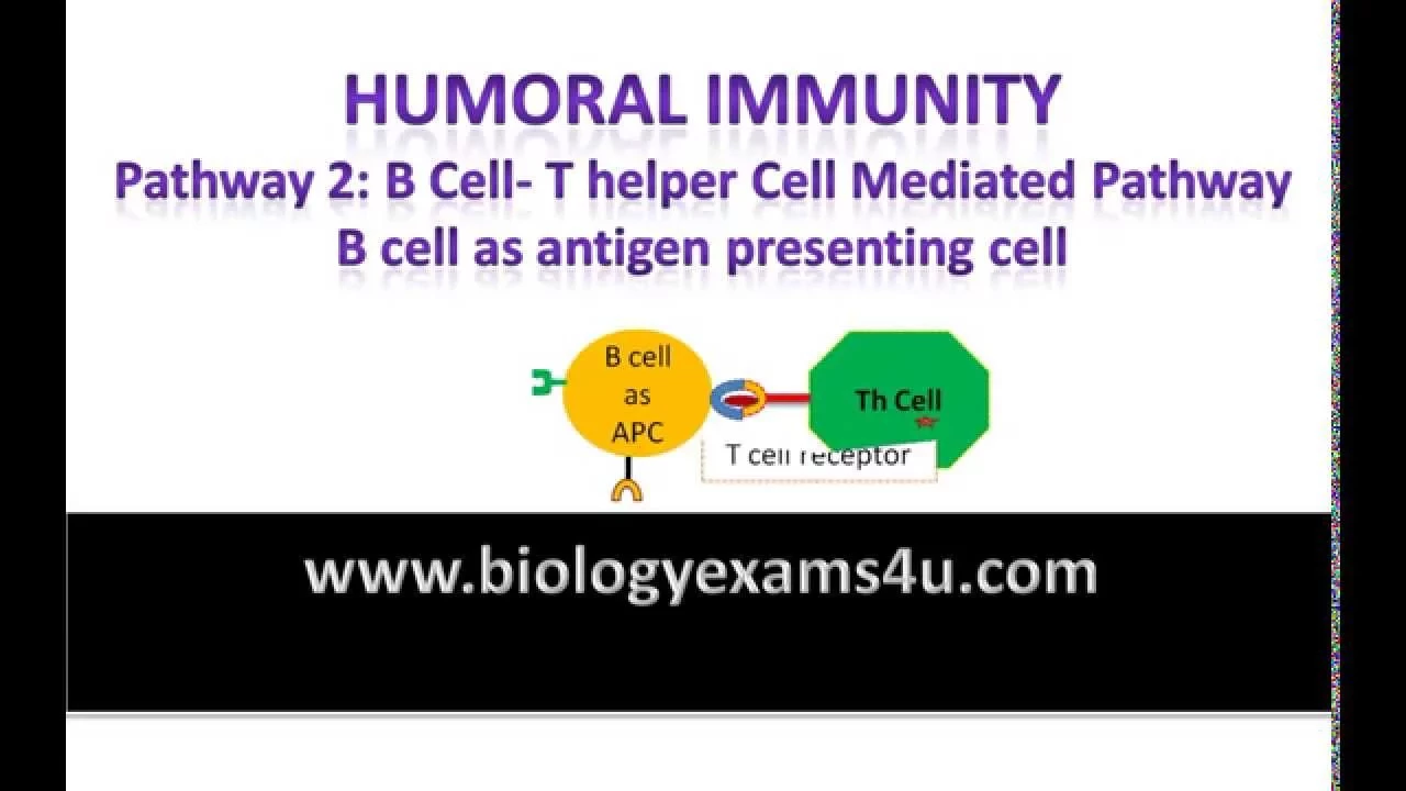
So the the cytotoxic tea cells kill uh infected cells and then we have the regulatory cells to look at and here we have again an antigen presenting cell that wants to activate a T- cell. You can see there is a peptide presented and an co-meatory molecule presented there also some cytoines which I was too lazy to put into the slide and this uh cell would get activated because it gets gets all three signals and if we imagine that this is for example an autoreactive T- cell that would then start to run around and mindlessly kill our own cells in our body then we do not want this and this is where also the the regulatory T- cells come into play. They recognize the same peptide. It's again like the pink liver peptide from from the liver cells and they uh recognize it a bit differently.
We will look at this also later and they also bind to the antigen presenting cell showing the peptides on on the MHC molecules. But now it transmits a deactivating a suppressing signal to the nucleus of the antigen presenting cell.
And these uh these molecules that they use to transmit the suppressing signal are co-inhibitory molecule and they are somewhat the the contrary molecule to the coilatory molecules. We already learned about the coilatory molecules.
These are the orange guys that the antigen presenting cell use uses to activate the T- cell. And here I put some example like CD40, CD8, CD86 where CD means cluster of differentation. This is just a way of of different delineating that this is a molecule expressed on on an immune cell also a non-immune cell and they bind to to certain counterparts certain receptors and they are called for example CD40 ligant in case of CD40 or CD28 in the in the case of CD80 and CD86 co-stimulatory molecules result in the activation of the cell and then uh it can fulfill its function. But they're also co-inhibitory molecules. This is like uh the purple one and they are called for example CTLA4 or PD1. PD1 is for example short for program death one and they bind to to ligans like CD80 and CD86 and uh PDL1 or PDL2. And you can see that there's a certain overlap. I I directly addressed this before one of the two ladies asked me the question.
CD80 and CD86 can either be co- stimulatory molecules or they can also be co-inhibitory molecules. It depends what they bind to and how strong they bind to it. So there it's a diff it's it's a question of of aidity how how strong it binds. If it binds very strong to its counterpart, this is usually a coin inhibitory signal. about it uh if it's binding not so strong or if the counterpart like the uh CTLA4 is not present on the other cell it is a co- stimulatory signal and this results then in T- cell inhibition though this T- cell will not be activated and start killing our own cells in addition the the regulatory T- cell which is a subtype of the uh CD4 positive helper cells as we learned can also produce cytoines So small soluble messenger molecules and they are also suppressing the T- cell activation. They do this into two ways. First we have suppressive cytoines and examples are interlocking 10 or transforming growth factor better TGF beta. They just act on receptors on the T- cells and they suppress here this activation of the T- cell. So if they get get triggered with these suppressive cytoines they just cannot be activated.
So this also happens not only from the regulatory T- cell to theector T cell, this also happens from the regulatory T- cell to the antigen presenting cell.
This basically shuts down the antigen presenting cell because it does something stupid here. It tries to to initiate for example an an autoimmune response.
Then we have another set of of soluble molecules. So the regular tort cells can also produce perfor and granzyme. And as we just learned this will just lead in in the T- cell that it gets it gets confronted with these molecules to undergo apoptosis. So uh then the cells is not there and this is the different and in the upper part the cell is still there but it cannot be activated. This is what we call energy. So this is a non-reactivity even if they become stimulated by the next antigen presenting cell in the next lymph node.
Whereas the second the lower part is is called deletion. And there the the autoreactive T- cell is is not there anymore. And this this is just a a important distinction. And Hannah has a question. Yes.
>> Yes. My question is is energy energy um reversible or >> it is yeah it is reversible. So if for example a certain amount of time elapses or the cell gets very strong signals to become to become activated it can be reactivated.
But I know this is going to be the next question. You probably ask me why is this the case? Nobody knows at the moment because one would say okay if if the cell is autoreactive the best way is just is just to shoot it in the head and get rid of it. Why do we keep the cell around that that has no function? It is the case. We notice this can be very elegantly shown in in animal models. In humans, it's very difficult to show this because we it's it's it's difficult let's say for for ethical reasons and technical reasons and but we know this is the case and we do not understand it at the moment.
Okay.
Do you think this is due to epigenetic modifications that we turn the energy so >> yeah I think so epigenetic modification means that the genes are are modified so there are ways it's not it's not a genetics lecture here we also not geneticists sorry I don't want to lie I am a geneticist but I've stopped working in genetics like 20 years ago um there are ways to to like constantly over long periods of time modify the the way how the genes are are used in these cells. That's what epigenetic means. Everybody has the same set of genes, but we can we can modify how they are used, how they are activated, how they are not activated.
And this is probably something that happens because these these cytoines that induce energy, they bind to receptors, they trigger a lot of signaling in the cell. And this has of course of course influences on the genes in the cell. And this is how this is probably manifested over long periods of time because we will learn this also later on. These tea cells once they turn into memory tea cells they can live for decades. So this is something needs to be maintained for very long periods of time. Okay.
>> Thank you.
>> You're welcome. So let's let's go on. So uh we have completed our trio of of tea cells. So we have the regulatory T- cells that are able to suppress to shut down both helper cells and and cytotoxic tea cells in order to I always tell my students also the best immune response has to come to an end at some point ideally once we have fought the pathogen.
So now I want to turn to the question how antigens are presented to lymphocy receptors. So how do the T- cells and for the B cells is very similar recognize the antigens that they need to react to? And to understand this I have to say it's it's very complicated as always we we are doing this in in a simplified way but in order to understand this you have to understand that there are two different types of pathogens. So we have intracellular pathogens. These are pathogens that have chosen to infect cells in our body and then they try to multiply to grow to replicate inside the cells of our of our body. And this has certain advantages because they can hide or at least try to hide from the cells of our immune system and they're not so easy for example to eliminate via antibodies.
And then there are extracellular pathogens. So these are pathogens that do not infect cells. They live outside uh of our cells. For example, in our blood, in our stomach, in in our gut uh which is outside of our body of course, but for example in our tissues, in in our brain, but not inside the cells. And this is this is as you can imagine, these are two different problems the immune system has to face and has to solve. And it is done or is coordinated by the antigen presenting cell that we already know and we will start with the left side first. So we have intracellular pathogens and the antigen presenting cells swim around everywhere in our body and they just let themselves be infected for example by this virus.
So they're just passively infected by just wandering around in an infected tissue.
And then this virus triggers the activation of of cells of the antigen presenting cell by recognizing the pathogen via the path recognition receptors. This triggers again the production of of many different proteins including cytoines co- stimulatory molecules and parts peptides from the virus proteins are presented on the surface of the antigen presenting cell in a complex with so-called MHC molecules major histoility complex proteins of type number one and then we have all three signals to activate a T- cell we have signal one which is the pep peptide with the MHC we have signal two which is the co- stimulatory molecules and signal three which are the cytoines and this now results in the the activation of the CD8 positive cytotoxic tea cells and they then start to search for uh normal body cells if this is again our liver cell on top that shows virus peptides the blue ones as part of their MHC molecules and if it finds one cell for example the lower left It then kills or it induces the cell to kill itself via apoptosis before it can produce more more virus proteins. And you can see this is very efficient because only the one cell here that uh shows the blue virus peptides gets killed. But the one on the right side that uh shows normal liver peptides does not get killed.
Okay. So um there are these MHC class one molecules that are used to present peptides that have been generated inside our cells and they contain a alpha chain. This is the the green one and a beta 2 microlobylene and this is a small globular so round protein that stabilizes the fold of the alpha chain uh and uh the alpha chain forms a peptide binding groove where the peptide is sitting. So how do the peptides get on top of the MHC plus1 molecules? So here we have a antigen presenting cell and what this cell does uh it's the same for for all cells in our body. They make new proteins all the time and once the proteins are old they must be must be replaced by new ones and also protein production is a very complicated process. uh as you might might know a lot of proteins uh do not correctly fold into their very very complicated uh structure. Around 30% of all proteins are directly defective from the start because they do not find the right folding and these old damaged used or misfolded proteins have to be have to be degraded and this is done by a system uh that is called ubiquitinulation. So there is a ubiquitine tag added to these proteins that should be degraded. This is done by a structure called the proteosome. This is like a big uh a big barrel where the proteins get defolded on one side and then the the the the peptide chain is fed through this barrel and inside the barrel are uh active sides of proteasis. It's like like a scissor and they cut this long polyeptide chain into small peptides. So it's like a a wood uh wood chipper where you put pieces of wood on one side in and smaller pieces of wood come out on the other side. And these peptides are then transported into the endopplasmatic reticulum by a molecule called TP transporter associated with antigen processing. And there they find empty MHC class1 molecules. They bind to them and then they are transported by the GI operators via exocytoic vesicles to the surface of the cell where they bind to the T- cell receptor of the CD8 positive T- cell. And this not only happens for misfolded proteins but also for virus proteins that are produced for example in the the cytoplasma in case of an infection and it also happens with proteins that were ingested by the antigen presenting cell and then transported to the cyto cytool. This only happens in antigen presenting cells because they have special mechanisms to make sure that also proteins that were taken up from outside of the cell. We looked this in the second second case can be can be fed into this MHC1 loading machinery to make sure we can also make these CD8 T- cell responses against these extracellular antigens.
So we sum this up. MHC class one molecules present antigens which are internal in origin. So they are generated inside the cell or they're generated from proteins that are generated inside the cell. The peptide degradation happens in the cytool and these MHC molecules are presented on all nucleated cells. So all cells that have a nucleus. So there are some some red blood cells which do not have a nucleus.
They do not have MHC plus1 expression.
That's why you can also make blood transfusions very easily or more easily than than organ transfusions. And the function is of these MHC monlecules to show on the surface of the cell what happens inside the cell. And the effect of the cell presenting these MHC molecules with the exception of the antigen presenting cells is that the cell gets killed because it shows virus peptides.
So if we come to the second case, we have the extracellular pathogens. they are a bit different in how they are fought. So there the pathogens are taken up actively by the antigen presenting cell. I said the antigen presenting cells like PhD students they eat everything including the the the pathogens also. Then they are inside the cell. They trigger again the activation of the cell again production of for example cytoines co- stimulatory molecules and peptides derived from the pathogens are presented on MHC molecules. now on MHC plus2 molecules and this are again all three signals uh cytoines co- stimulatory molecules and peptides in combination with MHC molecules that are necessary to activate a T- cell now it's a CD4 positive TH cell and once this gets activated it produces cytoines that then recruit for example fagocytes that eat up the pathogen or ant B cells that produce antibodies that can then help eliminate the pathogen. Okay. So there's there's a second class of MHC molecules which are called class 2. They consist of two chains, an alpha chain and a beta chain. There's no beta 2 microglobal found here. And here both alpha chain and beta chain together form the peptide binding group that binds the peptide. How do now the peptides uh end up on MHC class 2 molecules?
As I said, these are peptides generated from proteins usually that are taken up by the antigen presenting cell from from the outside. So these then end up in endoomes. So endoomes are small bags inside the cell where the material is is collected that the cell has taken up from from the outside. And these are special endoomes. These they are called endolyzosomes because the the bags fuse with other bags. They are called lizoomes and they contain a mixture of uh proteasis. So proteins that have specialized in degrading other proteins.
And these endo lizoomes are a very hostile environment for proteins because of all these proteasis which results in the proteins being degraded being cut into small pieces and then we have the peptides. That's that's cool. Now we have the peptides.
Now we need to load them onto MHC class 2 molecules.
The problem is that these MHC class 2 molecules are also produced in the endopplasmatic reticulum. And if we would just produce the the MHC molecules in the endopplasmatic reticulum, they would be directly loaded with peptides coming from the MHC loading pathway. So this wouldn't help us because uh once the the endo the peptides from the endzoome reach the MHC molecules they would already be loaded because this happens within within fractions of a second in the endoplasmatic reticulum.
Therefore we need to block the peptide binding roof of the MHC plus2 molecules in um the endopplasmatic reticulum and this is done by a so-called invariant chain. uh this is abbreviated with a capital I and a small I and this is like a chain that that hugs the MHC molecule on one side. Then it reaches over the peptide binding group and it puts just a placeholder peptide in there that just blocks the binding groove. These molecules are then uh um separated from the endopplasmatic reticulum and they fuse with the endolyzosome and this invariant chain and this this blocking peptide are very sensitive to these polyasis in the endzyosome. So this gets rapidly degraded which leads to the peptide the placeholder peptide just swimming away because it is not really specific for this this molecule. It binds very very weakly and it's mostly held in place by the invariant chain and then we have the peptide binding group available to load it with peptides and the peptides are loaded transported again to the surface of the cell and can be presented to the CD4 positive T cells.
So if we sum also this up, uh the peptides presented on the MHC class 2 molecules are um generated from proteins that the cell has taken up from from its outside. They are generated the peptides are generated in these endolyosome these endoccytoic vesicles and the MHC class 2 molecules are only presented on professional antigen presenting cells.
These are dendritic cells, macrofasages, B cells and some epithelial cells in the thymus and some endothelial cells which is not so important for us. And the function here is not that the cell gets killed but the cell that gets activated and this is the reason because only the antigen presenting cells present these MHCl2 molecules and they try to educate or find out which T- cell is useful and activate this T cell. Therefore, this has the function of activating these cells that then coordinate the immune response against the extracellular pathogens.
There also difference in peptide binding to MHC class one and class 2 molecules.
Here we have a MHC class one molecule and you can see that the peptides are 8 to 10 amino acids long and 10 is the maximum already. It's more like eight or nine. And the peptide binding happens via biochemical interactions between the the amino acids of the MHC molecules and the amino acids of the peptide at the end of the peptide. In contrast, uh the MHC class 2 molecules can bind peptides that are much longer 13 to 18 amino acids. And this is the case because this peptide binding group is not so narrow but it is more wide. Therefore, the peptides can can uh go outside on on both sides and they can just hang over on both sides. Therefore, they can be a bit longer. And you can see that the peptide binding happens via interaction between the peptide and the MHC class 2 molecules over the whole length of uh the peptide sequence. So, this is an important difference.
So what one can do if one has a lot of time and energy and money, one can can take antigen presenting cells, pulse them with with a with an antigen, then isolate the MHC molecules. As said, if you have a lot of time and energy and money, isolate the MHC molecules and then isolate from these MHC molecules the peptide that are bound and then you can sequence these peptides. And if you do this, you find peptides that you have the sequence of and they are very similar. So for example, here you have uh peptides from the first isolation. You can see they all have the same length. It's the same number of amino acids. And in certain positions uh you have very similar amino acids. For example, in the fifth position you have a tyrosine, a fine lanoline and in the last position you have a leucine. And these are the the peptides or the amino acids in the peptide that make then the interaction with the MHC molecule.
If you do this for a second isolation, you find peptides that are maybe a bit longer, but you find again on position two and the last position, you find amino acids that are very similar in their biochemical characteristics or even the same in all the peptides because these are the the points of of contact if you want to call it like this with the MHC molecules that need to be similar.
Okay. So uh now I'd like to introduce another term. We talked about MHC molecules all the time but there's also a uh another term which is called HLA.
It is the abbreviation of human lucasite antigen and uh the thing is MHC uh is a term that is used by people that work mostly in the mouse system. So a lot of basic research is done on mice and what people have done in the the 1930s and 40s of the last century they took mice and transplanted skin from one mouse to another and then they checked if this this skin graft is accepted. So it it stays on the mouse or if the immune system of the white mouse attacks the brown skin and then it gets gets rejected and that's how these major hystocompatibility complex proteins were discovered. So these are the proteins that are important to to decide if uh a skin graft gets accepted or rejected.
Similar results or similar experiments were done in in humans. Of course there you do not just wildly graft skin. That would be not so nice. But what you can do is you can take blood from different humans and you can just take small drops of of blood from different persons and mix them and then two different things can can happen. If the blood is compatible, it will it will stay liquid.
If the blood is not uh compatible, it will coagulate. So it will form uh aggregates and it will turn from a liquid into something that is that is more more less liquid and and forming these these clumps. And the antigens that are involved in this reaction were called human luccoside antigens. And once once uh one could could study and could clone and isolate and identify these genes, one found out this is the same. Yeah. So the genes that are responsible for for uh transplantation success in in mice and for the success of blood transfusions in a way in the end in humans are the same and therefore people that work in the mouse system will talk about MHC molecules and people that work in the human system will talk about HLA molecules. So in the following I will just talk about MHC molecules to make it make it more simple. If you looked at the genetic organization of these MHC genes they are found uh on chromosome number six and there we have uh the MHC molecules and there we have three different copies of the alpha chain which are called HLA A B and C. They are not not in the correct order. I know this has just to do with their uh discovery. A was discovered first and then B and then C before knowing where they are located in the genome. And with a certain distance on the same genome, we have a whole set of genes. Uh for MHC molecules, you see they consist of a alpha and a beta chain. Uh and the MHC or HLA molecules in in the human system are called DP, DQ and DR. And they always have an alpha and a beta chain coded by an A and a B Gene. And for the DR molecule, we have even two B genes. One of them is already deactivated in in um in some of us already.
So we have these these genes here. Then we have accessory proteins. There we have for example TU. We know TUP already. This is one of the pink ones.
This is the transporter that transported the peptides into the endopplasmatic reticulum to be loaded onto MHC molecules, MHC class1 molecules. And then we have some chaperones. Chaperones are are molecules that help other molecules to find the correct folding.
And chaperone is a is a term coming from the last century. So the the young ladies like like Hana and and Alex were not allowed to go out alone uh and to make uh make forbidden stuff with with with male persons and they always had a chaperon with them. This was usually a old older woman that that made sure that that nothing inappropriate happened to them. And here chaperon in the biochemical sense is a molecule that makes that makes sure that no inappropriate foing happens with these molecules. Okay. So after looking at how they are organized on on the chromosome, if you look at the bigger picture at the whole population of of humans, you will find that there are different variants of the the genes that form uh the proteins for the MHC molecules. And you can see here that for the MHC class 2 molecules, there are as I said three different uh variants DP, DQ and DR. And among these variants, there are at least 350 to 1,200 different variants of the beta chain. And a lot less variability exists for the alpha chain. So these are just different variants uh in the human population that are able to bind different peptides. And it's even more pronounced for the MHC molecules. So in in total, there are more than 6,000 different MHC class1 molecules. This allows the the whole population of of humans to uh uh present different peptides from different pathogens more or less efficiently and form immune responses against these these pathogens more or less efficiently. Sorry always persons in a in a population that can present for example uh I don't know uh influenza peptides very very well and do not get sick and other persons can for example present HIV molecules very efficiently and do not get sick from HIV. This is a very very small percentage that that get HIV infected but do not develop develop AIDS. They are called elite controllers or master controllers. uh and this is just a way to broaden uh the protection against different pathogens not on on the individual level but on the the population level.
So if we look where this this variation comes from, we can sequence a lot of different MHC molecules and we can check which amino acids differ in these different different variants. And you can see we can uh divide the MHC class one alpha chain into an alpha 1, alpha 2 and alpha 3 domain. And you can see that most of the frequence variability happens in the alpha one and two uh domains. And if you look at the beta chain of the MHC class 2 molecule, you can see this is a beta one domain on top and a beta 2 domain at the bottom. You can see that the sequence variability is much much higher for the better one domain. And if we color the respective amino acids in blue that are very variable, you can see that these are exactly the amino acids that form the peptide binding group. This makes a lot of sense to have sequence variability in the the regions that bind to the peptides and to keep the rest of the molecule more or less conserved. So this allows make sure that it's able to to form the correct three-dimensional structure.
Yeah. And as I said this this polymorphism so the the fact that there are many different variants of these molecules that can bind different peptides makes sure that on a population level we are able to recognize a lot of different peptides from a lot of different pathogens.
So two different uh phenomena come together and this is usually where a lot of my students have for some reasons problem with. The first one is called polymorphism. This means they have more than one variant of a gene. you have a a blue variant of a gene and a green variant and they can be expressed at the same time uh co-ominantly.
So then you have green and blue molecules on the surface of your cell.
Then you have polygeni. So this means you have more than one gene for for a certain function. You can have a red one, a yellow one and a green one and they are also expressed at the same time. Therefore you have green uh red and yellow molecules on your cell. And if you combine both uh mechanisms polymorphism and polygeni you can have even more. So you can have for every for every gene you can have different variants and this results that you can have six different uh six different variants of of one kind of molecule with three different uh genetical loi because we have as you might have know we have two chromosomes of of most chromosomes one from the father and one from the mother. If we look at how these genes are inherited from from one person uh to the next uh one has to keep in mind that they are all on the same chromosome and they are all very close to each other.
This means that they get inherited as a package all of them together. The far further genes are away from each other on the chromosome the higher there is the frequency of re combination. because these genes are so close together on on the same chromosome that the frequency of re combination that splits them is is very low to basically non-existent. So this one set of genes is called a hloype and every one of us has two hloes. One is from the biological mother and one is from the biological father. And now uh if these this theoretical biological mother biological father have children their hlo types get also split among them. So you can see the first child has here both upper hlo types from mother and father. The second one has the upper hlo type from the mother and the upper the lower hloype from the father and so on. So there is a 25% that children from the same biological parents have the same habypes and that's why when we have the need for one of these children to get an organ uh transplant that we first look at at the siblings because there the chances is much higher to find a compatible compatible donor for example for a kidney or for a bone marrow transplant and only if this doesn't work we will look at more distant relatives like mother, father, uncles, aunts and if there's no no match then we can we can look at these these registries where thousands or millions of peoples are registered and Alex please you have a question >> but the mother will never or the father will never have the exact same um MHC molecules because we have from mother and father so it's only 50% Right.
Uh maybe usually yes I would say usually yes but there are also mothers and fathers who realize that they are very closely related afterwards. So this can also happen and there is only I said there are 6,000 uh different uh MHC class one molecules but there are also billions of people on the planet. So just by chance you might have might have the chance that they have maybe out of these uh there's six MHC class one molecules and six to eight MHC class 2 molecules maybe five of them fitting is is already enough for transplantation but of course it is much much more uh um prominent to find this in in siblings than in the parents.
>> Okay thank you. Yeah. But but crazy things have happened as brothers and sisters have have have produced children without knowing. Yeah. This is Yeah.
Okay.
So what what these uh MHC molecules do is they present peptides in the peptide binding group to the T- cell receptor. And you can see that the T- cell receptor not only recognizes the peptide but it also recognizes structures on uh the MHC molecules. And this is something we will look at in in the next video. And the T- cells are trained to recognize uh peptides only when they are presented on our MHC our own MHC molecules. So my T- cells are trained to recognize uh peptides on my T- cell MHC molecules whereas the T- cells of Alex and Hana are trained on different MHC molecules.
So um as I said before already we have three different types of T- cells and they are also characterized by their expression of either CD4 and CD CD8. I said is this the T-helper cells are CD4 positive the cytotoxic T cells are CD8 positive and the regulatory T cells in the end are a subtype of of the T- helpper T cells so what is notice the CD4 and CD8 CD4 and CD8 are co co-ceptors to the T- cell receptor here you see them uh the the CD4 is more like a a longer molecule whereas the CD8 is a more compact uh global So like my fist is is is the folding molecule and they are co-expressed on on either CD4 or CD8 T- cells. That's hence why they are named this way and they uh are found next to the T- cell receptor and they uh do not bind to the peptide binding group. So they have nothing to do with peptide specificity, but they bind to the sides of either CD4 to MHC plus2 molecules or CD8 to MHC plus1 molecules. And they stabilize the binding of the MHC peptide complex to the T- cell receptor. And that's that's why we have them to make this binding more more stable, more longerlasting so that the T- cell can stick long enough to the target cell or to the antigen presenting cell to become activated or to fulfill its effective function.
Alex, yes, >> you said that all immune cells produce MHC1 which have a nucleus.
Um and you said also that only antigen presenting cells can activate CD8 positive T- cells.
So maybe just give a little um highlight why only APCs can induce the immune response in CD8T cells and not other immune cells because other immune or other cells also produce MHC1.
>> Yes. So the question if if I got it correct is basically all cells have MHC molecules but only the antigen presenting cells are able to activate uh initially activate the T- cells. Once the T- cell has has been activated by the antigen presenting cell once it is fully authorized to form an immune response.
It can start around and kill all the the virus infected cells for example that express virus proteins on the MHC plus1 molecule. The difference between antigen presenting cells and normal body cells is both present signal one MHC plus peptide but only the antigen presenting cell and only if it has found a pathogen uh expresses signal two and three only the antigen presenting cell has co- stimulatory molecules and cytoines and therefore only the antigen presenting cell is able to activate the T- cell.
This is also something we will look at in the next lecture. Does this answer >> this was the question?
>> Yeah.
>> Yeah.
>> Thank you for for asking this clarifying question. So as I said um the T- cell receptor not only recognizes the peptides but also structures on the MHC molecules and they are trained on our MHC molecules. How this works we will see in the next lecture. And this leads to the T- cell recognition of antigens being so-called MHC restricted. So this means they only uh recognize peptides if they are in the peptide binding group of MHC molecules and only in those uh MHC molecules that belong to our body. Here you can see three different examples. In the first one we have the correct peptide for our T- cell receptor and the correct MHC molecule. The T- cell will get activated.
The second one in the middle, we have the exact same peptide, but it gets presented on a different MHC plus one molecule that this T- cell was not trained on it and it does not recognize that. So there's no T- cell activation.
And in the third example, we have the same uh the correct if you want to call it like this MHC class one molecule, but a non-matching peptide. So this peptide does not fit to the T- cell receptor and also in this case the T- cell does not get activated. So the T- cell only gets activated if the peptide matches and uh the correct MHC is there to present the peptide.
So how do the specificities of the lymphocy receptors now are generated? We talked about it in the beginning already that there are millions of different of different T- cell receptors and if we look at uh the number it is approximately 1 * 10 to the 8 different T- cell receptors this is around about 100 million of them and the the sum of them is called the so-called lymphocy repertoire this is the all these 100 million receptors form our repertoire and this determines what we are able to recognize. Uh so I said it's 100 million roughly and now I'll ask the two of them if they know how many genes are in a human genome. I already mentioned it in the beginning.
Alex, do you know?
>> Yeah, you said 19,500 to 23,000 genes.
>> Yeah, exactly. So Alex at least is is listening to me. Hana also, of course, I know. uh and if we put these numbers in our graph you will see that this this is like way way way lower than these 100 million so it cannot be that there is one gene for every T- cell receptor so there must be a different mechanism and this mechanism was discovered by a Japanese scientist in the 1980s that was uh Mr. Tonaggava and he looked at the T- cell receptor actually looked at Bisa receptor but it's the same uh and we are talking about T cells today therefore changed it to T- cell receptor he looked at the T- cell receptor and looked at the gene and his initial theory was okay there is one gene for every T- cell receptor and then he he sequenced the T- cell receptor there was a new technology in the in the 1980s that you could sequence the genes so determine the sequence of of of the genes how they are made made up of which bases they are made up and he found that that there's not there are not different genes there's a set of of segments uh in the genome uh of the T- cell and these are like Lego bricks that can be built together uh to form the T- cell receptors that there are pink ones and green ones and yellow ones and uh the cell picks one of these these uh these segments and puts them together in different combinations and that's how these different receptors are made.
Crazy. So, how does this look? So, we have the genomic DNA. So, this is the DNA in the the chromosome and there you have uh if we look at uh the the first first chain of the T- cell receptor, the alpha chain, it is encoded by the so-called alpha lucose. There you have a variable and joining segments. That's just how they were called. A big capital V for variable and a capital J for joining.
And they're called V alpha for the variable genes of the alpha locus. And there are 70 to 80 uh B alpha segments.
They have like a black leader sequence that is needed for their um for their expression. And then there are 61 uh J segments. And now the cell by mechanisms which we will look at in detail in the next slides picks one of these uh these segments just randomly by chance. So here the third uh V alpha segment was chosen and the first uh J segment but could also be the the 71s uh V alpha chain and the 60s J segment and they are put together and you can see that the that the sequence between them. So everything from V alpha 4 to uh to the last one gets deleted. It gets cut out by recombination events. We will look at the mechanism in the next slides. And this leads to a irreversible loss of genetic information. Meaning the cell cannot go back to the initial state and try it again because all this the sequence information in between was lost.
Then uh also uh the other parts that are not needed are cut out. This gets transcribed into an MRNR that's gets uh spliced to get rid of the inrons and then it gets translated into a a protein and this protein then folds and forms the the alpha chain of the T- cell receptor and you can see there is a constant part which is called C alpha that forms like the the lower base of the receptor. This is always the same because this is a complex molecule that somehow needs to fold. If we randomly put everything together, it will never form the correct folding. But the tip of the T- cell receptor where it recognizes the the peptides with this is then made from these different segments. This is also true for the other half of the receptor which is called the beta chain.
There we have also V beta and J beta but also D better. So D stands for diversity and they also put together and therefore by these this set of segments that are put together randomly we form different uh T- cell receptors in every cell and so this is a a random random event.
How many how many segments do we have?
So for the typical T- cells uh these are alpha beta T- cells expressing a alpha beta T- cell receptor we have 70 to 80 uh alpha chains alpha V alpha chains and 52 in the beta chain we have no diversity uh segments in the alpha chain and two in the beta chain and we have 61 joining uh segments in the alpha chain and 13 in the beta chain there are also Additional T- cells they are called gamma delta T- cells. Uh they are very special T- cells. It's not completely understood what they do and how they do it. Uh you see that there are many less uh segments and it's not completely also clear if there are certain segments that that participate in actually forming receptors. That's why there is a question mark after the four. And these gamma delta t cells are important in the gut for example to fight uh pathogens but they for example do not need uh MHC molecules presenting peptides to get activated. So it's not completely understood. Don't ask me why and how it is. Uh but they seem to recognize mostly lipid antigens. So antigens derived from from fatty uh fatty acids fatty molecules that are part of certain certain pathogens. And uh this is just a second type of of T- cell receptor and T- cell T cell T- cell type. But the vast vast majority of T- cells are these alpha beta T cells expressing the alpha beta T- cell receptor.
So how do we now uh combine these segments and this is this is very complicated. It will take us several slides but I will do my very best to explain it. and we will look at one variable gene segment and one joining gene segment that we want to combine. So this is the the VI one is the the pink one and the J1 is the yellow one and they have so-called RSS sequences. So recombination signal sequences in front or behind of them and these sequences are recognized by a molecule that is called recombinase activated gene one two. This is abbreviated RAG12 and this has binding sides for these RSS sequences. And what happens this rack molecule has two binding sides. First it binds uh the the blue uh RSS belonging to the V segment and then it binds uh the green RSS belonging to the chain segment. And this results in both segments that were chosen to be combined to be located very close to each other and be fixed to each other in a certain uh threedimensional orientation. So that here the the genes can be can be cut and put put together. This this uh bringing two RSS uh stretches together is called forming a sinapse.
So from this sinapse the the rack molecule then cleaves uh the RSS off and the RSS gets stay attached to the to the rack rack gene rack protein sorry rack protein and uh the the segments that we want to combined in this process uh are separated and now we have we have a problem because we have cut our chromosome into three parts. Yeah, the middle part that we want to get rid of and the first part with the V segment and the second part with the J segment.
And if we now do nothing, everything will flow into different directions and our chromosome will be destroyed. So this would be a catastrophic event for the cell. The cell would die and uh this we need to need to prevent and this is prevented in the next step. But uh what we need to keep in mind and what we need to make ourselves aware of is that the the DNA strand here is is cut uh to get the the RSS out and it's cut it's it's cutting results in in different uh endings of the DNA. For the the V and the J segment we have coalently closed DNA hair pins. So a DNA consists of of two strands and these two two strands in this double helis and these two are joined together at the top so that the that the ends cannot cannot flow in two different different directions and the other ones are just open but this is not a problem because they are fixed to the rag anyway. So now we we continue this is basically the same and now additional molecules bind to the DNA. They are called KU78 and 80. 78 and 80 is the the molecular weight of these proteins. They put them on a on a gel. This is just how big they are, how how heavy they are. And um these now take uh on the one side the the RSS sequences and phosphorolate them. So they add a phosphate. So they put energy in there. And on the other side uh they bind our two loose ends uh so that they cannot cannot flow away. So they are fixed also in position and then to these uh Ku molecules we um attract a mot molecule that is called DNA protein kynise.
This has binding sides for these KU molecules. And this just results in these two loose ends of our chromosome being fixed there in position and again fixed and in positions so closely together that we can put them back together. Yeah. So this just is is the way of putting them in the right orientation to each other that they cannot flow into different directions and that our chromosome uh is not destroyed. And what this molecule now does it opens the hairpins. So you can see that there's a little maybe cut that is made in the the DNA sequence that we have now two two separate DNA strands again and then uh another molecule is is recruited as uh being part of this complex and this is called the terminal this oxyonucleio transferase TDT and with the other with the other segments with the RSS. Nothing happens.
So, let's just put it on on the right side that we do not lose it. Then this TDT molecule processes the DNA ends. It puts them together. So, it connects the the pink V segment with the yellow J segment and liates the two strands again together. And you can see now both ends of the chromosomes are again fixed to each other. But they look different.
Yeah. because uh because if you look there is not just the the pink V segment and the yellow J segment there is also a red uh green and blue sequence in there that was not there before. So there has been some some sequence information has been added on this the other side our our um RSS sequences are also liated together. So liase is a protein that connects DNA molecules and uh at the end we have formed our coding joint and our signal joint. So the signal joint is if you look at the original germ line configuration is just the RSS sequence together with all the stuff that was between them. This forms like a circular plasmid structure that gets lost during uh following uh DNA or cell cell uh cell divisions because it gets diluted out.
And on the other side we have now the V segment and the J segment very very close to each other basically directly next to each other with a little bit of of sequence information um put put in the middle between them and this is called the the coding joint because this is then coding for the T- cell receptor forming the the protein of the T- cell receptor and this is imprecise. Yeah, the the right side was precise. You can see exactly the blue and the green moment. Nothing else, nothing more, nothing less, but uh the coding coding joint is imprecise because there was information added or even subtracted as we will see. Okay, just answer two. They got it. That means somebody else can also get it. It's complicated.
Yeah. Yeah. Hana.
Um so the somatic rearrangement and the rack dependent rearrangement only happens for T- cell receptors and B cell receptors not for the MHC molecules.
>> No the MHC molecules are just taken as they are from from their genome. They are also not not encoded in the genome as as segments. they are fully uh finalized functioning genes that just get translated into a protein put on the surface of the cell. Here there is no there is no finalized functioning T- cell receptor in the genome of the cell.
It exists only as these segments and they first have to be put together and as as Hannah just rightfully said this process of putting it together is called a rearrangement uh process and because here a V and a J molecule were put together it's called VD VJ rearrangement if there's also a D segment involved it's called a VDJ rearrangement Alex >> I would like to add that um this kind of rearrangement. We also see in DNA repair. So if you're into DNA repair, maybe you know already this mechanism.
>> Yeah. But they the I mean in the end what we do is we damage the DNA by by cutting it by trying to cut the middle part of asset. It's a big problem to have three one chromosome turned into three fragments and then we need to put them together. This also happens of course and other processes. It's part of of normal life that damage occurs and uh therefore these these dam repair process are also of course relevant in other scenarios but here it is like a a damage that gets intu introduced on purpose in order to be able to to recine these these these these two fragments in the end >> in the end this is a cleverly used mechanism that already exists when you have a >> reuse reuse of resources Yeah, in in the end. Yeah, >> exactly. So now I would like to look at this this final part in a bit more detail to understand or for you to understand how and why this this this coding joint is imprecise in the end and how this additional uh information ends up because this is very important. And if you would just combine these few hundred um segments randomly with each other, we would reach a certain certain amount of certain number of T- cell receptors, but we would never reach the 100 million.
Therefore, it's not not enough segments.
So there must be a process to add even more um variability in the T- cell receptor. So if you look again at at um the stage where the the two RSS uh stretches are bound to rag 10 or two.
This looks like this. You have the pink V segment and the yellow J segment. And you have some additional um sequence information or part of the sequence information of these genes.
And then we form these these hair pins at the at the coding ends. And you see that there's there's stuff missing on both uh on both segments. These are the RSS stretches that are removed. And now we have these these coalently closed DNA hair pins. Then uh as I said these hairpins are opened and when a hairpin is opened then this this the sequence just folds over and uh we have a long long longer uh strand on one side and one that is a little bit shorter on on the other side of the DNA molecule and what we generate here are so-called palindromic pucleotides. P is just the abbreviation of palendroic and a palendrrome is a short DNA sequence that repeats itself.
Uh because we have like A binding always to T and G always binding to C. And if you fold this over, if you think about it, this will always uh result in in like a mirror. If you put like a mirror between the the first two bases and the the second two bases in this case the sequence on both sides of of the mirror is is the same. This is what is called a palendrome.
Okay.
Then uh we have the TDT molecule and this is a crazy molecule that just adds randomly some bases uh to to the end of these palindroic nucleotides.
And this is purely by chance. Yeah. Puts an A, puts a C, puts a G in there.
Doesn't care what. It just takes what it finds and puts it in there. So we have some some new sequence information added that was never there before. Yeah.
only the palendroic uh information was there before but the nucleotides so these are the the non- template nucleotides that were not coded in the DNA before are added randomly by chance okay then we try to fit these two strands together this this works rather nicely because that's that's the example I made up and you can see that from from these nucleotides that were added there is a stretch of three nucleotides this T A GC and A T that fits together but the A on the end of the J segment doesn't fit and this is then removed by uh endonucleazes. So there are proteins that that cut off um these non-fitting nucleotides and then we have a partial overlap of these two strands but it's not not a complete double helix is is there because uh they are just just holes in there and this is now filled up by the DNA li4 XRCC4 molecule. This this fills up checks. Okay, there is an A at the end of the V segment. So we put a T, then we have a T, then we put a A and then we have an A, then we put again a T and so on. So it fills up all the holes and then liates together both strands at the end. And then we have then we have uh our segments joined and you can see we have on one end you have the V segment on the other hand you have the J segment on in the middle you have the N nucleotides the non-mplate nucleotides that were not coded before they are new genetic material or new new material in these molecules new sequence information in these molecules and then we have uh the palendroic that sequences that result from from opening the happens. So that's why the the coding joint is imprecise. And with respect to the V and the J segments, this is there's a sequence information that results in a functioning uh protein but the the other stuff in between is more or less random. Yeah. And in most cases or in many cases this results for example in premature stop codons inpoint uh mutations or the the repair mechanism doesn't always have to work. So this is not guaranteed that this always results in a functioning uh functioning molecule functioning protein.
Okay. As I said, as this is not always guaranteed to work, the cell has basically two chances to do this because it has one set of of segments on one chromosome and another set of of segments on on the other chromosome. If it always always starts with only one chromosome trying to put this together in this rearrangement process and if this is successful it transmits a signal to the second set uh that stops um uh the rearrangement of the segments. So it's not completely understood but it was shown that for example the leader sequences and DV alpha sequences are highly methylated afterwards and this just prevents for example the binding of of these rack molecules and this then prevents that a second uh variant of a T- cell receptor is put together and this is called aic exclusion that only one set of of genes is always recombined if it doesn't work in the first chromosome, the cell can try again with the second one. But if it doesn't work with the second one, then the cell has in German we say peskhat it didn't work and then the cell usually dies and then we have to try again with a new cell. That's why we make so many many tea cells. So there are 100 million T- cells made every day in your thymus and most of them uh do not form functioning T- cell receptors. So 95% of them are uh not functioning in the end.
And Alex has a question.
>> You said that um we make like 100 million tea cells in a day in our >> thymus.
>> Thymus and um you said that the most of them is waste. So this is a loss of resources, isn't it?
>> Yes. And uh we will also learn in the next lecture that maybe out of the 5% of of T- cells that make it through the end a good fraction of them is even autoreactive. So it's like even more uh more dramatic. But the thing is it's it's a compromise between uh losing or wasting a lot of cells and material but it is like the only way at least the only way that that our body has come up with with generating 100 million uh different receptors from uh a few hundred gene segments. Yeah, this is just a compromise and you just take it for granted that most of the the the stuff you you put out is junk. Yeah, this is it's just the way how to do it.
There's no other way. Otherwise, you would have I don't know thousandfold more genes for T- cell receptors. Then then you have other genes and uh you know how how many mutations happen over time in all of us which can lead for example to cancer or stuff and if you have thousand thousandfold more genes you will have thousandfold more mutations. There's no way around it. So this is just a compromise in the end.
>> Okay thank you.
Okay. So from this allelic exclusion results that um one T- cell T- cell and also one B cell only expresses one type of T- cell receptor. So this is this is the question that Hannah asked in the beginning. Yeah, there are of course many many several thousand several 10,000 T- cell receptors and B cell receptor on every cell but they are always the same because of this alyic exclusion.
So where does lymphocy development happens? So as I said uh we have this hematopetic stem cell that develops in the common lymphoid progenitor. This then turns into the T- cell progenitor.
And here in the thymus, we have prot cells that develop into pretel. You see they have like half of a a T- cell receptor already because they did this rearrangement on the first first chain.
Then they if they successfully do this, they get some some survival signals that they can recover a little bit. Then they try on the second second uh chain of the T- cell receptor. And if they have also successfully done this formed a functional fully complete T- cell receptor, they start to express both CD4 and CD8 and then they get checked uh if they bind to to MHC class1 molecules better than to MHC then they allowed to keep the CD8 expression if it's the other way around. If they bind better to peptides presented on MHC uh class 2 molecules then they're allowed to keep CD4 because in the end the T- cell receptor is a randomly generated structure. And now we just look at what we generated and does it bind better to one or the other type of MHC molecule.
And then they leave the thymus as mature T- cells. And the same also happens in the bone marrow. So we have a prob cell that first uh puts together uh the the heavy chain of the the the antibbody.
It's basically the same or from a same progenitor as the T- cell receptor but developed into a different direction.
And then we do it for the other half of the B cell receptor and then the mature B cell is allowed to leave uh the bone marrow. Just for for completeness sake we have also a proenk cell that develops into a mature enk cell but this happens neither in the thymus nor in the bone marrow but in the periphery meaning the rest of our body.
So the T- cell receptor complex is consisting of not only the T- cell receptor but there are many other molecules also involved. So there is CD3 molecules there are epsilon delta gamma chains and they bind to the outside of the T- cell receptor but they also have like uh sequence um motives inside the cell that then transmit the the information that a peptide has bound to the T- cell receptor into the nucleus of the cell and there's also the CD3 seta uh complex protein that also is very important for signal transduction. So the T- cell receptor consists of a variable region that is different because it is made from these segments that are randomly combined and this is needed for antigen recognition and we have a constant region that is the same for all tent receptor that is necessary for folding of the T- cell receptor in terms of the T cell receptor by itself and also for signal transduction in the T receptor and all the the accessory proteins.
So um if now the T- cell receptor recognizes a peptide on a MHC molecule as I said the T- cell receptor is not an isolated molecule but part of a complex with all these accessory proteins and if the cell also the antigen presenting cell presents co- stimulatory molecules to the T- cell this results in uh an activation of the T- cell and what basically happens is that calcium ium uh ions are allowed to come into the cell.
They bind to a protein called calcinorine. This then goes into the nucleus of the cell, activates a transcription factor. A transcription factor is a protein that binds to many different spots in our genome and activates or shuts down many genes. And this transcription factor is called NFAT.
that is an abbreviation for a nuclear factor of activated T- cells. So we know where it is relevant already by name and this triggers then uh the activation of many genes and one of them one example the most prominent example is the interlocking 2 gene. So this gene gets activated we make an interlocking to mRNA interlocking 2 protein. this protein gets uh gets uh secreted outside of the cell and this is a cytoine that binds to its receptor. So interlockin 2 binds to the interlockin 2 receptor and this triggers a molecule called mtor mamalian target of rapamy and this is a another regulator that drives then the division and the activation of the cell.
So this allows all the processes in the cell that that the cell needs to to divide into daughter cells and um to form all these millions of of daughter cells that are needed from the T- cell to fight the pathogen.
The problem now is as we just discussed also with Alex is that these T- cell receptors and without spoiling you it's the same for the B cell receptor are generated randomly also generating autoreactive T and B cells. So T and B cells that recognize with the receptor our own cells and tissues and they would inevitably uh kill us if we wouldn't have mechanisms to control them. And this is what we are going to talk about in the next lecture. So we're going to talk about tolerance in the next lecture and we talking about NK and NKT cells.
So if we sum up what we learned today, we looked at T- cells and we learned that they are one of two types of cells in the adaptive immune system. So they are also B cells which we will look at in the next next lecture. And we distinguish between three classes of T- cells. We have the helper cells that orchestrate the immune response by producing cytoines. We have cytotoxic tea cells that directly kill infected cells or cancer cells. And we have the regulatory tea cells that are able to shut down immune responses if we no longer lead them or if we make them for example against our own cells and tissues.
We learned that the T- cell receptors are not coded one by one as as final complete molecules by different genes but they are coded by by segments that are randomly uh combined with each other also adding some some information by the summatic rearrangement. We also learned that um the T cell receptor binds to peptides in combination with MHC molecules and it recognizes both the peptide and the MHC molecule. And finally, we learned that the T- cell receptor consists of a variable region that is different for every T- cell receptor and is responsible for binding different peptides and a constant region that then transmits the activating signal. And we also learned that the T- cell receptor is not not an isolated molecule but part of a really complex of different different proteins that together transmit the activating signal.
With this we had at the end of the second lecture and I would ask the two of them again if they have any additional questions.
>> No, I asked all everything already.
>> Okay. Thank you, Alex. Thank you Hannah.
Uh really appreciate that you joined me for this again three-hour long recording session. But uh if you dear listener have uh additional questions just put them in the comment section of this video and we will try to get back uh to you with an answer as as fast as we can.
Other than that uh we thank you for watching. Uh we hoped we could bring a little bit of light in this complex topic of of T- cells and how they are and what they are and how they get activated. We will continue as I said with the next video on uh tolerance and NK and NKT cells and we hope to see you all again in the next video with this.
Thank you. Stay uh stay hungry for knowledge and see you in the next video.
Bye >> bye.
[Music] >> Cuz Alana is more warm. I'm somewhere in the middle and you look like a ghost.
>> But this is already the warm line because I can also take this one and now I'm I am a ghost.
>> Yeah, because Oh, side.
>> Oh, oite.
>> Oh, side. Uh-oh.
>> Uhoh.
>> We have learned that teaser.
>> Okay, that's not Alexa.
[Music] Are we ready as as ready as we can get?
Probably.
>> I can take another coffee. But >> yeah, go take a coffee before we start.
>> What about a pee pee break?
>> Yeah, then we make a pee break. I take coffee and we start afterwards.
>> We can manage to dis >> Yeah, that peep peep synchronization >> to 30 30 minutes. Okay. So So 4 minutes.
It's not It's not the old guy that wants the PB break. Yeah, I just want to >> make this clear. But okay, >> I just wanted to say um it's not the old guy who wanted the people break, but it's the old guy who takes the longest and he has recorded that. Yeah.
>> Now we have a break. Maybe we make a Pippy break.
>> Pippy break again.
>> Pippy break. Okay. Alex has Check your prostate. Check your >> prostate. I don't think that I have that. I just have a high consume of coffee.
>> Yeah.
>> Then I can eat my banana.
[Music] >> Not on camera.
[Music] We will look at that in a distant lecture in the past.
>> Time has is not not the same concept for me than it is for you. I'm a trans transcended being as a supervisor.
>> Where do I start again? Where do I start again?
[Music]
Up Next
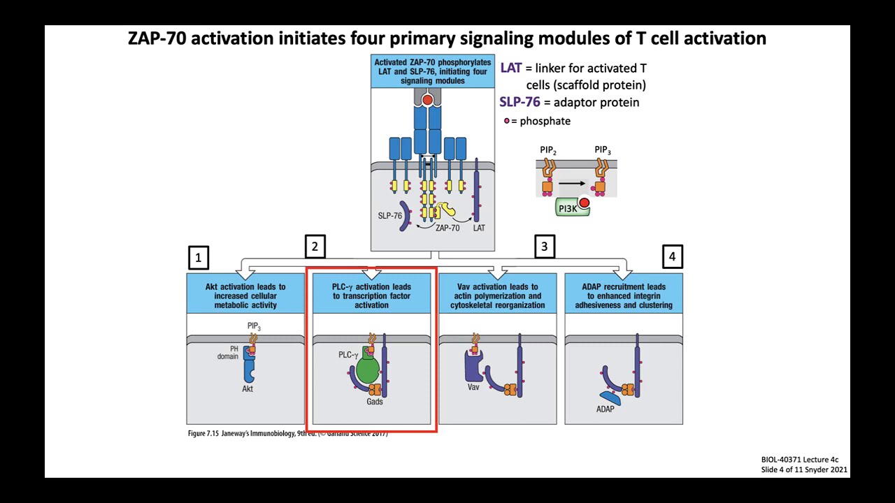
T Cell Signaling and Activation | Immunology Lecture (UCSD Extension)
@annelisesnyder4364
22.6K views•2021-03-17

Bacterial Communication: Quorum Sensing and Biofilm Formation Explained
@JHUAAP
12.6K views•2011-06-28

Enteric Nervous System Explained: The Gut's Brain | Neurobiology Lecture
@alumniu6029
438 views•2018-09-12

Bacteriophages: Earth's Deadliest Killers and Future Antibiotics
@kurzgesagt
34.6M views•2018-05-13
Related Study Plans & Knowledge Roadmaps
Structured learning paths in Biology