

















![★★초음파 유도하 정맥관 삽입술 Axillary v, femoral v, PICC/ MC 길은미 [2020 SECCI 온라인연수강좌]](https://i.ytimg.com/vi_webp/ukcyhZgUAcE/maxresdefault.webp)





The RUSH protocol is a systematic three-step algorithm for evaluating critically ill patients in shock, organized around the principles of 'Pump-Tank-Pipes' (or alternatively 'HAI MAP'): assessing cardiac function (the pump), evaluating preload status and fluid responsiveness (the tank), and examining major vascular structures including the aorta and deep veins (the pipes). This protocol enables non-invasive bedside assessment to differentiate between types of shock (hypovolemic, cardiogenic, distributive, obstructive) and guide appropriate therapeutic interventions.
RUSH Protocol: Rapid Ultrasound in Shock Evaluation
Added:Classification and pathophysiology of clinical shock, including cardiogenic, hypovolemic, distributive, and obstructive shock states.
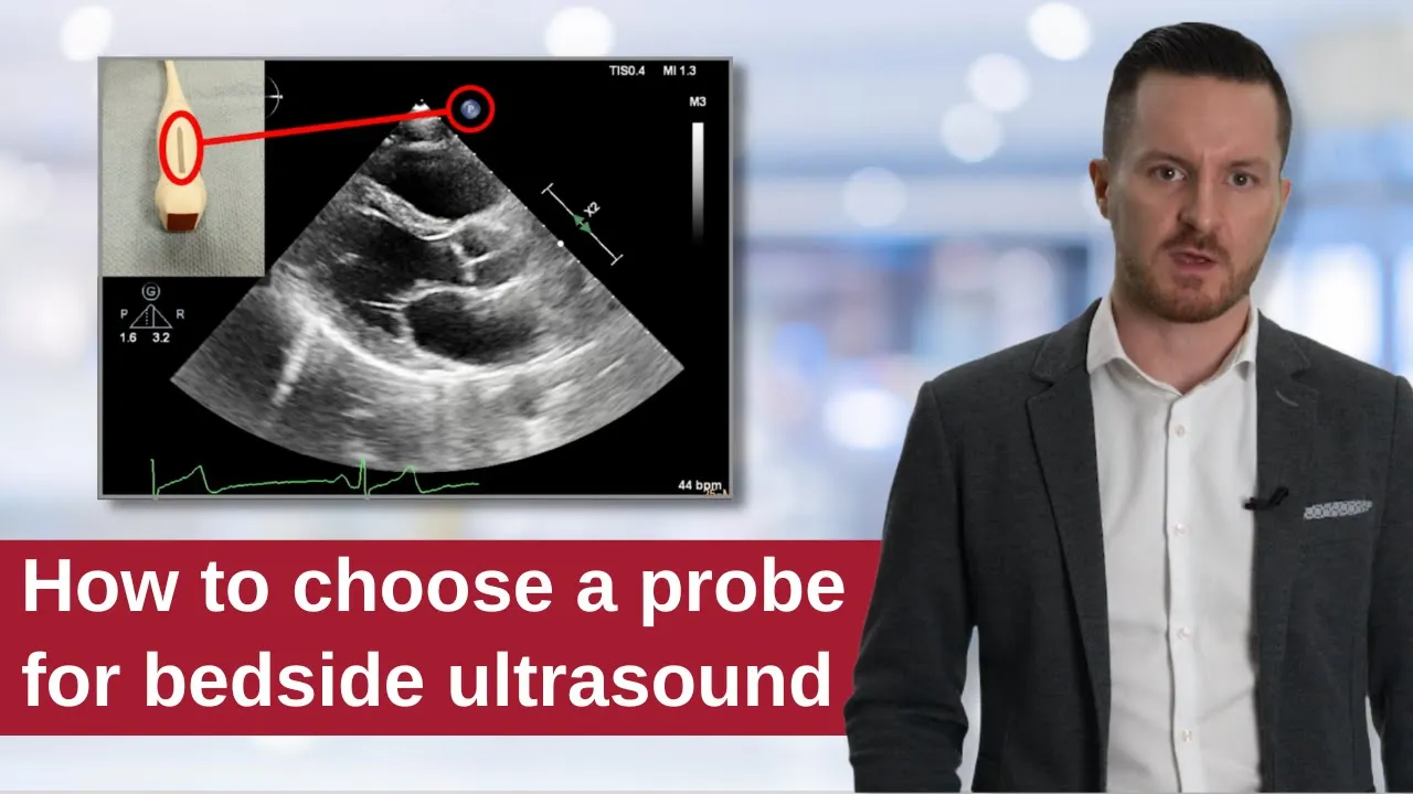
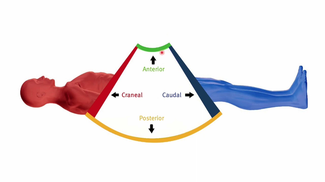
Basic physics of point-of-care ultrasound (POCUS), including probe selection, image orientation, and fundamental knobology.
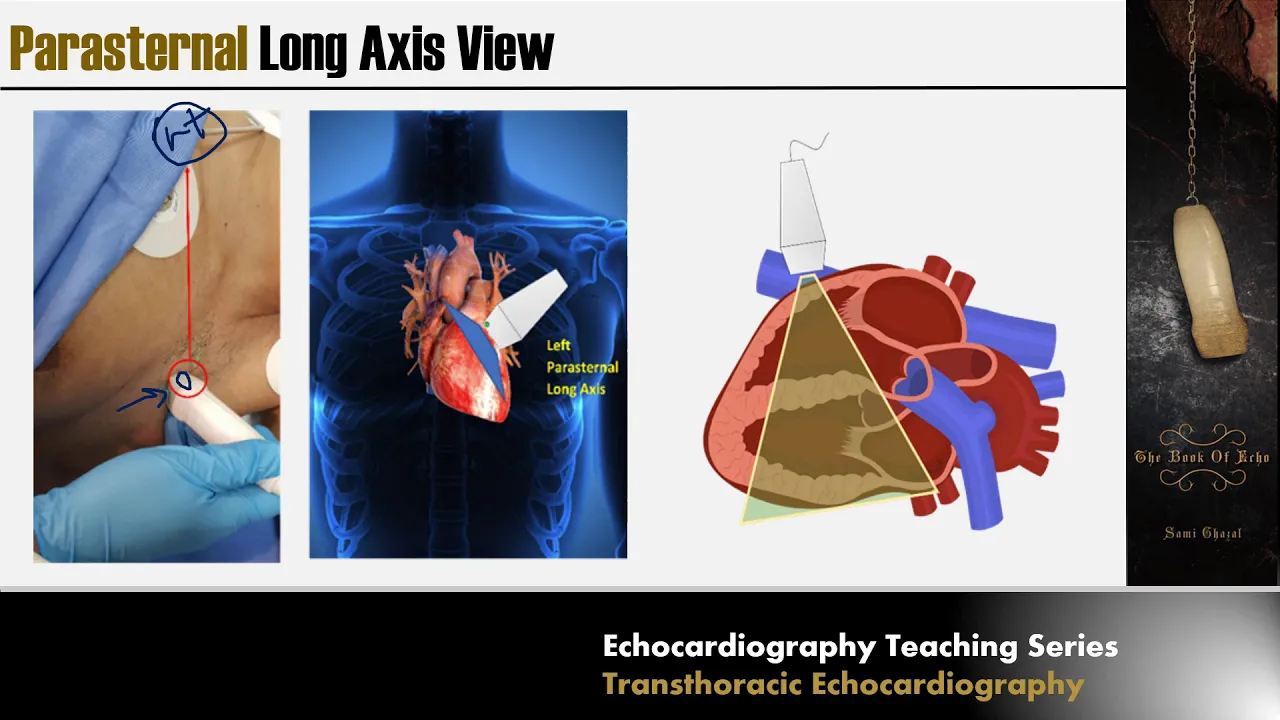
Standard anatomical landmarks and echocardiographic windows, specifically the parasternal, apical, and subxiphoid views.
The clinical utility of the FAST (Focused Assessment with Sonography for Trauma) exam, as it shares anatomical overlaps with the RUSH protocol.
Methods for assessing volume responsiveness using ultrasound, such as inferior vena cava (IVC) collapsibility index and passive leg raise trials.
Integration of RUSH protocol findings with target-directed fluid resuscitation and vasoactive medication titration.
Ultrasound-guided vascular access techniques, including central venous catheterization and arterial line placement under dynamic visualization.
Advanced echocardiography in the intensive care setting to differentiate complex mixed shock states and evaluate right ventricular strain.
122.5K views1.6Klikes35:32Original Release: 2013-05-18
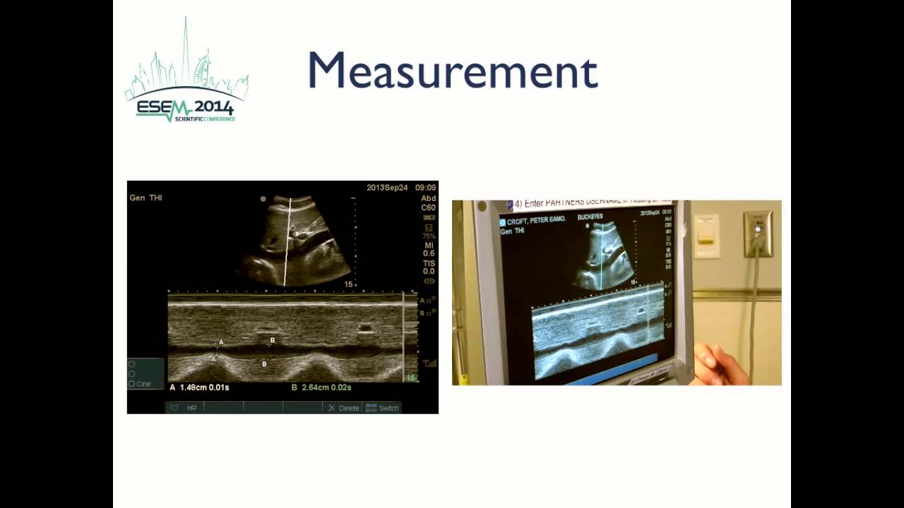
critical care ultrasound more than just lying placement evaluation of shock objectives are to discuss the role of bedside ultrasound in the assessment and treatment of critically ill patients in shock there's a strong case for use of bedside ultrasound in the clinical arena recently the American Society of echocardiography created a consensus statement with American College of Emergency Physicians they clearly stated there's a focused cardiac ultrasound has become a fundamental tool to expedite the diagnostic evaluation of the patient at the bedside the fate protocol has been described as the focused assess transthoracic echo there are several goals to the bay protocol which has been described in previous recording the goals are to exclude obvious pathology to assess wall thickness and chamber dimensions also known as the static parameters of our to assess contractility or the dynamic measures and to image the pleura bilaterally but the key is to relate the findings into the clinical context in the original article by dr. slaw that all the results of incorporation of the fate protocol into the intensive care unit or as follows the fate protocol provide provided useable images that contributed positively to evaluation in 97 percent of patients more than 37 percent of patients their echos revealed new information that hadn't previously been suspected and in more than a quarter of the cases the imaging was decisive in evaluation in terms of deciding a therapeutic path along which to send the patient let's talk a little bit about PA catheters PA catheters can measure central venous pressure pulmonary artery pressure wedge pressure calculated cardiac output calculated systemic vascular resistance and other phenomenon such as tamponade the valuable issues these days we typically see the PA catheters being used in a few situations one of which is the evaluation of the undifferentiated shock okay catheters as we know are invasive and the clinical results we get depend on the knowledge and experience of the user the questions we try to answer with PA catheters or why is the patient in shock also what type of shock or we use it in respiratory failure to determine cardiogenic versus non cardiogenic pulmonary edema and we also utilize PA catheters where we need to monitor our therapeutic interventions such as volume resuscitation or pressors bedside ultrasound one of the questions we can answer with bedside ultrasound well these should look familiar pretty much the same questions that we try to answer with PA catheters are the ones we can answer with bedside ultrasound there are a few exceptions and what can we measure with bedside ultrasound well we can estimate the central venous pressure from the inferior vena cava collapsibility we can estimate pulmonary artery pressure from a truck us tricuspid regurgitation pulmonary capillary wedge pressure cardiac output can be calculated assistant in vascular resistance can be calculated once you know the other parameters and we can also assess for tamponade and valvular issues pretty much the same things as pulmonary artery catheter and this has the added benefit of being non-invasive and we're going to talk primarily today about the rush protocol rush protocol is an acronym for rapid ultrasound in shock in 2010 this was a protocol that was described about how to approach a patient who is critically ill and in shock and essentially describes a three-step algorithm which will I will organize into three areas the pump or the heart in terms of contractility of the ventricles and effusions the tank how full is the tank is it leaking are there things been pinching upon the tank and preload returned such as pneumothorax and the pipes the major pipes of the body in terms of the aorta as well as deep venous thrombosis rapid ultrasound and shock you can also think about it as hai map which is the goal when a patient's in shock is to hit a higher map this would evaluate a patient who's in shock by thinking them in terms of the heart the IVC Morrison's pouch a order in veins and the pleura and essentially this is the same thing as pump tank and pipes it's just a different way of organizing your approach to the patient and in the end it doesn't really matter which method you choose as long as you approach the patient in a systematic way a brief review of the categories of shock we all should be familiar with hypovolemic shock which is absolute hypovolemia of hemorrhage dehydration or GI losses but there's also a relative hypovolemia sometimes described as distributive shock where the volume hasn't changed where the tank got larger and this could be in sepsis adrenaline sufficiency neurogenic shock conveys a pledge of states there's also cardiogenic shock there's diastolic dysfunction which we see in patients who've had long-standing hypertension and also tachycardia but more often than not when we're talking about shock we're talking about systolic dysfunction global cardiomyopathies takotsubo cardiomyopathy regional wall motion abnormalities due to ischemia or structural issues such as valve failures finally there's obstructive shock the types of things that can impair cardiac output from impingement either from without outside the heart or within include hypertrophic obstructive cardiomyopathy with systolic anterior motion of the mitral the health massive pleural effusion and ascites tamponade tension pneumothorax and pulmonary embolus so let's focus on the pump first when we're thinking about cardiogenic shock we can evaluate the left ventricle size and function qualitatively and the eyeball method has been well documented as being very very accurate when you compare it to quantitative methods so here we have a normal left ventricular function where you see the walls thickening and coming towards the midline as compared to a moderately depressed and a severely depressed left ventricle the division of the ventricles is a separate discussion but just briefly when you look at the left ventricle you should see an imaginary dot in the middle of the ventricle all walls thickening and coming towards that dot and the chamber size to change by 30 or 40 percent just on the eyeball here's a larger view here we have the left ventricle and the right ventricle I notice how the walls are all uniformly thickening and coming towards a imaginary spot in the middle of the ventricle we can also assess the metrical from other views this is the pair external long access view and here what we're looking for qualitative qualitatively is the mitral valve hitting the septum this is essentially seen typically in normal life and regular function states or hyper dynamic as well the other areas we can lock is back in this chamber we look here to see if the chamber size is changing we also look to see if the walls are thickening for the septum and for a lateral wall this would indicate normal and left ventricular function keep this view in your mind when you move to the following parasternal long axis view and you can definitely see the difference even without a lot of training you can see that this mitral valve is not coming up to hit the septum and if you focus in this back area of the ventricle there's not a significant amount of thickening of the walls and there's not much change in the chamber size so this is a poor cardiac function here's another example from the apical for chamber view focus on the septum here the lateral wall is showing some thickening but the septa seems to be swinging and it doesn't seem to be coordinated so this is really a dyskinetic septum it's going in the opposite direction from where you would expect during systole and likely this is a is down or dead from ischemia many times when you see cardiogenic shock you also see additional findings that support your diagnosis one of a plethoric IBC full a BC without much respiratory variation and also pulmonary B lines which are the ultrasound equivalent of curly B lines people can become shocky not just from left ventricular failure but also from right ventricular failure and how do we assess the right ventricle well in general the right ventricle is expected to be two-thirds the size that electrical that's normal the right ventricle is also more triangular in shape and it contracts differently here you see where the tricuspid valve attaches to the lateral wall and what you notice is that tricuspid valve is moving towards the apex in this direction about two centimeters in each system systolic excursion we call this taps a tricuspid annular plane systolic excursion and that's normal about a two centimeter movement we also focus on the fractional area change or the change in the size of the triangle of the left of the right ventricle besides looking at the overall size of the ventricle and the movement of the tricuspid valve and a change in the size of the triangle we also look at the apex and the apex should be predominated predominated by the left ventricle compare that to the second view apex is being shared by the right ventricle and the left ventricle the tricuspid plane is moving some but not a nice two centimeter excursion like in the normal picture and the right ventricle is enlarged so this is a bright particular failure picture in a patient in shock in this final picture you can see poor tap see you can see enlarged right ventricle same size as the left ventricle which you also notice that the left ventricle is struggling in this picture and this is a example of by ventricular failure a closer view in the Paris from a short axis view we can assess the right ventricle fairly easily normally the right ventricle is a crescent upon on top of the left ventricle and the left ventricle should be circular in this case you can see significant right ventricular enlargement and you also see paradoxical septal motion meaning the septum is going towards the left ventricle during diastole when the mitral valve opens and should be going the opposite direction and this is due to pressure overload from the right side you also have the D shaped ventricle v-shaped septum due to the flattening for the pressure of the right side all of these things are indicative of right ventricular dysfunction so that's primarily the pump we will talk about pericardial tamponade in the obstructive section the now let's talk more about the tank in the pipes the main question we usually have in our shocky patients is will they be volume responsive and for that we need to determine if they are on the steep part of the frank-starling curve but they're kinda tropic might a triplet function so if a patient has poor systolic function versus a normal systolic function there's a narrower window where they're on the steep registering curve but don't forget that they can still be on the steep part of the Stirling curve so a patient who comes in with who has baseline congestive heart failure still could be hypovolemic and we do need to still assess for that and provide the appropriate volume challenge if it's indicated typically we've looked at static parameters historically including central venous pressure and IVC diameter but we don't encourage that anymore as it's been shown to not be predictive of volume responsiveness dynamic parameters instead we focused fun in ultrasound this is a picture courtesy of annsofi perot from Stanford University here we see a increased into throw a stick pressure situation will decrease the venous return and in a spontaneously breathing patient this would be during exhalation and in a ventilated patient this is during inhalation on the right you see when there's a relatively decrease in two threats of pressure this enhances venous return so if you think about frank-starling curve which secured Ickx for a change in preload that you will change your stroke volume or cardiac output if you have a patient who's breathing is affecting their stroke volume then they are on the steep part of the Starlin curve and how do we tell that well there are a few waves we look at IVC distance ability index Delta velocity times integral Delta V Max passive leg raised the qualitative sign called the kissing papillary sign subcostal AVC assessment I've simplified this down here there can be more detailed to this chart but in general if you have a small inferior vena cava with more than 50% collapse you have a central venous pressure probably less than 10 if you have a larger inferior vena cava cave a diameter with less than 50 percent collapse your central venous pressure is probably greater than 10 and here's some qualitative assessments this IBC is small and collapsible now here over here each one of these marks is a centimeter so this is a possibly 2 centimeter with more than 50% collapse but the CVP here in this patient is less than 5 if you use n load which is motion though and play some line across your area of interest you can watch everything that happens along that line over several seconds here when you have a relatively elevated into thoracic pressure they see the IVC is two point seven nine centimeters and here where they enter threatened pressure is relatively negative you can see a collapse of the IVC down to one point nine nine centimeters but if you put this into the calculation this is a 30% change so this isn't greater than two percent greater than two centimeter IVC less than 50% change and this patient had a CVP who was in the 10 to 15 range my comparison here you have a large inferior vena cava with minimal respiratory variation and hepatic veins here are not distended central venous pressure is probably greater than 10 much greater than 10 but maybe not greater than 20 which is what we typically the situation where we typically see hepatic veins dilated just a reminder central venous pressure does not predict volume responsiveness except at the extremes your central venous pressure is 0 to 2 then you can pretty much assume the patient will benefit from volume if they were in shock and likewise that the central venous pressure was greater than 20 then the chances that they benefit for volume are low but in between your guess isn't those mine how do we measure the stroke volume what we're interested in is does a change in preload affect the stroke volume and the change in preload comes from the changes in into threats of pressure during during tidal breathing here we're going to talk about Delta V Max so if we measure if we place a Doppler gate across the aortic valve in the outflow tract and we measure the deflection the downward deflection on this Doppler curve represents a stroke value across the air without going away from the probe and here you can clearly see even without measuring that there's a significant difference in stroke volume across the respiratory cycle we can measure V min and V Max and put it into a formula V min might be maximized Neiman mean and if this is greater than 12% then it's been shown that these patients tend to be volume responsive in an obvious case where you can eyeball this and see that it's clearly greater than 12% the measurement is not absolutely necessary another thing we can do is is trace the velocity times integral both the minimum and the maximum and also put it into the same formula and then max minus min over the mean and in this case if you have greater than 20% volume changed then those patients tend to be volume responsive the distance ability index describes basically the change in the inferior vena cava over the respiratory cycle here are the examples again of a collapsible IVC and a non collapsible IVC and is one that is in the interim situation and here if we put the IVC max minus min over the mean that number of 12% comes up again it's been shown that they if you haven't I received distance feeling index greater than 12% those patients tend to be volume responders qualitatively we can also look at the papillary muscles in two different views the first view here on the left is the apical for chamber view and the view on the right is the parasternal short axis view if you identify the papillary muscles post your median in the anterolateral what's your meaning anterolateral you can see that essentially during systole they are touching each other and so your blood reading the cavity and this typically represents hyperdynamic states such as hypovolemia and distributive shock we under let you ttle eyes the passive like rays often but in a patient especially a patient with any arterial line it's very useful let's say you have a patient with no arterial line you want to see if raising their lives will increase their cardiac output raising the legs from a 45 degree angle down the body down to supine with a 45 degree angle of the legs help passively a one provides a one-minute Auto bolus of 300 to 500 CCS what this low-risk it's reversible it's been validated in diverse populations including atrial fibrillation ventilated and not ventilated and here if you find a 12% change in the v-max or the vti from sitting up to laying flat with the legs elevated then that predicts a 15 percent change in stroke volume with a fluid challenge now if you're incorrect and there's no significant change in V Max or PTI and before-and-after then no harm is done you just put the legs down and raise the body you haven't challenged a patient who has a full tank with an additional fluid bolus and cause any issues so we've assessed whether the tank is full by looking at the heart and the inferior vena cava we also want to see if there's any leaking going on these are some of the views of the fast exam focus assess transthoracic echo the view here is Morrison's pouch correlating with V on the chart here is the diaphragm where you can see that thoracic cavity and the abdominal cavity where they meet on the right we have the Salina renal recess or C and the bottom picture is D or their retro vesicle pouch pouch of Douglas depending on the the sex that you're discussing these are normal examples by comparison when there is free fluid such as in a trauma patient where the assumption is that pre fluid equals blood you can see that on these views here's an example in this patient here's the kidney the right kidney and the liver and this trace this triangle of black fluid in between it's not within a viscous it's outside the viscous we know this from its sharp corners that in a trauma patient would be blood until proven otherwise on the bottom picture an example of free fluid behind the bladder is right here you can clearly see the wall of the bladder and the fluid within the bladder but then there is a black stripe of fluid on the bladder extra outside the bowel and again in a trauma patient this is blood until proven otherwise so in a shock patient this is a very important picture to obtain what other issues we talking about we've talked about the pump and the tank we need to not forget about the pipe I think this is the step that is most often neglected especially in the ICU there's issues of overlying bowel gas issues of obesity and body habitus but with patients and with consistent pressure you can often get a good view of the aorta here's a view spine here a Orta IVC and what you want to do is find the aorta near the xiphoid entrace to trace it all the way down until the bifurcation and you're assessing to see if there's any outpouching zorse ocular aneurysms or dissections even now this is not the definitive test for any of those but this is a screen again in shock patients and if you see something that seems abnormal then the appropriate follow-ups the test is called for we typically look for the aorta to be around two centimeters and again the centimeters are on the side there's some examples of some abnormalities this initially was seen there's some opacity inside the lumen so we're turning in along access you can more clearly see that there is a flap throughout this aorta so in the shock patient that you find this in the appropriate clinical situation you know what to do this particular patient we also did a bedside echo of the heart and this is perished on a long access view and what we're seeing here there's signs of aortic insufficiency a order for vegetation you can also see a very wide a or DeGroot but if you pay attention you would also be able to see a dissection flap and that descending aorta the other pipes that we need to think about the femoral vein popliteal vein the places where we would get deep venous thrombosis so if you have a shock patient say who has signs of right ventricular strain and appropriate clinical scenario and you look in the legs and you find a non compressible femoral vein or popliteal vein then this very much helps you in your diagnosis and your therapeutic decision-making obstructive shock the processes that physically impaired cardiac output again we're going to talk about a few of these pneumothorax pericardial effusion pulmonary embolus abdominal hypertension compartment syndrome massive pleural effusion and hope of exam you don't have example examples of each of these but these are the types of things that we can say with ultrasound and you would need to be thinking about in a patient with shock that you're performing the rush exam on here's an example of pericardial tamponade so this is a subcostal view with the liver at the top of the screen in the heart and in the subcostal view you can see the layers of the pericardial sac separated by stripe black fluid you can also see a hint of diastolic collapse of the right ventricle pneumothorax here you can appreciate in an upright patient the line of the pneumothorax but in our supine patients it's very common that we do not find a pleural line and that anterior air is often missed on the chest x-ray here's an example of what ultrasound pneumothorax looks like this point the specific points called the lung point and it's on a percent predictive and positive for pneumothorax on the left of the screen we have a pneumothorax we can see the skin and soft-tissue it see the pleural line and we see no activity or sliding at that pleural line and then we have these air artifacts or a lines whereas right adjacent to it in the same floral space you can see sliding of the visceral Bridal pleura and you do not have the a lines in fact you have some minor B lines the only thing that can do that is where the lung drops away from the wall in at the point of ending the thorax if you were to do end load across these two sides you would have a stratosphere sign or an old-fashioned barcode sign on the Left bruise on the right you have the seashore sign where it looks like waves coming into a sandy beach again have been studies many studies that show that for supine patients trauma patients are typically supine on a backboard chest x-ray is on a bed most $0.75 sensitive for picking up a pneumothorax and some studies as low as 50% result or sound is in the high 90s for sensitivity and negative predictive value massive pleural effusions can also create shock here we have a situation where you have massive pleural effusion compressing the lung against the heart this is a short axis view of the heart and the heart is actually on the right side of the thorax it's being pushed over so far so when this patient had their fluid drained their shock improved abdominal compartment syndrome can also cause the same thing here we have a cirrhotic liver and we have the diaphragm and the fluid around the liver and this patient had a bladder pressure in greater than 30 and they were in shock and when a large volume paracentesis was performed their blood pressure improved here's an example of what a pulmonary embolus may look like again you can see the right ventricular dysfunction it's large compared to the left ventricle you also have the tricuspid annular plane not moving a nice 2 centimeters towards the apex you don't see much of a change in the size of the chamber and you see something called McConnell sign which is a hyper dynamic apex with right ventricular lateral wall is fairly a kinetic and then some in this study the original study from a Mitch McConnell 77% sensitive and 94% sentence specific for PE was this sign however it's important to keep in mind that patients with right ventricular in first excluded from that study and that can also appear such as this and this particular patient PE was suspected so deep venous thrombosis evaluation was performed quickly at the bedside and with downward compression there was a non compressible femoral vein and a DVT and since the patient was too unstable to go down to the CT scanner we were able to initiate treatment at the bedside quickly here's an example of something we don't see often and you need to be thinking about in certain situations we often miss this diagnosis so how come with Sam this is a slowed down version of a heart that has a very small like particular chamber and disproportionately large septum especially approximately so in patients like this often little old ladies who have long-standing hypertension have gotten dehydrated when they go into systole their anterior leaflet the mitral valve can become adherent to the septum there's another view of the same patient and at the beginning of systole if you freeze the picture that and release of the mitral valve is kind of suctioned against the in the septum which impairs the aortic outflow tract and instead creates a situation where you have mitral regurgitation such as here these patients will often present hypotensive with pulmonary edema due to the acute mitral regurgitation and the natural tendency is when you have a patient who has pulmonary edema is to give them lace six however in the situation this situation was set up because the patient was hypovolemic and their chambers the walls of the chambers became too closed during systole so the therapy for this type of pulmonary edema is to provide vollis's of fluid until the walls of the septum and the walls and the anterior for the mitral valve did not approximate each other so closely and therefore the mitral regurgitation will go away this patient also showed diffused pulmonary edema and seen in the right image and it completely collapsible IVC and this is often a combination that you'll see so anytime you see a patient with pulmonary edema but of very other indications of hypovolemia consider that they might have a situation of acute mitral regurgitation from this physiology so to summarize the rush protocols and rapid ultrasound and shock it's just a suggestion that when you have a patient in shock that you approach them in a protocol eyes away you think about the pump think about the tank and you think about the pipes you evaluate the patient systematically and you look for sources of shock you could also look in terms of hi map heart IVC Morrison's pouch a Horta veins and flora and again it doesn't matter which method you pick as long as one worse for you and that you undergo it systematically a few cases case number one 65 year-old man with a known poor EF have been in a nursing facility with cluster deem difficile colitis multiple days of diarrhea and poor P O intake presented with hypotension and dis man the ER Bowl is two liters of fluid thinking that the SI difference of predominant etiology and there have been no improvement in his map and his source of breath was about the same so you're called to and ask how much more fluid should I give this patient to resuscitate them and what comes to mind should be what kind of shock is this is this hypovolemic shock truly there's a cardiogenic shock septic shock you would treat them all differently so in evaluating this patient you can appreciate the poor EF which was known as a baseline but when you look at the inferior vena cava it's completely full without any collapsibility this is after the patient's gotten two liters of fluid you can also see diffuse pulmonary edema so the answer to this question of how much more fluid is needed for this patients fluid resuscitation is no more fluid so the patient was actually admitted to the CCU for congestive heart failure exacerbation diary stone was placed on very well k-stew 50-year old woman shortness of breath and home complained to her daughter who soon thereafter found her on the bathroom floor unconscious paramedics arrived and she was APNIC with thready pulse and went into cardiac arrest in the ambulance in the emergency room these were the images that were obtained this is a subcostal view left ventricle and right ventricle the right atrium right here you can appreciate as a free-floating thrombus right there this side you have this is the eighth there is a parasternal long axis viewed which is angled to get the right ventricular inflow tract and then atrium right here with the free-floating thrombus you can also appreciate both views that the right ventricle is very enlarged invisible promise in the right atrium right ventricle enlarged pulmonary embolus patient arrived to the ER and but she's soon thereafter died and this was a parastone a short access view very enlarged by ventricle flattened septum left ventricle that's essentially empty case number three a one-hour bedside lesson for medicine house staff was given one afternoon in the ICU we practiced obtaining the sub cost of cardiac view and we really only focused on what a normal heart should look like we did not go into any pathology but that night one of the residents who was in that lesson was called for a patient who was hypotensive and decided to get an image this is the image he got he didn't know how to record the image but he took an iPhone video of the screen and texted it to me and asked me is this what I think it is and indeed he was right it was pericardial tamponade initially the patient was thought to be septic and had been started on antibiotics and given fluids but within 20 minutes this view was gotten in the appropriate path was corrected and cardiology fellow came in to address the situation the patient did well so the take-home point here that side ultrasound can rapidly assess patients with safely non-invasively we can be doing this and we should be doing this according to our professional societies we'll be doing this this is the way the medicine is going so now it's the time to get comfortable if you have any questions please feel free to contact me
Up Next
7:54
DVT Screening with Two-Point POCUS: A Resident's Guide
@ConanLiuMD
3.7K views•2022-08-06

48:47
Graphic Medicine: Comics for Collaborative Healthcare Communication
@nationalpatientadvocate
189 views•2023-12-04

10:28
Neuroanatomy: Central and Peripheral Nervous System Divisions Explained
@AKLECTURES
136.2K views•2014-09-20

3:05
Stages of Labor and Vaginal Birth | Childbirth Animation
@nucleusmedicalmedia
52.1M views•2017-08-18
Related Study Plans & Knowledge Roadmaps
Structured learning paths in Medicine
medicine
Abnormal LFTs: Diagnostic Workup & Pathology
01Liver Anatomy and Basic Physiology
02Introduction to Liver Function Tests (LFTs)
03Hepatocellular vs. Cholestatic Patterns
+ 3 more learning modules
12h6 modules
Explore Roadmap
medicine
Analyzing Blood Smears: RBC, WBC & Platelets
01Introduction to Blood Components & Smear Preparation
02Normal Morphology of Blood Cells
03Erythrocyte Pathologies & Morphological Variants
+ 2 more learning modules
12h5 modules
Explore Roadmap
medicine
Mechanical Ventilation: Waveforms & Alarms
01Anatomy and Physiology of Breathing
02Introduction to Mechanical Ventilation & Core Modes
03Ventilator Settings and Waveform Interpretation
+ 2 more learning modules
12h5 modules
Explore Roadmap
medicine
Neuro Exam: Localization & Stroke Syndromes
01Foundations of Neuroanatomy and Vascular Supply
02The Neurological Examination: Step-by-Step
03Principles of Neurological Localization
+ 3 more learning modules
18h6 modules
Explore Roadmap