


![RA-22 EPITHELIAL MESENCHYMAL TRANSITION MD Pathology |RATS [GAME] |DR GSS |EXAM PG](https://i.ytimg.com/vi/9coWqd7OhTM/hqdefault.jpg)






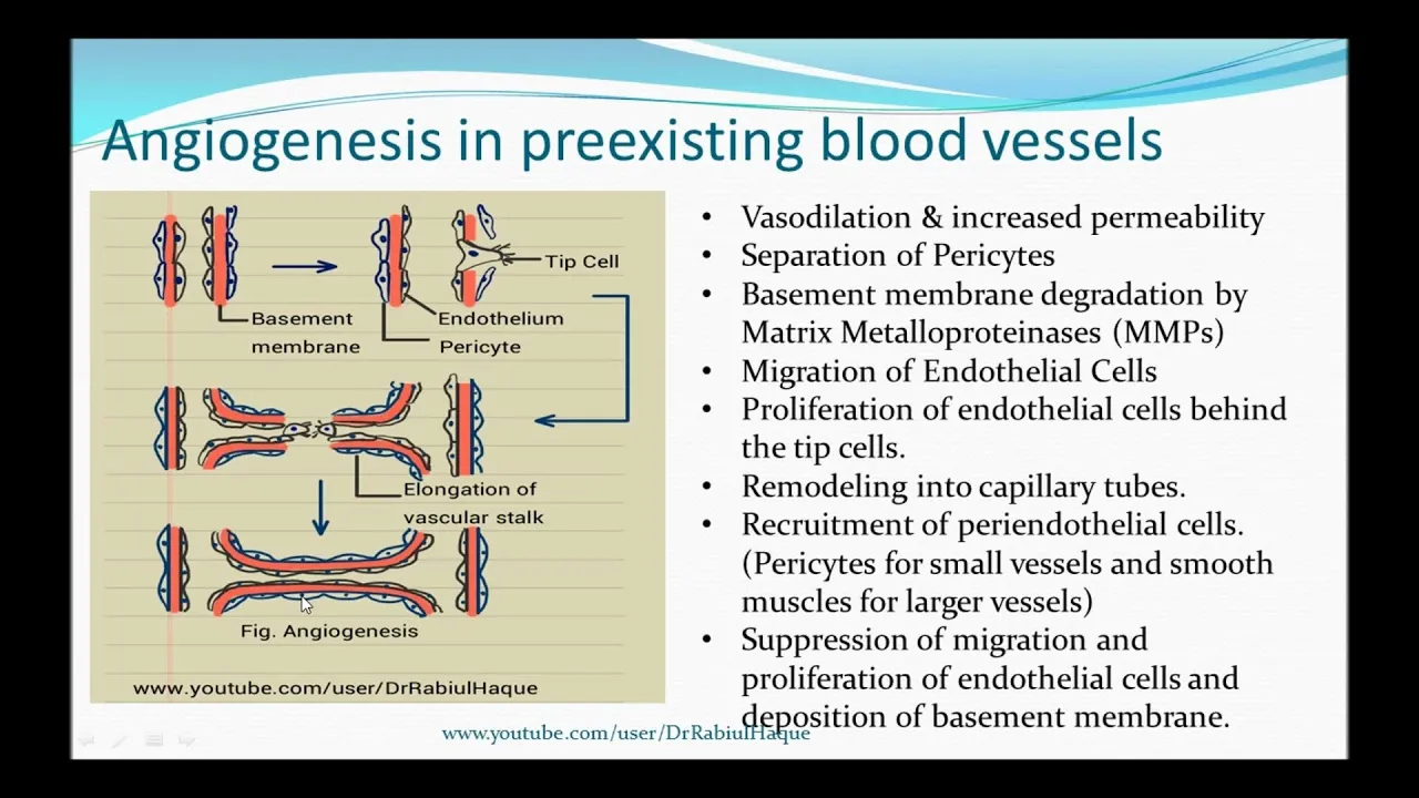












Neural crest cells undergo epithelial-mesenchymal transition (EMT) to migrate and differentiate into craniofacial, cardiovascular, and enteric nervous system structures, with precise timing controlled by post-transcriptional regulation of draxin via p-body-mediated degradation; similarly, endothelial cells sense mechanical forces through integrin-based adhesions and cell-cell junctions, where DLC1 acts as a downstream target of YAP/TAZ to control focal adhesion dynamics and angiogenic sprouting, while paxillin 2 regulates asymmetric junction remodeling during collective cell migration.
Mechanisms of Cell Migration: EMT, Adhesion & Angiogenesis
Added:Basic cell biology of the cytoskeleton, focusing on how actin filaments, microtubules, and integrins mediate cell adhesion and motility.
The fundamental structural and functional differences between epithelial and mesenchymal cell phenotypes.
Core principles of RNA biology, including post-transcriptional gene regulation, non-coding RNAs, and mRNA stability.
General concepts of vascular biology, specifically the physiological process of angiogenesis and blood vessel formation.
The pathological role of EMT and aberrant cell migration in cancer progression, invasion, and metastasis.
Therapeutic strategies targeting angiogenesis (such as anti-VEGF therapies) and small-molecule inhibitors of EMT pathways.
Advanced mechanotransduction pathways, focusing on how physical shear stress and extracellular matrix stiffness translate into biochemical signals via YAP/TAZ or integrin signaling.
Applications of cell migration and vascularization principles in tissue engineering, biomaterials, and regenerative medicine.
1.2K views8likes1:13:00Original Release: 2020-09-29
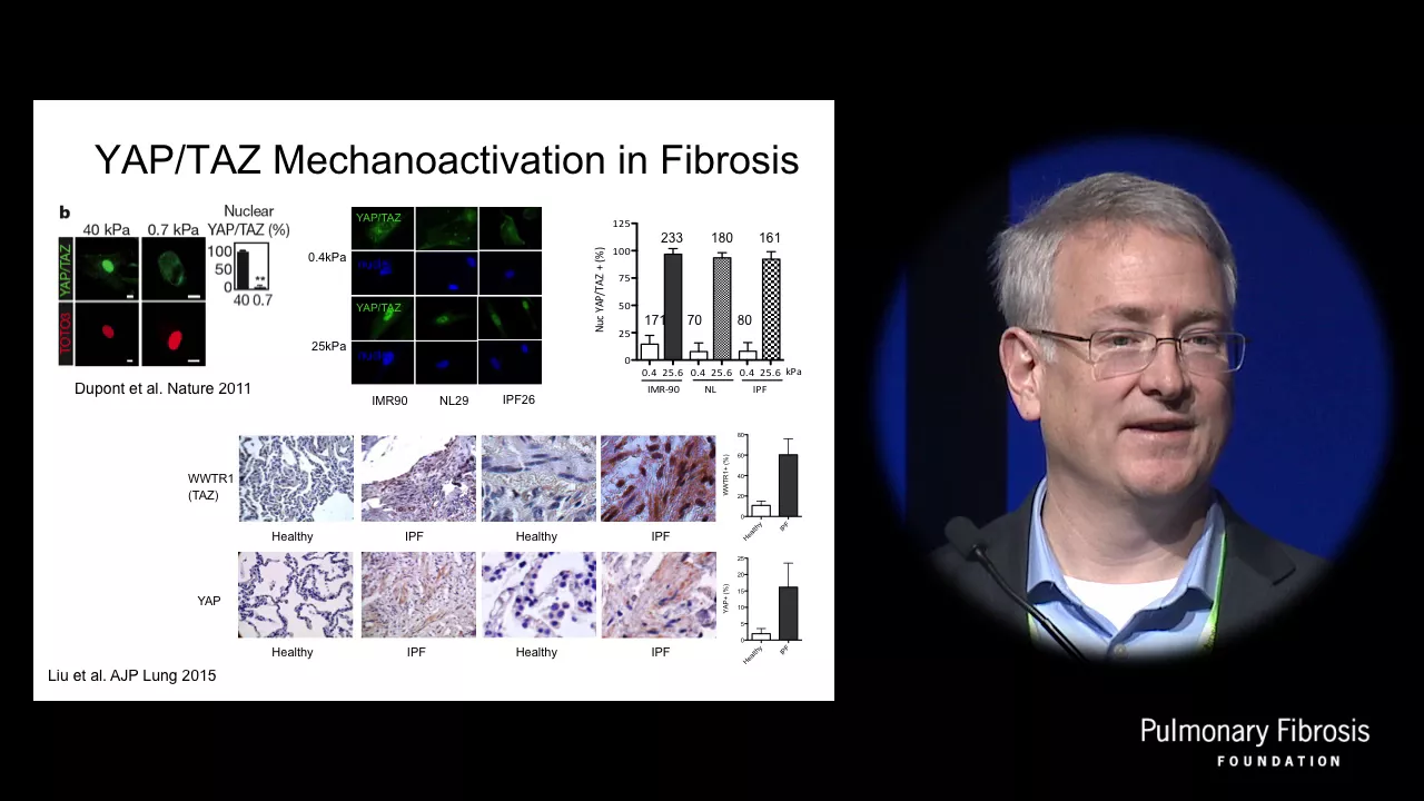
oh nice enough to tweet out the link all right we should be live on youtube and um welcome everybody that's here already thanks for showing up early um we'll get started just about right on time in two minutes the activity working well okay i've always thought that we should have some kind of like background music for this part while we're just chilling and waiting like when you're on hold at the call center exactly but like maybe less terrible than that music i don't know better music for sure i'm not sure if there's a good way to do it though over zoom like if i just play it from my speakers it'll probably sound like crap right so yeah there should be a way to stream that as well i've seen a few people doing online teaching saying that they'll have music going in the background oh you can share computer sound thank you thorsten so that's great you can try i think i'm the only person who doesn't have spotify i still listen to i use pandora remember that i st are you on myspace as well [Laughter] um i do have a myspace account clearly haven't logged into it and i haven't logged in in like i don't know a decade but i don't have facebook which i think um i was on a good bandwagon before it was a bandwagon of not having facebook i'm pretty proud of that you want to get started on time today so that's about so we're not running over yeah i mean being being on time is antithetical to my personality but um i think that's a great idea okay so hi everyone we are week 19 i'm tempted to say um so we've got two speakers today uh same thing as always which is if you have any questions you can put it in the chat on zoom or on youtube um okay so we'll get straight on with that so our first speaker is erica hutchins erica did her phd in the lab with ben sarro at the university of albany and uh she's been a postdoc in marianne broner's lab at cal tech since 2014 um looking into uncovering the molecular mechanisms of emt for neural crest cells and is now working to understand post-transcriptional control mechanisms of emt and cell migration so if you want to show the screen and take it away and hopefully you all can see that um thank you adam jennifer and rebecca and everybody working behind the scenes to make this really wonderful seminar series happen and i'm thrilled to be able to be here virtually with you all today to talk about work that i've been doing most recently as adam said as a postdoc in marion broner's lab and in the broner lab we are interested in this wonderful dynamically migrating cell that you see in my title slide here the neural crest and so um for those of you that are unfamiliar with neural crest cells and maybe even developmental biology systems i work in the chicken embryo so you're going to see a lot of these um what i think are very beautiful chicken embryo images and the neural crest cells which are labeled in this magenta color with immunostaining for this neural crest marker pac-7 and the neural crests are specified at very early stages of development when the embryo is actually still even a flat sheet of cells and at the lateral edges of the neural plate which will give rise to the future brain and spinal cord as those um neural folds begin to elevate and the tube starts to close neural the neural plate border becomes specified and that is the future site of neural crest so once the neural tube closes and becomes sort of focused at the midline the neural crest cells actually become internalized and reside internally at the very dorsal aspect of the neural tube as part of the neural epithelium and in a very tightly precisely regulated manner undergo a epithelial to mesenchymal transition which i'll be referring to as emt throughout my talk and are very dynamically migratory and as you can see with this um live uh confocal imaging that we've done where we can actually label neural crest cells with a neural crest specific enhancer driving fluorescent protein expression these cells are very highly migratory and that emigration from the midline and that emt leading to neural crest cell migration is very tightly regulated in an anterior to posterior fashion throughout almost the entire axial the entire axis of the embryo and so that emt and migration is really critical to human development as well as many other organisms because the neural crests are actually a multipotent stem cell population and that emt is critical because they leave the dorsal neural tube and migrate extensively throughout the body where they will then differentiate into a whole host of really critical cell types ranging from parts of the craniofacial skeleton parts of the cardiovascular system and even parts of the enteric nervous system and so in order to actually reach those destinations to become these really essential derivatives mineral crest cells undergo this emt and migration and when there's any kind of defects in the timing or regulation of emt and subsequently cell migration um you end up with congenital defects that we call neurochrostopathies which just broadly refer to anything that's sort of neural crest related congenital defects and actually in the adult because these are a stem cell population and they're highly invasive they are prone to metastasis in the adult so um there's really important i think implications both in development and disease for understanding how this ent and migration is regulated and so when i joined the lab that was the sort of question that i wanted to answer which is what controls that timing that precise regulation of neural crest emt and so um in order to discover that um the lab has these really thorough beautiful data sets rnac data sets for a number of um axial levels and developmental time points and so i took a um comparative transcriptomics approach and just sort of mind the data sets that we had looking for differentially expressed genes in the cranial neural crest and one of the genes that identified as being differentially expressed is a gene called draxin and so this is a fluorescent in situ hybridization along with an immunohistochemical staining for the migratory neural crest marker hnk1 and again this is a top-down view of just an embryo head for the chicken embryo and you can see that at the pre this is a pre-migratory stage of neural crest development we have this very beautiful um expression of draxon that's actually localized to the neural crest and at the onset of neural crest emt which you can see in these um these the staining for hnk1 where the neural crests have left and migrated away from the midline there's a rapid down regulation of drax and transcript levels and in work that is now published and so i didn't really want to spend too much time on today i found that draxon is this really critical throttle for the timing of emt and migration and so as you see that it's down regulated i wanted to see what happens if you apparently maintain its expression and so you can see in this cross section through a migratory stage chicken embryo midbrain level that these neural crest cells which are labeled in the cyan color on the side that have draxon over expressed when it should have been endogenously downregulated you see this really major defect in emt and cell migration so they can't migrate away from the midline and so what i want to impress upon you from this work um is that it's really critical that draxen be downregulated for neural crest migration to proceed in the head and so i became really interested in how draxen down regulation is controlled and so when we think about how gene expression is regulated we all know very well this sort of central dogma of gene expression where dna is transcribed into rna translating the protein but that's a wildly oversimplified version of events as we know there's a myriad of controls that are regulating transcription and beyond transcription there's also a number of levels of regulation at the transcript level of rna and so rna has to be spliced it has to be exported from the nucleus and then once it's in the cytoplasm it's subject to a whole host of further regulations from subcellular localization to translational regulation to even its turnover and decay and a lot of that um post-transcriptional regulation things that occur at the transcript level is regulated via the three prime untranslated region of the three-prime etr of an rna transcript and so i wanted to see is draxen perhaps being post-transcriptionally regulated um and so in order to test that i designed these reporter constructs and so i'm going to talk to you about my control construct first and so this is a ubiquitous promoter driving a destabilized gfp with just a control utr that we use for all of our transgene expression constructs that generates just a very long lasting stable rna and my experimental construct is essentially the exact same construct and the only thing i've changed is i've swapped out that control three prime utr for the endogenous draxon three prime utr um i am then bilaterally electroporate these into early stage chicken embryos and then develop them to either the pre-migratory stage when direction is on or the migratory stage when endogenous traction should be turned off and then i assess what happens to the gfp expression and so first looking at pre-migratory neural crests so again this is where the neural crests labeled in pac-7 here are still residing within the dorsal neural tube at the midline and on the left side of the embryo which has my control construct you see this very nice robust gfp expression and for the draxon 3 prime utr containing construct we actually see very nice robust gfp expression as well however just about one so might stage difference so that's about 90 minutes to two hours of developmental time we see something drastically different and so in the context of the control utr we still have very robust um uh expression of the gfp however with the draxon theory premium chair we see this rapid down regulation and so this leads us to conclude that actually um yes draxen is likely being post-transcriptionally regulated via three-prime utr at that switch from pre-migratory to migratory neural crest stage and so i wanted to get more of a handle on how that might be happening and so to do that we turn to this mrna in vivodynamics visualization system and so i'm going to just sort of walk you through the logic of how this experiment works and so i've modified my utr containing reporter constructs to now have these ms2 stem loops in between the stop codon of the fluorescent protein and the beginning of the three prime utr and what those um ms2 stem loops are there for is they are bound by an mcp a cap protein that is on a different construct that has fused to it um some fluorescent proteins as well as a nuclear localization signal and so this mcp in the absence of this ms2 containing construct because of this nuclear localization signal will just entirely localize to the nucleus in the presence of an ms2 stem loop containing transcript it should bind those stem loops and then be exported from the nucleus with its transcript so any signal that we see in the cytoplasm allows us to infer what's happening and where these these specific transcripts are localizing and so um the constructs that i use again everything is identical the only thing that's changed is just the nature of that three prime utr whether it's a control utr or the draxon 3 prime utr and again i co-electroporate these things into an early stage chicken embryo and the chicken model system i think is really great for this kind of approach because we can develop the embryos to pre-migratory stages and then dissect out the dorsal neural folds which contain our pre-migratory neural crest and then explant them into just cell culture media and the neural crest will actually still undergo emt migrate out of the neural tube and we can actually perform confocal time-lapse live imaging to see what's happening to the rnas in an in vivo context and so just to give you an example of what you're going to be looking at on the next slide this is one zoom in of one single cell with the nucleus that has the mcp label and a cytoplasmic signal which allows us to infer where the transcript is being localized and so i'm going to show you a couple movies so the first movie i'm going to show you is with our beta globin utr control construct and so i'd like to focus your attention on these very large intensely fluorescent cytoplasmic granules so you see a number of nuclei that are labeled with the mcp construct and as these neural crest cells begin to migrate we see that those granules for the control construct they stay really intensely fluorescent and bright and stable this is in contrast to what we see with the draxon 3 prime utr and so i'd like to just focus your intention on these small granules here and what you'll i hope observe is as the cell is migrating within the span of two hours those granules actually fuse and then dissolve away and disappear and so the nature of that fusion of those very small granules um is actually indicative of a liquid liquid phase separation and what's interesting is that they're sub-micron in diameter and also rapidly degrade within the span of two hours and so those um three factors suggest that perhaps it's actually localizing to um cytoplasmic rna granules which are liquid liquid phase separated and so there's a variety of rna cytoplasmic granules that either store or process rna in eukaryotic cells and in particular i was focused on p bodies because p bodies in particular um are very well known to be involved in processing and degradation of rna and so i wanted to ask is jackson actually being localized to these peabodies and migrating neural crest cells so um one of the first things i wanted to know is are there peabody components actually expressed in migrating neural crest cells and so these are a fluorescent in hybridization chain reaction in c2 images of again dorsal down view of chicken embryo heads and we have a neural crest label here in green and what i hope you can appreciate in whole mouth as well as in cross section is that a number of these peabody components um very specifically co-localized with neural crest as well as they're just generally enriched in neural tissue they are ubiquitous which is not unexpected but there's this enrichment in neural crest in the neural tube which i think is really interesting and so um in particular i focused on dcp1a because dcp1a really nicely localizes to peabody specifically whereas some of the other factors are more broadly expressed and so this is a zoom in of one single cell that i've collected with my mcp and draxon ms2 ut containing construct and express the fluorescent protein tag version of dcp1a and so this is a zoom in of one single nucleus and zooming in on this granule in the cytoplasm nearby the nucleus the dcp1a labeling the peabody is labeled in magenta here and what i hope you can appreciate is there's this really nice sort of overlap with the draxen cytoplasmic signal and so this is highly suggestive that perhaps yes draxen is localizing to peabodies and so um in order to determine um or to sort of solidify that evidence uh we also want to see what happens if we perturb peabodies do we then alter that subcellular localization of the ms2 containing utr construct and so in order to do that we used uh crispr cast 9 to knock out ddx6 and ddx6 is an rna helicase that is essential for peabody formation so it's been well established in other systems that when you knock out ddx6 um you dissolve pea bodies and they can't form and so um for again these are all draxon utr containing constructs with mcp and so in the control crispr these are fixed cells just to remind you um direction focuses into these small cytoplasmic granules when we knock out ddx6 and thus disrupt pea bodies we actually see this drastic change in the subcellular localization of those draxon transcripts where it becomes just sort of broadly diffusely cytoplasmic and so um i thought this was really interesting but i wanted to then know what happens to endogenous jackson because these are all um essentially exogenous reporters so what happens to endogenous draxin when we perturb ddx6 and peabodys in vivo and so i have um again these are zoom in of chicken embryo heads dorsal down the left is control crisper and the right is ddx6 specific crispr and we have done a fluorescent in situ for the neural crest marker ap2 beta as well as draxon and so drax and i've pseudo-colored here based on fluorescence intensity and what i hope you can appreciate is so on the control side remember um significant down regulation at the onset of emt you can see the neural crests have very nicely migrated away from the midline on the ddx6 knockdown side what i what you may notice right away is that the neural crest migration has been basically disrupted and if we zoom in um what i hope you can appreciate as well is there's actually an up regulation of drax endogenous draxon rna at the emt stage so this is when draxin should have been downregulated and indeed on the control side is much lower in expression and so when we quantified the um area of neural crest migration for the ddx6 knockout we um we found significant decrease in the area of neural crest migration indicating that disrupting pea bodies through ddx excuse me ddx6 crispr is blocking and disrupting neural crest cell migration and this actually phenocopies what we observed in my uh previously published work with drax and overexpression where we see this significant defect in neural crest migration and so to summarize the story that i've told you today um i have previously identified this gene draxen as a throttle for neural crest emt and migration and it's expressed in the pre-migratory stage and has to be down regulated in order for neural crests to delaminate and leave the neural tube and then begin migrating and that down regulation is accomplished through post-transcriptional regulation um via targeting of draxon to peabody is where it is subsequently degraded and so um taken together this suggests i think for the first time i want to i want to say that p bodies are controlling an essential developmental emt program and it's mediated through a post-nutritional target degradation of a specific target in which in this case is the draxon rna and so to acknowledge my post-doctoral advisor dr marianne bronner this work was done in collaboration with another postdoc in the lab dr michael piacentino i've also had help from technicians and students with various cloning projects related to this i also would like to thank these individuals in their labs who made essential reagents available on ad gene um also my funding and um i am available on twitter and via email if people have questions or would like to connect about this work any further um thank you for your attention i'm happy to take questions great thank you thank you um it looks like roberto mayor has our first question if you want to um unmute and videoify yourself uh yeah can you hear me yes yeah very nice talk very nice image and movies so uh you show i mean some of the of the data that you show were done in this very nice system that you haven't checked in that where you can remove the neural tube and culture in vitro um so it is known that emt can be controlled by the by stiffness of the subject so i wonder if some of your result could be uh affected by the stiffness of the substrate because you are capturing the nuclear crescent in a very hard subject so have you tried to to modify the stiffness or to compare for example i was thinking in the in the in this assay that you show with the ms2 system to analyze the stability of the rna is that dependent on the stiffness of the satchel do you have any idea about that yeah i it's so funny that you bring this up because i was just thinking about this um last night uh and i actually do suspect um i think that's an excellent point that you bring up i do suspect that stiffness may have um may play a role on perhaps the either localization or rate of decay or or something to do with pea bodies and the reason i sort of suspect that is as you point out stiffness not only has important implications with emt and regulating cell migration but i think it actually can affect differentiation and what's interesting is that um if you perturb peabody's you essentially alter the ability of stem cells to or fibroblast cells to reprogram and so i think there's this really critical role that peabody's and sort of full-scale rna decay play in these sort of um cell state transitions and so anything that is um sort of affecting cell state transitions i think is going to be intricately linked with p bodies and i and i fully expect and think that yes indeed stiffness is one of those factors and i'm actually really interested in um as a future sort of direction um figuring out how how that is accomplished and how the cell is able to sort of sense that stiffness in order to possibly i still need to verify this but possibly affect um either the rate of decay or the localization of various transcripts to peabodies depending on stiffness so i think that's an excellent an excellent point it's something i've i've been thinking about and would really like to to pursue thank you i think that would be very interesting to explore further thank you i agree thank you a quick question from me before we go to the other questions but i was wondering um if you've tried enhancing uh the draxin degradation to see if you can get early empty and maybe migration starts early yeah so actually um i didn't think i had time to talk about uh so my published work i found that um so draxen is expressed in this really brief pulse but it's essential because if you knock it down prematurely the neural crest basically just get pushed out of the neural tube too soon and so they retain that pac-7 label so that they're sort of primed as like neural plate border but they then down regulate neural crest specifier gene expression so there's something about draxon maintaining the neural crests within the dorsal neural tube so that they can commit i guess to a neural crest specification fate that if you knock it down yeah you get premature delamination but it doesn't facilitate sort of like sooner migration it actually still disrupts migration okay so the loca the localization at the at the border is really important for that emt they can't be pushed away yeah cool thank you um we also have another question from rachel who can go ahead and um unmute and start your video if you want hi eric i actually had a very similar question to roberto so are you practically absolute good joining my training but it was a really lovely talk oh thank you so thanks thank you you need to get on that stiffness angle now we've got a question from c chang uh is traxxan the only rna involved in europe's dmt that is degraded via pea bodies yeah sorry to my view yeah that's me sorry i was gonna say that's a great great question um i don't know and it's something i'm actively pursuing um using uh transcriptomics so the assumption is that if we disrupt peabodies via ddx6 knockdown not only drax them but any other rna at that stage that's required to be degraded in p bodies is also then going to be up regulated so that's that's on the agenda to figure out what other rnas are um degraded by pea bodies absolutely yeah uh second question is does disruption of peabody it's a similar line but does disruption of people interfere with other rnas and a third one is can you rescue peabody disruption by draxin rescue with utdl minus rna oh utr minus iron um so so with the peabody disruption um draxon is sort of apparently upregulated so what we're what we're going to try is actually to rescue with a timed downregulation so um as i mentioned it's sort of tricky because if we knock out draxon too soon um it also disrupts emt and cell migration um but we're hoping that we can use the crispr cas9 system to more temporally control when we knock out draxin in in combination with knocking out ddx6 in the chicken system um because it cast 9 is so big and it it's kind of um laborious for it to sort of transcribe translate fold and actually be functional so there's this lag uh for neural crest uh development when we do plasmid-based crispr so my hope is that that lag is sufficient that we might actually be able to rescue it but that's that's definitely something i'm gonna try great thank you and um this will be our last question unless um anybody else has more if you do go ahead and put them in the chat from aj kittness when the cells delaminate prematurely do they adopt a particular fate i don't think that they do i actually think they're just sort of stuck in the stasis of of primed but not committed to anything they do retain um the pac-7 label so they're they're still sort of like pre-neural crest but they like i said they down regulate the sort of like a later neural cross specification genes so i actually i think they're in this sort of like purgatory where they don't really know what to become um but yeah that's that's it's a really interesting question i'm not quite sure what happens to those cells um at like later stages of development at least um at the emt stage they don't die they they just sort of like hang out there my assumption is that later they might die but i i don't i don't know thanks thank you so much thanks aj um a quick one from me i was just wondering um do you think that uh jackson is important in other types of cell migration or do you think it's this really sort of specific to you know neural crest and fates and and all that yeah so i it's a um so i didn't really mention this it's a canonical wind antagonist um and that's it's sort of like main function at least in emt um but what's interesting is it was actually discovered as a repulsive exon guidance molecule so it's a secreted factor that is i think inhibitory in a lot of different ways so um because it's secreted and acting externally i think it's perfectly it's reasonable to expect that it's likely acting on other cell types depending on sort of what's nearby um but in terms of yeah what what else it's doing in development i'm not quite sure in terms of cell migration itself but certainly in terms of axon outgrowth it appears to have effects on like growth cone directionality and sensing great very cool um thank you so much that was really wonderful um all right we are going to move on to our second talk of the hour um so we have stefan who veneers he got his phd from the netherlands center at the netherlands cancer institute at leiden university um and that's where he got interested in meccano transduction and then as a postdoc at the hubric institute he decided to study cell cell junctions and how inflammation-induced forces affect the endothelium and blood vessels and now stefan is an associate professor at the university of amsterdam where he studies how tissue remodeling is controlled by cellular interactions with the extracellular matrix and neighboring cells so we're really excited to have him as our second talk today and go ahead and take it away stephane yeah thank you jennifer for the introduction um yeah so so what i'm going to do today is show you uh two stories at least about how and how the endothelium responds to mechanical forces and one of the mechanical forces that i will start off with is the amount of stiffness that the endothelium senses from the vascular micro environment so i thought it was a nice bridge with the with the questions that came up uh during the last the previous start um so but we are studying uh how and how the endothelium basically adheres to the vascular matrix and how they adhere to each other as uh with their cell cell contents and of course the endothelial cells like many other cell types they they sense continuously their environment they bind to the extracellular matrix through the integrands which is very important for stiffness sensing they relay their information to the ectomyosin cytoskeleton and that in turn is remodeling the cell cell contacts in which in the case of the endothelial cells is being built by vegetarian-based adherence junctions so normally the endothelium is doing fine despite all the mechanical forces that are at play within blood vessels if you think about blood flow the contractility of the the mural cells for the muscle cells and the differences in blood pressure of course but things go a bit wrong when you look at older vessels then the vascular wall starts to stiffen and then the whole mechanotransduction pathway inside this endothelial cells is perturbed in the end leading to increased permeability and to the onset of inflammation which is for driving the development of cardiovascular diseases so if you now take a closer look at how the endothelium first starts to sense the stiffness uh the stiffening of the environment of course this takes place uh for a large part at the integrand based adhesions and that the focal adhesions and uh like many of uh like many of you we studied them in vitro so if you look at the focal adhesions and the endothelial cells they are usually quite beautiful partially because the endothelial cells are very flat cells so you don't see all the cytoplasmic background or the cytoplasmic stains for instance of pexillin or thinking but what is also important to realize is that these structures are very much present also in vivo so if you look inside blood vessels themselves and if we open them up and then do a normal imaging of the endothelial wall and this is a picture from a human artery then you can recognize the intriguing-based adhesions that are anchored to the effect in stress fibers and we see this particularly in the atro or at the arterial side of circulation compared to the venous side and we think that this is partially due to the fact that the arteries are a little bit stiffer than the veins so we we study how the focal regions in the endothelial cells are turning over and formed and we got interested in a protein called deleted liver cancer which is a well-known grow gap protein which has gap activity towards several of the road gdp aces and it is a focal adhesion protein because it binds to several members of the integrand-based adhesions and when we started doing some stainings and looking at the amount of dlc1 in in the blood vessels and we see an upregulation of dlc1 expression in atherosclerosis which is correlating with more stiffer with a more stiff microenvironment and we also see increased expression of this protein in endothelial cells that we have from pulmonary hypertension patients so we set out to investigate what dlc1 might be doing and indeed we see that in endothelial cells it nicely co-localizes with excellent so it's also in the endothelial cells a focal adhesion protein but when we got very much intrigued by dlc1 if we started comparing its expression in different if we culturally endothelial cells on different stiffnesses so we see a relatively low expression on soft matrixes and on stiffer substrate we see an up regulation of the amount of dlc1 protein that is being expressed and this is the same for if you look at mrna levels so this is being controlled at the transcriptional level and this of course provoked us to ask the question whether dlc1 might be functioning somehow downstream of japantos which are well-known transcriptional co-regulators that cycle between the cytoplasm and the nucleus depending on mechanical cues that they receive most notably stiffening of the ecm but also disturbances in blood flow or stretch of endothelial monolayers and angiogenic growth factors which all stimulate the activation of the up and downs and when we started looking at the transcriptional start style of dlc1 and specifically the isoform ii that is being expressed in the endothelium cells we we found a a tiat motif which is the transcription factor that binds to the arpeggios and the based on available databases we also find and we found that there is a peak in binding of tiat to this transcriptional start site and indeed making it possible that dlc1 is a target of the open test directly so we set out to investigate this and we looked at the promoter region and but we cloned the promoter region and mutated the tiat binding site and we see reduced expression of the dlc from the dlc one promoter and if we do a knockdown for yup we see a reduced expression of dlc1 just like other target genes and we have the same findings if we knock down tusks from the endothelial cells and vice versa if we over express a constitutive active mutant of yap which is uh always in the nucleus because it's syrians are being mutated so it cannot be degraded anymore then we also see an upregulation of dlc1 expression which then becomes independent of the extracellular microphone microenvironment so we see it then also on soft substrates so it seems to be that dlc1 is a direct target of the and that activation of the open test is sufficient to drive the expression of dlc1 and what is next is that we try to find out what dlc1 might be doing and so we did knockdowns we have multiple short hairpin rnas that that efficiently target dlc1 and then we got rid of the protein and then looked at how the endothelial cells are functioning and the first thing that we notice is that there is a lot of more affecting fibers that are present in the basal plane of the endothelial cells and the next thing we looked into is how the intercom-based focal adhesions are being organized so what you see here is a turf movie uh from control cells uh uh or dlc on cells and they are part of an endothelial monolayer it's just that you see only one cell because that's present or that is positive for the taxiderm gerry expression and if we look at the turnover of the focal adhesions in the absence of dlc1 we see a lot more focal lesions that are being formed upon dlc1 depletion and if you compare it to control cells you see much more turnovers so you see a continuous assembly and disassembly of the focal adhesions whereas in the absence of dlc1 there is sort of a stabilization of the focal adhesions so they have a longer lifetime and the reason is that we see that there is a decreased disassembly rate of those focal adhesions and we also measured the amount of traction forces that the cell can exert on the extracellular matrix and then we see that in the absence of dlc1 we have massive upregulation of traction forces so dlc1 controls the dynamics of integrand-based adhesions of course the first thing that we next analyzed is whether it also affects the function of the integrity so we did that by doing scratch wound essays and as you will see in the in this movie is that despite the fact that i have more focal adhesions and they exert more traction forces if you do a scratch wound essay you see that they are less sufficient to close the wound if you compare it to control cells and we also compare that directly and this is one of the movies i i put on twitter to to kind of uh motivate you all to come and see the talk but if you compare the green cells which are the knockdown cells you can see that these cells are much less efficient in sensing where the wound is and and basically all the red cells which are the control cells they are much more efficient and in the end those are the cells that are first closing the wound and this is because in the absence of dlc1 their cells have a lack of directionality and they failed to polarize so they don't put the golgi in front of their nucleus and we think that this is related to the fact that the integrand-based turnover is is not normal compared to control cells so because yap and us are very well known players in the vasculature for instance the angiogenic growth factor is known to uh to drive the ex to activation of japan tiles and thereby drive a transcriptional program that should support angiogenic response surprisingly we do know we do not know a lot about the the downstream targets that are uh really involved in the endogenic program but we know a lot more about also about the endothelial specific activation of yaptas in the vasculature and how it's needed uh to for angiogenesis and the control of tip and stock cells so the leader and the follower cells in the angiogenic process so we set out to investigate what dlc1 might be doing in angiogenic sprouting and upon knockdown of dlc1 we see a massive reduction in the number of sprouts that are being formed in this essay so this is a 3d sprouting endogenesis essay where you can look at sprouting in collagen gels based on steroids that are composed of like a thousand epithelial cells so in the absence of dlc1 we don't see angiogenic sprouting vice versa if we increase the amount of dlc1 by overexpressing it we get increased sprouting activity which indicates that the level of dlc1 expression is really important for the angiogenic activity of the endothelial cells [Music] well uh so we now know that dlc1 is involved in endothelial migration and that it's uh also involved in engineering sprouting but of course now it becomes the question whether uh what is the contribution of dlc1 in the downstream pathway that japan does are activated so to study this more in detail what we did is we knocked down yup from the endothelial cells efficiently which leads to a reduction in the expression of dlc1 and these cells they they have difficulties closing the wound again and that's also again because the integrand-based adhesions they are matured they've failed to turn over and this has been shown by the lab of joe bruco previously and now if we restore the expression of dlc on in these cells so we have a yap knock out cell or the up note down cell and then if you restore expression of dlc1 we get more or less levels that are comparable to the control situation and if if we then look at those cells we see that uh now the these cells basically turn are turning over their focal adhesion much better and they are they are sensing the scratch wound so they are orienting in the right direction and we get a partial rescue of cell migration but what is more prominent is that if we now turn to the energetic sprouting essay is that the defect of those yap knock down cells to angiogenically sprout is completely rescued if we only express its downswing target gene dlc1 so we think that dlc1 is a is a very prominent target of the japantos activation program we know it's being activated by ecm stiffening and that vegf also drives the expression of dlc1 we suspect that the flow patterns are also important for activating aptos and dlc1 but we have to further investigate this and then once dlc1 is expressed it probably functions in inside the endocrine based adhesions and helps to turn over the focal lesions for instance in stiffer environments and this appears to be important for directional cell migration and sprouting angiogenesis so more details you can read in the in the paper which we published earlier this year and now i want to turn to the second part of the story is that's because not only the mega transduction pathway is functioning at the cell extracellular matrix adhesions but it's also uh very active at the cell cell contents and that is of course due to the fact that the atomizing activity inside the cells is pulling on all the structures that to which it's anchored to and we have been studying this for years uh by now uh and mostly in the context of the vegetarian complex which anchors to the effect inside the skeleton through the bound catheters that bind to vegan hearing and if you look at the turning over of the fecation-based adhesions you can roughly distinguish several subtypes of junctions we have stable junctions which in which vegatarian is oriented in a vector is is filling the contact interface between two cells and it's flanked by parallel actin bundles as you can see here and in in those stable conditions there's a lot of dynamics going on but in the end what you see is that the junction is staying intact throughout the time and then there are junctions which we call focal adherence junctions because they look a little bit like integral-based athenians because of their focal nature and they are being pulled apart by radial actin bundles which pull on the vegas hearing complex and basically remodel the vehicle based junctions and this site those focal adherence junctions are the active sites where a lot of mechanotransduction activation takes place and we have looked into this we have compared to two states so we have compared the linear and mature junctions to the focal adherence junctions and what we know is that winkling for instance is present in the focal adherence junctions but not in more relaxed state but mature junctions and that is because the ectomyosin activity is pulling on alpha-catenin and exposing a cryptic binding site for thinking here and we know that vinculin recruitment to those junctions helps to protect these junctions during their force-dependent remodeling but we also know that there are a lot of more proteins that are specifically recruited to those force-dependent junctions and the last part of the talk i will be discussing a new member being present at those junctions and that protein is called taxing two and paxin two is an a bar protein so as a dimer it can recognize like many of the other bar proteins it can recognize curvatures in the plasma membrane and what we observed is that if we stain for this protein paxing is that it's specifically recruited to the focal adherence junctions as you can see here and it's not present in other type of junctions which where there's no pulling on the vehicle hearing complex taking place as you may already notice is that if we would stay for winkling in this case we would see vinkling all over the focal adherence junctions but pakistan is not recruited in a similar pattern as fingerling is so what we see is that it's recruited to the trailing ends of those junctions so at the terminal ends and it's only decorating the junctions halfway so and we know now that it's recruited from one side to the junction and we got further interested in this also by a publication from the tobias meyer lab in stanford is when he showed that if you start pulling and pushing on the plasma membrane that this on itself is already a sufficient trigger to recruit bar proteins to the plasma membrane so what we think we have now is that if by staining for paxing we recognize those curved plasma membranes specifically at the force-dependent junctions and indeed if we look at super-resolution level then we see that vaccine is not binding to vegan hearing but it's rather decorating around the terminal ends of the remodeling focal adherence junctions so i don't have time to show you all the data but what we know is that if you look at directional migration and if you then look in scratchmoon's essays is that you see that if there is directional migration then this is the front of a junction and this is the rear of a junction and unpacks in is recruited to the trailing ends so to the rear of uh of those junctions so we we use it nowadays as a staining to to to show which junctures are asymmetric in their buildup and we know that there is polarized trafficking of wikiterun from the front of the junction towards the rear and that at the rear the vegetarian complex is being internalized so what we next is is to investigate the role of those asymmetric junctions and the function of accident in it and we studied that in the in during collective cell migration uh we did a knock-downs of pakistan too which occurs very efficiently it doesn't affect the level of vegan hearing expression it also does not affect the amount of cell cell contact so the cells look relatively normal at first glance but if we don't do scratch essays we see that the pecs and knockdown cells are very much preserved in collective cell migration so they have a reduced speed of migration and most importantly they have a reduced correlative length so they basically are not migrating as a cell collective anymore in the absence of paxi which would make sense if you think that vaccine perhaps helps in guiding the turnover of the junctions in asymmetric junction sites so of course one of the major collective processes that is being mediated by endothelial cells is sprouting angiogenesis and and we have been collaborating with the lab of marcus ploman to study the function of paxil in the mice and this is these are two pictures from from a retinal angiogenesis essay in which after birth you can look at the embryos in the mice and then follow the development of the vascular network in the eye and as you notice is that in the absence of paxon there is still a vascular network so blood vessels are still being formed however this seems to be a slightly different organization of the vascular network that's being deposited or being formed and this especially is clear in the angiogenic front if you compare the the the controls the control angiogenic front to the energetic front of taxi nocatee mice you can see that there's an accumulation of cells taking place there and if you take a look at this in a in a higher resolution and then you can see that in the wildtap cells we can easily distinguish the leader cells or the tip cells but in the absence of paxin there is an accumulation of the endothelial cells in the leading sprouts here sometimes it's even difficult to by eye to pinpoint which is the the leading tip cell on which one is the follower cell so we think that the paxon is needed to coordinate the endothelial cells are the energetic sprouts and we have also quantified this confirming our observations so um so what might paxin be doing then and as i already told you that we suspect that pakistan controls the trafficking of vegan hearing we already showed before that if you deplete vaccine you have an increased amount of turning internalized economic vesicles inside the endothelial cells so we think that paxin locally functions during the trafficking of vegetarian so here you see a the vegan hair complex labeled in red with alpha-catenin m cherry and there is pectin being recruited to these sites and you can see these red dots here or it's maybe better to visualize here in the in the grayscale picture these are the the dots of the wicked heron complex that is already being internalized and pexin is specifically basically decorating the interface between what is still part of the junction and what is part of the part that's already being internalized and what you can see is that pakistan disappears from the junction and there is another internalization process taking place once it's gone and then after that texting is being recruited again right there between the boundary of the junction and the internalized parts so next we try to figure out how pakistan might be helping to or how it might control the trafficking conflict here we uh we ruled out some uh pathways some canonical pathways that have been shown by andrew kowalczyk already in which piano 20 continue takes a major role and the next thing that we did is we looked at the domains of paxin and paxon has a bar domain which recognized this curved membranes but it also has npf motifs which it uses to bind to hd proteins which are well-known proteins involved in protein trafficking and if we look then there are four isoforms or four forms and we have checked for each d1 and eg4 which bind to paxion 2 and if we then look at where egd4 is localized we can see that it clearly is being recruited to the asymmetric junctions right there where paxil is being present and i don't have time now to show you all the data but what we know is that if we do a knockdown of paxon 2 we lose the recruitment of each g4 so we think that this interaction is really crucial to recruit eg4 and help to control the trafficking of wikiteer at asymmetric charge so what i want to finish with is that i hope that aside from the integrand based stiffness sensing i also hope you realize now that the junctions themselves if they are sensing forces from the ectomize inside the skeleton that this could be direct forces on the vegan hair complex which for instance lead to recruitment of thinking and this is when there is uh global pulling on the cell cell junction so when there is like a an inflammation or something this leads to uh overall globalization and contractility in the endothelial cells and there's a massive formation of focal adherence junctions and fingerling recruitment but then there are the more subtle force dependent activities that include local differences in cytoskeletal derived forces for instance during leader and follow ourselves at the interface between the leader and follow ourselves and we then think that there is force dependent regulation that takes place at the junctional plasma membrane so next to the digital based structure and we think that this helps to guide vegetarian trafficking and to assist in the collectivity of the collective migrating cells the final part is of course to thank the people that did most of these beautiful imaging experiments in particular sveta and anna who have been working on together with informa who have been working on the bar protein project and the role of paxil in angiogenesis and i want to highlight also misha who has been working on the dlc one project in the past years and some of the collaborators in particular marcus ploman for sharing with us his his work on the pax and knockout mice and sharing the model with us and with that i'm happy to take questions if there are early thank you that was that was really great um i we do have a number of questions in the chat and people should keep posting but um i wanted to ask one first that might just be my own uh ignorance but so when you first showed the movie where you had the focal um adherence junctions what i was thinking was like wow it would be really bad if endothelial cells in vivo actually looked like that like it looked like they were just so stretched and almost like there were you know big gaps between the cells and so you know i think of of the endothelium is something where it's so important to maintain you know that that tightness and not not be leaky and it looked to me like those junctions would be really really leaky and so i guess i'm just wondering about sort of like in vivo would you expect to see anything that actually looks like that or or is this something that that shows up in vitro because of stiff substrates and anything like that yeah i think i think the structure is really emphasized in very stiff environments uh so we that's why we see it so easily in vitro and we kind of use that now to discover which molecules go there to protect force-dependent remodeling of course when you start looking within blood vessels it's much more difficult to find them and there's much more of the stable junction form which protects the endothelial barrier and and prevents vascular leakage just for where we find most of those junctions being formed in the same organization i would say is at the shoulder regions of atherosclerotic plaques so not on top of them but on the shoulder regions and we know that there's a lot of collagen deposition there um but there we do see them being formed and we also know because we did tracings with nanoparticles is that uh there there's leakage not not the massive leakage like with where the red blood cells actually enter the tissue but we do see that small molecules can easily more easily transfer the endothelial barrier there so that's most likely where it's also relevant i guess thank you i appreciate that cool so uh we got a question from arbit uh object dead boy sorry i hope you're announcing that wrong um since it is a row a guef i think that means the dlc one could these effects be contributed to increased raw activity and subsequent rock myosin activation yeah yeah yeah of course this is a very good question and we looked into this so it's a gap for roe we don't know for sure which row isoforms are actually regulated by dlc1 we tried to address this directly so we did biochemical assays and we don't see differences in global role a activity or rho c activity so um so if even if we deplete dlc1 the the global role levels are still the same and if we treat those cells with thrombin then we can still raise row a activity so even the regulation seems to take place at least dlc only is not it's not taking part in this this signaling route what we of course cannot rule out is that and that's actually the direction we are thinking now is that it's doing something very local so that it might be regulating raw gps locally and that would then in this case be the integrand based adhesions but we have no direct evidence for it yet thanks uh we have a couple questions over from the youtube live so from akshata kadpikar uh wants to know what is the stiffness of the substrate on which the wound assay was done because um substrate stiffness influences yap localization which in turn controls dlc one and um yeah so they're wondering about that interplay yeah so that was in a completely non-physiological relevant stiffness environment being uh fibronectin coated glass coverslips so indeed i think also maybe even relates a little bit to your first question jennifer is that this is of course a environment that we use a lot but it's not definitely the same as as the dogfest culture uh however as i told you uh in the beginning is that this is um we do see those uh the endothelial cells in vitro look very similar to what we see in arteries so we don't think even though we have such a different mechanical environment somehow they are very comparable but this is a neat uh on glass thanks michael bachmann asks could dlc one mediate its adhesion and migration effects by stabilizing active talon and thereby acting binding by talon this could be a possibility because vlc1 of course binds to tailing and other groups have already shown that the binding of dlc12 tailing is depending on the it actually binds to a lubin tailing that is force sensitive as well so somehow there must be some uh some mechanism there taking place in four cents for sensing um so but i did the the real answer is that i don't know whether it's affecting further activating tailing or not we don't find differences major differences in downstream signaling and focusing kindness of shark but of course we don't know for taylor all right we've got another one from uh youtube um jarraj muther says in the spheroid invasion assay is there the presence of vegf and are the cells just invading on their own yeah no i forgot to mention that this is uh driven so there's veggie added to it although we've also performed the essay without veget and then we did see that if you then overexpress dlc1 that the the invasion or the sprouting already takes place so we do think that the anti-endothelial cells can actually drive the process from themselves as well but it's much more accelerated in the presence of badger um a question from atul who says thanks for the nice talk uh phosphorylation of yap is necessary for its nuclear translocation and retention in your studies constitutively active yak mutants leads to the activation of dlc1 do you think the mutant gap remains somehow phosphorylated in the nucleus type regulate dlc1 yeah so we prevented its phosphorylation and hence it is more retentive it goes to the nucleus so it's not degraded anymore that's why it's more active yeah it's been published already uh by several other groups disputes thanks from louis hodgson would over expression of a rogue gap dead version of dlc1 recapitulate the migration and focal adhesion turnover phenotype um yeah we have tried that um and we so what we know is that if you over express the role get that mutant then you do not rescue those protein energy analysis essay we have not done the migration essays yet so we still need to do that but my guess is that that the gap activity is important for this function uh kevin harkin uh is asking what happens to the beta katina and when the v e cadherin is recycled or internalized yeah that's a good question i wish i knew uh so what i do know is that they they are being internalized together so vegetarian uh batter cathedral not pre plutonium but better coutinho is there as well and alphacatine so the first internalization step takes place with the whole complex um and that's why we also suspect that it's the real pulling forces that that somehow trigger the pinching off of particles from the junctions uh and then after a while they they segregate so i don't know what happens with beta catinia whether it's involved in signaling uh intracellular signaling consequently i think a lot of people are very much interested in this so because there there might be of course a crosstalk with the wind pathway there yeah okay great uh we have a question from yutaka matsubayashi who would like to know how the follower cells are mobilized and how they join the collective migration and the scratch assay both in the control and the pax and knock down and also are the knock down cells not affecting the movement of the leader cells yeah this is an excellent question um so what we know is that it does not affect the migration of the lingua cells that's why we did the uh thank you for your question by the way that's why we did the mosaic experiment so there you see that the the control cells still lead equally fast uh has to be controlled of course the migration is a little bit slower because it's half the population that has knocked down cells in the heart of other control cells but if you track individual cells we don't find a massive reduction in speed no no i think you have something more i wanted to ask something a bit different yeah what can i say oh yeah uh you are scratcher say we can only see several rows of sales but if you have a lower magnification image or connected images many things can you see uh front several road cells immediately start migration and then next road next cells start migration next next next this kind of wave is affected by your uh pax taxi no knock down no so yeah so i wouldn't know actually i think this would be very interesting because this is these waves of migration of course the signaling uh that takes place to be able to mobilize the whole model layer to migrate is very much dependent on waves now now we know most of the analysis that you've seen is in the real front so this is the leader cells and then the next four or five cell rows that we have analyzed and there we see that there is a difference in the in the polarization of the endothelial cells to be honest i have not looked at the deeper regions of the monolayer what we did do is we did look in the pectin nukat mice and there also if we there look at the we can look at the polarization of the endothelial cells and we think that the polarization is perturbed in the absence of pakistan um and i think what we should do now is also check in deeper in the region uh so in in the more mature vessels whether the polarization there is different or not thanks for the question thank you thanks uh before we move on i just want to check um stefan are you okay to stay on for a few more minutes we're past the hour point sure no problem okay great go ahead adam cool uh so we got a question from tony tsai he says thanks for the talk if i remember correctly you showed past skin two knockdown does not change ve catering level can you comment on the role of pascal 2 and internalizing the criterion but not affecting its level yes so maybe i was not clear on that part so what we know is that the total amount of vegan that is expressed in the endothelial cells is the same we also know that the surface levels are the same so we did facts analysis for this we see the junctions being formed so on first glance we thought that there was no effect on vp trafficking but if we then do close labelings and then track where the vega hearing goes we see much more internalizing here in the absence of vaccine so when we count the amount of vegan positive vesicles impacts and knockdown cells that they are much increased actually so there we think that there is something really taking place in the turning over that somehow either occurs very locally so that you at first glance don't see it but we do think it's important actually for the collective behavior of the endothelium thanks from raphael petra-san says thanks for an interesting talk is there a good way to directly measure the forces between cells that are transduced via the ve cadherin complex um [Music] i think there is and i think we cannot do it that's my that is still my frustration but i think savier terpat has a very elegant mole to actually measure the amount of forces across the cell cell junctional interface and then of course to directly analyze if it goes through vegan iron those kind of essays if you were able to combine it with the vegan hair and fret sensors then this this would be a way to actually directly look into this but we are not pursuing this ourselves so we we are very much happy if someone else is taking the effort know a couple of questions final three questions then uh david cardeward asked do you know how pasking association at junctions is regulated i know of pascan as a type two pack substrate packs influence epithelial cell junctions is pac-paskin influencing endothelial junctions we haven't looked at that yet it could be an interesting lead yeah based on what we know from tobias myers worked in his lab is that it's apparently sufficient to curve the membrane um and we think that the the the difference in pulling activity between leader and follower cells is sufficient to trigger the recruitment of pakistan and of course if we mutate the bar domain then we also don't get recruitment of packs into the junctions anymore but then what could be so i think that for the recruitment part it's sufficient to bind to the plasma membrane but then the in the downstream effect that back proteins could be involved as well yeah okay um penultimate question from anna pasapera did you see if expressing constitutively active yap in different st substrate stiffen sorry uh having trouble parsing this um constitutive active yap and different substrate stiffens nuclear localized dlc1 expression became stiffness independent um yeah we've looked into that so so what we know is that if you over express a consecutive active yet that you can also induce the expression of dlc1 on soft substrate so we think that then that that basically the activation of the upper dust is sufficient to drive the expression of dlc on and then it's in it's important and not important anymore to what the mechanical stiffness of the environment is um of course this is a very much artificial situation that would never occur in the endothelial cell so i think that there is several inputs that will control and keep the level of dlc1 favorable for cell migration thanks last question for today from christina bertocky did you check the effect of substrate stiffness or softness on pax in expression and localization um let me think we did try some things but i cannot so i don't think it's it it's it's regulated on protein expression levels uh so we do see similar level of action being expressed as far as i remember um but i have to get back to you on the recruitment of taxing two junctions in the different environments um we i have to and we didn't publish this it's been a while since we have done those experiments but i remembered that was an effect which would make sense because it might stabilize the optimize and satisfy different at the junctions depending on the environment great i think you're welcome yeah well done i think so thank you very much stefan and thank you erica um and the live stream and thank you guys so much we'll see everybody next week
Up Next
64:53
Cancer Stem Cells & EMT: Mechanisms of Metastasis
@salkinstitute
29.9K views•2016-04-15

50:12
Bacterial Communication: Quorum Sensing and Biofilm Formation Explained
@JHUAAP
12.6K views•2011-06-28

5:35
Enteric Nervous System Explained: The Gut's Brain | Neurobiology Lecture
@alumniu6029
438 views•2018-09-12

7:08
Bacteriophages: Earth's Deadliest Killers and Future Antibiotics
@kurzgesagt
34.6M views•2018-05-13
Related Study Plans & Knowledge Roadmaps
Structured learning paths in Biology
biology
RNAi: Biogenesis, RISC & Gene Silencing
01Molecular Biology Foundations: DNA to Protein
02Introduction to RNA Interference (RNAi)
03Molecular Biogenesis of miRNA and siRNA
+ 2 more learning modules
10h5 modules
Explore Roadmap
biology
Mycorrhizae: Signaling, Biophysics & Ecology
01Foundations of Mycorrhizal Symbiosis
02Molecular Signaling and Root Colonization
03Biophysics and Bioenergetics of Nutrient Exchange
+ 1 more learning modules
11h4 modules
Explore Roadmap
biology
Circadian Clocks: TTFL, SCN & Periphery
01Foundations of Molecular Biology & Gene Expression
02Introduction to Chronobiology and Actograms
03The Core TTFL: Molecular Architecture of the Clock
+ 3 more learning modules
14h6 modules
Explore Roadmap
biology
Population Genetics: Math, Drift & Selection
01Genetic & Probability Foundations
02The Hardy-Weinberg Principle and Baseline Models
03Mathematical Modeling of Natural Selection
+ 3 more learning modules
18h6 modules
Explore Roadmap