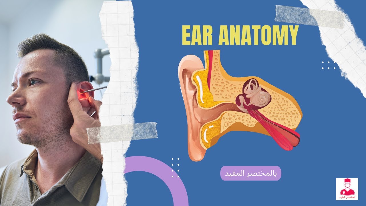
















The Organ of Corti is the sensory organ for hearing located within the Scala Media of the cochlea, containing one row of inner hair cells (primary sensory receptors) and three rows of outer hair cells (sensitivity modulators), with stereocilia embedded in the tectorial membrane that convert mechanical sound vibrations into electrical signals through bending, while the basilar membrane's varying stiffness along the cochlea's length enables frequency-specific sound detection.
Organ of Corti Diagram: Physiology of Hearing Explained
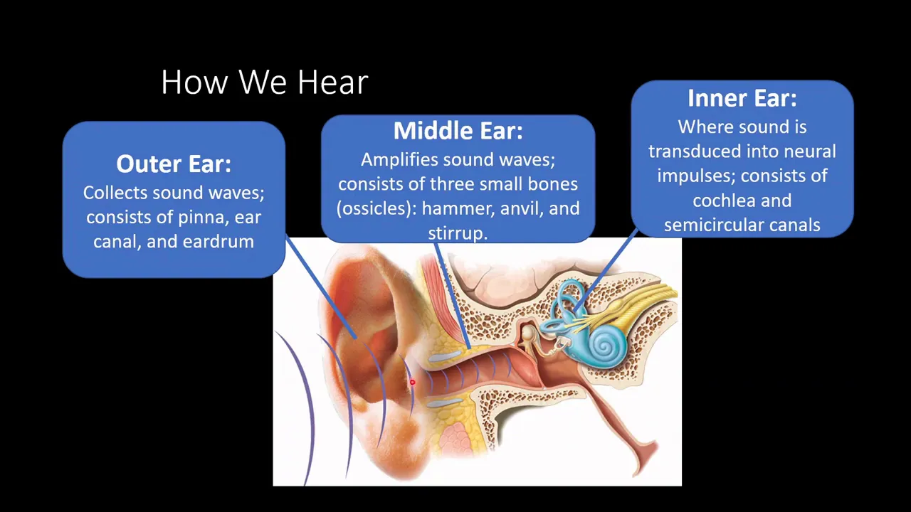
Added:Basic anatomy of the ear, specifically distinguishing between the outer, middle, and inner ear structures.
The physics of sound, including how sound waves travel and are converted into mechanical vibrations by the tympanic membrane and ossicles.
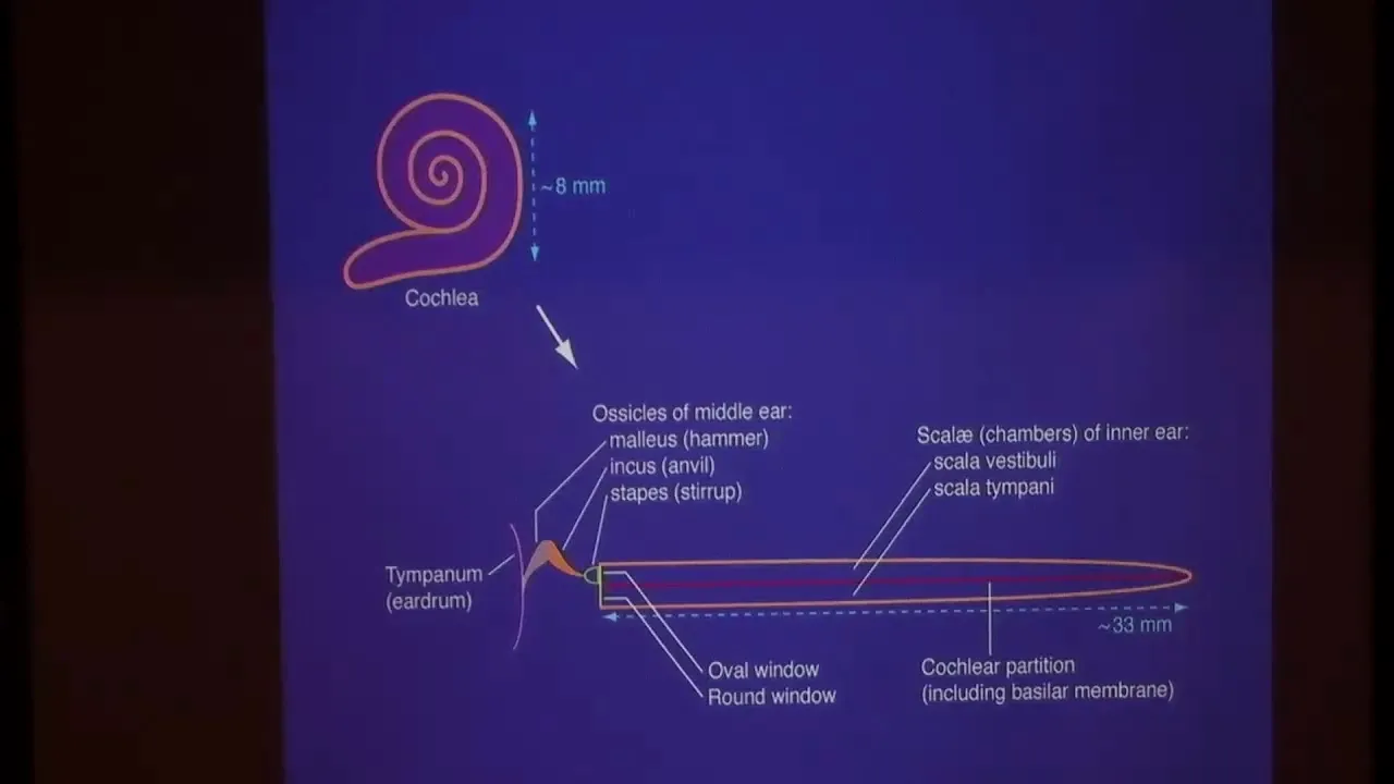
General anatomy of the cochlea, including the three fluid-filled chambers (scala vestibuli, scala media, and scala tympani) and the composition of perilymph and endolymph.
Basic principles of sensory transduction, particularly how physical stimuli are converted into electrical graded potentials in receptor cells.
The central auditory pathway, tracing how electrical signals travel from the cochlear nerve through the brainstem to the auditory cortex.
Pathophysiology of hearing disorders, such as sensorineural hearing loss caused by damage to inner or outer hair cells.
The biophysics and engineering behind cochlear implants, which bypass damaged hair cells to directly stimulate the cochlear nerve.
Advanced theories of frequency coding, including Place Theory and Temporal (Frequency) Theory of pitch perception.
42.8K views1.2Klikes13:48Original Release: 2023-07-30
Hello friends how are you doing today today we are going to talk about a very difficult topic a but a very important one also that is the organ of Corti so let's straight Deep dive into the topic so here is a diagram which is showing the cochlea now cochlea is like a labyrinth you see that it is taking some turns right so these are like two and a half turns cochlea takes and uh actually outside there is bony Labyrinth Labyrinth is anything which is very difficult to navigate okay so this is the Bony Labyrinth and inside this bony Labyrinth there is membranous Labyrinth so this outer walls which you are seeing form the Bony Labyrinth and inside we have membranes which divide this space inside the Bony Labyrinth into Scala vegetable and Scala timpani so there are two membranes actually so here what we see is a top membrane this membrane is known as a reasoner's membrane and then there is a bottom membrane bottom membrane is the basilar membrane and it is these two members brains which divide the cochlea into three parts where the top part where the first layer will be this is the Bony Labyrinth and the bottom will be the reasons membrane top part is known as the scalar vestibular how I remember up up is vestibular okay UV UV come together in letters right so up is Scala vegetable the below part which is again up Bound by basilar membrane and Below there is a layer of the Bony Labyrinth that is a scalar timpani and in between two compartments there is another compartment that is the scalar media now on this rela media is present organ of core type so that is the sense organ the hair cells which are there for hearing they are present in this organ of quartile now how this organ of Corti is arranged in this cochlea you see that this cochlea if it is running like this throughout the length of the cochlea in a transverse manner like this there is organ of Corti in the Scala media so here everywhere transversely there will be organ of Corti so please remember that throughout the length of the cochlea there is organ of cortiso sense organs are present throughout the length so this is a cut section of the cochlea where you see the various membranes and the organ of Corti and again as I was telling how it is arranged so this cochlea will continue like this and there is a cut section where it is showing the various compartments okay so top one is a scalar vestibuli bottom one is a Scala timpani and in between there is a scalar media and on the scalar media in the scalar media I mean you see these are this component is the organ of Corti so we will be studying this structure in detail so see how it is arranged it is in transverse manner so it will extend like this throughout the length of the cochlea so I just wanted to show you the arrangement of this organ of Corti that's why I had put this diagram so let's straight move into the organ of Corti structure so this is the basic structure of the organ of quartile now what is this blue line showing what will be the membrane we have seen that it is present on the scalar media and scalar media is bound on top by the reasons membrane and in Bottom by the basilar membrane so this blue line is showing the basilar membrane over which the organ of Cota is present with hair cells on the Top If You See I have not drawn reasons membrane rather there is another membrane tectorial membrane so let us go back to the previous diagram so this was a diagram which we saw you see this is the reason as membrane here this part is the reasons membrane and organ of cortide top portion it is not touching the resonance membrane here what I am talking is that there is another membrane and this membrane is the tectorial membrane fine so let us go back to our detail diagram of organ of Corti so what we see here bottom there is basilar membrane and top there is tectorial membrane so let us see what are the various other components so there are various kind of cells present in this organ of Corti so let us just enumerate the types of cells here there are pillar cells also known as the rods of Corti then there is hair cells and in this cases we have two different types there are inner hair cells and there are outer hair cells then there are supporting cells so all these green ones which you see these are supporting cells and they have different names hand senses three four types of supporting cells are there then the main part that there are hair cells which are protruding the part of the hair cells is protruding into the tectorial membrane so what are the types of cells there are pillar cells one there are hair cells which we have inner hair cells and there are outer hair cells and then there are supporting cells so these are the various types of cells then the membrane is basilar membrane there is tectorial membrane there is another membrane which is known as reticular lamina okay so what is the arrangement of these cells and membrane within the organ of Corti you see all the supporting cells are extending from the basilar membrane up till the reticular lamina so this is the reticular lamina and they are kind of supporting the hair cells We have basically one row of inner hair cells so this is just one cell showing here we cannot show you the row but as I told you it extends throughout the length of the cochlea in a transverse manner if a coffee has extending longitudinally then this row will be extending transversely throughout right so that is one row of inner hair cells and here we have three rows of outer hair cells so two types are there one row of inner hair cells and three rows of outer hair cells these air cells are supported by supporting cells and these hair cells have processes and these processes are known as stereocilia stereocilia which you see have certain characteristics first of all there is this reticular lamina and the stereocilia is piercing this reticular laminate is kind of piercing this reticular lamina and the top of the stereo Celia is embedded in this tectorial membrane so this is very important for the transduction process that is conversion of any auditory signal which will be coming as pressure waves in the cochlea this pressure waves are transduced to electrical signal by these hair cells so here cells are the transducing cells or The receptors of hearing so that is the structural aspect of hair cells also you see that there is a tunnel of Corti this is the tunnel of kotai which is basically Bound by the pillar cells that is the rods of Corti on either side so on the medial side of the tunnel there is his inner hair cell and on the lateral side of the tunnel there is outer hair cells and these hair cells they are innovated by neurons okay so there are neurons which are innervating both these inner hair cells and outer hair cells and medially they go into the part of the Bony part of the cochlea that is the modulus modulus and there is presence of spiral ganglion here that is the cell bodies of the neurons are there ganglion whenever we say there are cell bodies of neurons so inner hair cell present medially to the tunnel outer hair cells present laterally to the tunnel so that is the basic structure of the organ of quartile now what is the use of all this structure see whenever there will be a pressure wave that is here there will be reasonous membrane the pressure wave is going to travel and it travels via the end of the cochlea where that is the apex of the cochlea and it travels into the a Scala tympanic part of the cochlea so that is the pressure wave so it will cause the vibration of this basilar membrane so it will start vibrating okay now with vibration what will happen the all the structures which are present on the basilar membrane they will also start to vibrate so when the basal membrane basilar membrane starts to vibrate up this will cause the vibration of these cells also up and also the hair cells that will also move up along with that they will move to medial side the stereo sealer if you are seeing they are kind of fixed right in the reticular lamina as well as on the pectoral membrane so this tectorial membrane is kind of gel like okay so with the movement of the basilar membrane up and the hair cell up there will also be movement of this stereocilia towards the medial side right so all the stereocilia will bend towards the medial side and vice versa so if the basilar movement is down the hair cells will also move down and also outwards that is towards the lateral side so this movement of stereocilia of hair cells is responsible for transduction so these inner hair cells are responsible for transduction then what is the use of outer hair cells well these outer hair cells are responsible for changing the sensitivity of response of inner as cells means see these outer hair cells are innervated from efference ephrons from the central nervous system the neuron from the inner hair cell is the afferent it is carrying the information to the central nervous system but from the central nervous system the information is coming to the outer hair cells so whenever there is a depolarization of the outer hair cells these hair cells actually shorten in length actually there is a motor protein Within These Aces known as Preston okay so when that is activated these heses shorten in length and when depolarization is not there at the resting state these uh will lengthen or they can be hyperpolarization also and these can lengthen so when they shorten it is kind of making the structure stiff okay so information from central nervous system is making the movement of the organ of cortis stiffer and hence there will be less movement of these stereocilia of the inner hair cells and contrary will happen when there will be less signals to these outer hair cells that will cause them to lengthen thus changing the sensitivity of response of the inner hair cells so I hope in this short video of organ of Cortex I have tried to deliver the concept A difficult concept of the structure of organ of Corti how the cells are arranged and how outer hair cells are changing the sensitivity what is the importance of outer hair cells now remember one more thing here that this basilar membrane which we talked about its structure also changes throughout the length of the cochlea actually at the apex of the cochlea the structure is such that the membrane vibrates more in response to low frequency sounds okay and at the base of the cochlea that is where the cochlears is starting where the steps is attaching to the cochlea there this basilar membrane vibrates more to high frequency sound so there actually this basilar membrane is quite stiff and due to its stiffness it will vibrate only to high frequency sounds so the structure is similar rest of the structure is similar here cells are similar only the vibrating pattern is changing from the base to the apex of the cochlea and that is why there is a spectrum to which the cochlea responds from the base to the Apex Spectrum moves from high frequency to lower and lower frequency sounds this coding of information for sound I have dealt in another video on theories of hearing I will give the link in the description section below you can check out that video as well thanks for watching the video if you liked it do press the like button share the video with others and do not forget to subscribe to the channel physiology open thank you
Up Next

4:25
Muscle Fatigue: How Your Muscles Respond to Brain Signals
@TEDEd
5.7M views•2019-04-18

50:12
Bacterial Communication: Quorum Sensing and Biofilm Formation Explained
@JHUAAP
12.6K views•2011-06-28

5:35
Enteric Nervous System Explained: The Gut's Brain | Neurobiology Lecture
@alumniu6029
438 views•2018-09-12

7:08
Bacteriophages: Earth's Deadliest Killers and Future Antibiotics
@kurzgesagt
34.6M views•2018-05-13
Related Study Plans & Knowledge Roadmaps
Structured learning paths in Biology
biology
RNAi: Biogenesis, RISC & Gene Silencing
01Molecular Biology Foundations: DNA to Protein
02Introduction to RNA Interference (RNAi)
03Molecular Biogenesis of miRNA and siRNA
+ 2 more learning modules
10h5 modules
Explore Roadmap
biology
Mycorrhizae: Signaling, Biophysics & Ecology
01Foundations of Mycorrhizal Symbiosis
02Molecular Signaling and Root Colonization
03Biophysics and Bioenergetics of Nutrient Exchange
+ 1 more learning modules
11h4 modules
Explore Roadmap
biology
Circadian Clocks: TTFL, SCN & Periphery
01Foundations of Molecular Biology & Gene Expression
02Introduction to Chronobiology and Actograms
03The Core TTFL: Molecular Architecture of the Clock
+ 3 more learning modules
14h6 modules
Explore Roadmap
biology
Population Genetics: Math, Drift & Selection
01Genetic & Probability Foundations
02The Hardy-Weinberg Principle and Baseline Models
03Mathematical Modeling of Natural Selection
+ 3 more learning modules
18h6 modules
Explore Roadmap