




















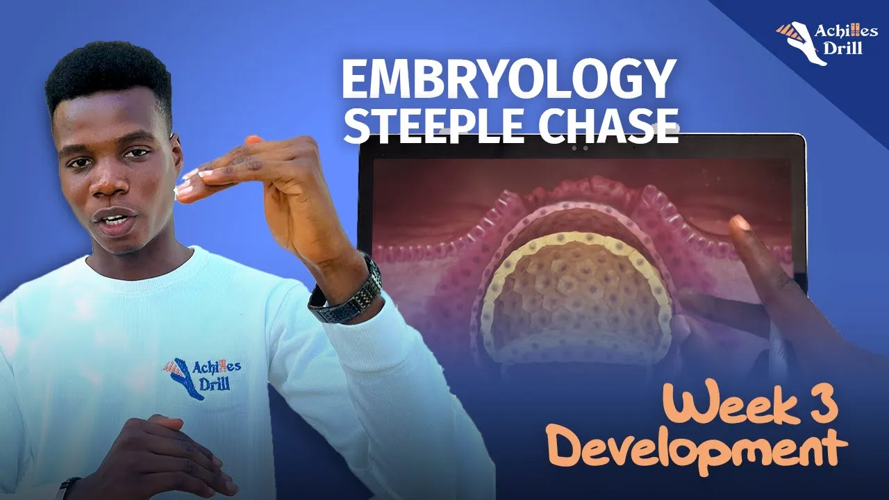
Gastrulation in Xenopus laevis involves seven coordinated cell movements—blastopore roof thinning/spreading, vegetal rotation, bottle cell formation, tissue separation, cell migration, and convergence/extension—that collectively transform a spherical blastula into a multi-layered embryo; these movements are driven by specific cellular behaviors such as apical constriction in bottle cells, planar cell polarity signaling for convergence/extension, and chemoattractant-mediated cell intercalation, with disruptions in these processes leading to developmental defects like spina bifida.
Frog Gastrulation: 7 Cell Movements in Xenopus Development
Added:The stages of early embryonic development, specifically cleavage and the formation of the blastula.
The definition and significance of the three primary germ layers: ectoderm, mesoderm, and endoderm.
Basic cell biology of the cytoskeleton, particularly actin-myosin dynamics and cell adhesion molecules like cadherins.
The concept of embryonic polarity, specifically the animal-vegetal axis in amphibian eggs.
Neurulation and subsequent organogenesis, detailing how the gastrula transitions into a neurula.
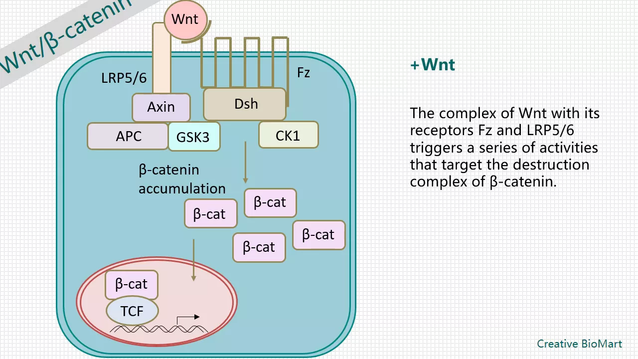
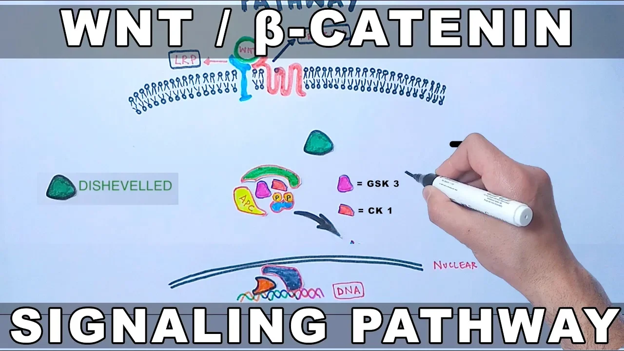
The molecular signaling pathways, such as the Wnt/Planar Cell Polarity (PCP) pathway, that orchestrate these cellular movements.
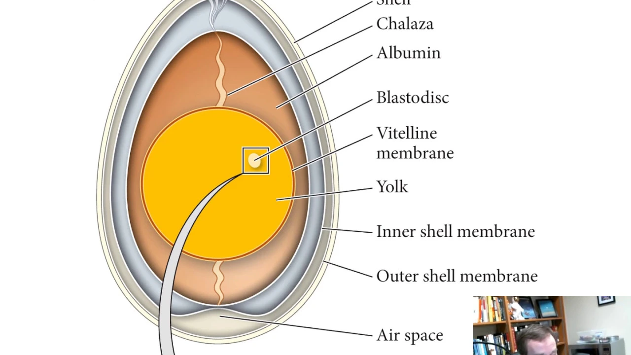
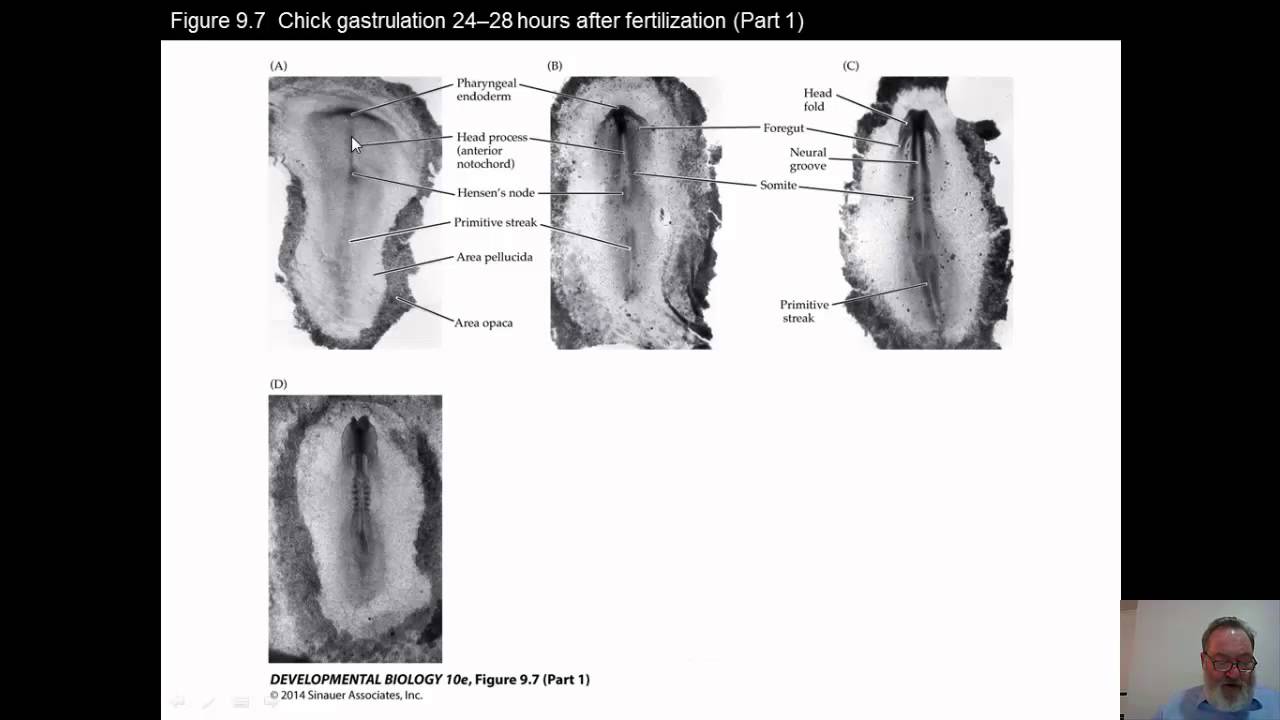
Comparative embryology, analyzing how gastrulation mechanisms differ in amniotes like chicks and mice.
The developmental and clinical consequences of abnormal cell migration during gastrulation, such as neural tube defects.
29.6K views474likes17:34Original Release: 2018-04-18
my name is Richard Holland and this is the second in the series of talks about Xenopus gastrulation and pass information' and in this short video I hope to show that gastrulation which is often thought of as a horrendously complex process it's actually fairly simple you just have to take it apart and look at the individual cell behaviors and they're quite a few different cell behaviors but these all conspire to get the whole mechanism of gastrulation to happen so let's first start with this schematic which we saw from the first video and so what we're going to discuss is all of the component movements that go into this complex process of gastrulation so we'll discuss for example the thinning of this roof the crawling of cells across the Blas sill roof and the other individual motions that contribute to the process okay so here's a schematic which shows seven basic mechanisms of gastrulation I'm only going to discuss six but we'll take them in turn so the first one is the apipa that is the thinning and spreading of the blaster seal roof so all of this blue tissue because the blue tissue has to spread and cover the whole embryo and recently there's been a lot of progress made in understanding how this may occur so a pibbly consists of this initial gathering of cells and these are freeze fracture electron micrographs looking through that blasted steel roof you can see at the beginning of gastrulation we have about three cells thickness of the whole surface but during gastrulation there's a thinning process and you can see these green cells here intercalating between one another and that of course is going to lead to a spreading so this is spreading not only of the green part by virtue of this rearrangements of cells the intercalation between each other but also up here you can see that these red cells begin to be a little stretched no longer quite so cuboidal it's subtle but it makes a difference in the overall process and below here it's represented schematically where you start with the three layers and then go through this intercalation and end up with a thinner but much wider array this is the work of Robert Mayo's lab what they found is that there's a signal that's made in the red cells it's actually a compliment signal and that tax as a chemoattractant for the green cells to compact against the red cells so that's thought now to be the major driver of this spreading process of the blasts Cecile Ruth let's move on to a second signal now vegetal rotation so vegetal rotation is the process whereby all of this yoki stuff you're a member has to constrict to move inside and so what we're going to see is there'll be a spreading of this floor of the blaster seal while at the same time the outside layer will have to collapse down essentially to a point to get enveloped by the rest of the animal now occurs by a fountain like movements of cells it's also slightly asymmetric such that this dorsal side the future head tissue up here is going to end up moved against the blast seal roof you can see this cleft here so unlike on the ventral side the dorsal side has this cleft the cleft of Brasher as it's known where there's a contact between the innards and the outer parts of the embryo so again let's watch what happens here you can see the spreading there of the blast seal roof it becomes quite a large surface area before it finally collapses the collapse of the blast seal is thought to be mediated mostly because these cells here are very loosely attached and the liquid moves from the blaster seal into this other cavity the arc Antrim which is the primitive gut cavity at the same time you can see that cells down at the bottom they initially covered a very large surface area but they collapsed down to a small point so again this is thought to arise as a result of the rearrangements of cells so that's a sort of fountain spreading of cells and this has been studied recently by Rudy Winkle Powell it's lab and here he's taken a slice through that yoky tissue and and put it in culture and he can see this fountain like movements of cells so these celery arrangements are exerting force and here you can see also the spreading of the blast sealed floor whereas the initial area was much much broader okay so that's thought to be the motor forgiving convergent this this vegetal rotation against the blasted steel roof the underlying mechanisms are not understood so well but at least the cellular behaviors can be described let's move on to battle cell formation we saw that as the initial pigment line which we can see from the surface of the embryo and actually we see these after there's already been quite a lot of action inside the embryo so these internal movements have already started to occur and indeed these buffle cells form slightly later and then as I mentioned in the first lecture they form a kind of inflection point so that when other forces extend this red mesoderm and crawling forces of the purple tissue occurred over the blasted civil roof and rather than going outside the embryo these will flip the corner and move inside the embryo so here's a light micrograph of a stained section done by Jen Lee in the lab and what she's done here is sustained the self the tubulin and actin and as is generally found with these apical E constricting cells they're called bottle cells because of the old-fashioned glass blown bottles that were use when these were first described in the late 19th century you see this apical constriction here the cells become elongated and actually a whole traitor's showed that these cells are quite invasively they really have a strong impetus to move inside the embryo while the apices constrict and you can see here there's some irregularity and how they go about this some of those were quite constricted whereas a neighbor is not so constricted but the act myosin construct contraction that's mediated their constrict these ABCs and leads to the beginning of an invagination movement on the outside of the embryo here's a movie that shows that from the outside so this is an embryo that's been labeled with a membrane targeted GFP and so what we'll see as this plays through is that up here there's not much action these cells are perhaps being stretched but down here where the baffle cells are forming you can see the constriction of the apical surfaces of these cells and it's by no means completely uniform there are some cells in between them that are not undergoing apical constriction and they're sort of passively stretched by the forces from the striction but ultimately all of these cells will start sway to click constrict and that will formulate this impetus for these cells to move inside the embryo and help the process of involution of the marginals and the equatorial zone of the embryo I've already mentioned this cleft here the cleft of brochet and of course this cleft could have to face one is that the tissues can remain separate or they could merge with one another and that's a real active process of tissue separation that these cells are maintained as different and that can be shown by these explant experiments again done by Rudy Winkle Bower and what he did was to take this blast seal roof this pale blue tissue prospective epidermis and invert it in culture then he can put onto that little groups of cells from different regions of the embryo and so for example what he can show is if he takes like tissue from the ectoderm and puts that on there those cells will dive in and just merge with the rest of the explant but instead if he takes this this red tissue or purple tissue either one he can put those on the blast seal roof and they'll maintain a separate identity and stay separate so this is an active difference in the cell fate of tissue separation that keeps these separate and allows this cleft to be maintained we're now moving on to cell migration and this is a real force generating process as well and this leading edge here of the endo mesoderm so the endoderm is yellow the maasdam is pink and red that endo mesoderm actively sticks to the overlying blasted steel roof it's just the first cell or two that does this and then those cells actively crawl across the blast seal roof so during gastrulation these cells have the ability to find their direction up towards the top now this has also been studied in isolated cells and again I'm going to use a similar assay where one has an inverted cap from the gastrula and take this purple tissue and put it on top if one does that with big pieces of tissue then that is a able to coherently migrated in a single direction but here what I show in this movie is that the individual cells they clearly have the ability to sit on the underlying blast seal roof but they're migrating they're migratory as individuals they migrate pretty randomly but when they're in a coherent mass there's an edge that provides a directionality to their migration oK we've done the first five and the final one is we're going to do convergence and extension and that is the process whereby this red tissue becomes very long this occurs not only in the mesoderm making the notochord but also up in the spinal cord the prospective spinal cord which is just up here this has been studied in detail again using ex plants and this particular one is the work of John Wallingford in the lab where he took x plants that have been originally made by Ray Keller the Keller X plant so over here as a whole gastrula and he's cut off with a pair of eyebrow nights this dorsal region here peel it back and chop it off so you have one piece of tissue that's just a few cells thick and this can be put nicely underneath a coverslip and squished down onto a slide it doesn't adhere to the slide but the cell movements and the cell behaviors in there can be visualized using this confocal microscope that's sitting underneath it so look at the behavior of the cells we we scatter label those cells by injecting one of the early blastomeres the early cells with a membrane targeted GFP such that we get some but not necessarily all the cells labeled now previous to this experiment Ray Keller had already described the behavior of these cells in particular that the onset of gastrulation these cells put out these lamellipodia these slabs of protrusion and all sorts of directions randomly but during the course of gastrulation those cells become very oriented media laterally so that they're putting out the protrusions on their size their left and right sides they then proceeded to crawl between each other so that a nurse Lien's for instance this array of four cells will become an array of four cells here that's in a line so it goes from a squat series of cells to a long skinny array of cells and this is a potent force generating mechanism in gastrulation that drives the elongation of the prospective notochord and the prospective spinal cord so this is that happening in a dish the technical reasons what's been done is to take two of those ex plants and sandwich them together and then they do this behavior very well even though they're sitting on agarose they have nothing to crawl on and so you can see here that the head end of this X plant does not undergo this behavior so much but this spinal cord area does and if we use molecular markers we can tell that this is all spinal cord here whereas down here we have the mesoderm the note respective notochord and head music oh now in this case we keep it flat underneath pressure from a coverslip whereas in the normal embryo all of this stuff down here would have crawled underneath and up the inside but this illustrates the autonomous behavior of these cells in this macroscopic view let's go to the the confocal view now and here we're going to discuss an experiment where we've manipulated planar cell polarity signaling so using a special internal deletion of the molecule dishevelled which is involved in planar cell polarity we're going to interfere and see what that does to these cells it had previously been shown by Sergei so-called and if you use this reagent in the whole embryo it prevents the convergence and extension behavior but the question we had was is this a very specific effect on the cell behaviors or is it a sort of a nonspecific toxic effect on the self behaviors now as you'll see we can resolve that clearly by looking at the behavior of the cells in detail so let's start this movie going and what you'll see is the one on the right actually doing something whereas the one on the left is doing very little this is the control and what this is showing these cells are aligned sort of from left to right and they have these extensions on their surface you can see these lamellipodia extensions so this is the phase where they're getting oriented they put out at least one extent that extension is very stable and so over the course of time it's thought that that can exert traction on the neighboring cells so that the cells crawl between each other this is the case where we put in that dominant negative reagent and you can see a very different behavior but importantly if this is a very nonspecific effect a toxic effect we would expect the cells to do nothing and not crawl between each other instead what happens is these cells are actually hyperactive they're putting out these protrusions and I think you can see on this cell for instance some of these protrusions are put out and then taken back quite quickly so the protrusions are unstable and therefore unable to sustain a force generation and the other thing is that if one looks at the shape of these cells they're much more randomly shaped the cells in the control actually become elongated whereas the cells in the manipulated case lose that orientation so we think it's that lack of polarity in these cells there and the lack of ability to sustain traction that causes them to essentially be non-productive they're like a bunch of kids with a DD the running around with lots of energy but not achieving a whole lot so instead of that sustained and disciplined behavior we have a very active behavior but without the polarity and the sustained a lamellipodia contact that allows this intercalation of cells we can the the consequence the embryo is as said is their lack of convergence and extension and we can see that in the whole embryo the top embryos are control that's undergoing its proper convergence and extension movements and here we see that particularly in the neural plate as the neural plate extends and comes together to make the neural tube in contrast the bottom embryo has been injected with a Stormin and negative reagent and you can see it although it can close the blastopore it fails to undergo much convergence and extension of the neural plate the consequence is that the neural folds although the neural folds try to form they never quite get close enough to touch so this mimics a human condition that is sadly quite prevalent in children that that of spina bifida very extreme form spiderific diffident in this case that results from the loss of that planar cell polarity signaling and indeed it's now known that one of the predisposing factors in human birth defects of spina bifida can be defects in the planar cell polarity components so those are the movements that I've summarized and as you've seen they can between them all get together to give this rather complex overall result but each individual cell behavior is relatively simple so in this movie the embryo ends up pointing at the sky but if we turn it over so that the anterior is now at the anterior end you can start to see that this is really a tadpole so we have a head over here we're going to have the spinal cord that's forming up here here's the primitive gut and the future anus and if that just goes along a little bit further with the extension of the axis we can see that it really it starts to form something that looks like an animal with the eye going to be forming over here the spinal cord this long stiff rod the notochord and then the gut beneath so as far as I'm concerned as an embryologist this is pretty much it the rest is just elaboration of this initial pattern and the growth of the animal into an adult so that's this short presentation on the cell movements in the next presentation we're going to discuss how the signaling events happen during early development to make the cells different from one another and give rise to the formation of the nervous system in particular you
Up Next

24:21
Gastrulation in Developmental Biology: Germ Layer Formation Explained
@shomusbiologyofficial
205.2K views•2013-09-23

41:32
Circadian Metabolomics: Sleep, Food Timing & Human Clocks
@tscnlab
359 views•2022-11-10
24:22
Genetics of Circadian Rhythms: PER2 Phosphorylation
@scicommlab
2.7K views•2017-11-20

7:08
Bacteriophages: Earth's Deadliest Killers and Future Antibiotics
@kurzgesagt
34.6M views•2018-05-13
Related Study Plans & Knowledge Roadmaps
Structured learning paths in Biology
biology
Circadian Clocks: TTFL, SCN & Periphery
01Foundations of Molecular Biology & Gene Expression
02Introduction to Chronobiology and Actograms
03The Core TTFL: Molecular Architecture of the Clock
+ 3 more learning modules
14h6 modules
Explore Roadmap
biology
Population Genetics: Math, Drift & Selection
01Genetic & Probability Foundations
02The Hardy-Weinberg Principle and Baseline Models
03Mathematical Modeling of Natural Selection
+ 3 more learning modules
18h6 modules
Explore Roadmap
biology
Animal Migration: Sensory Systems & Navigation
01Foundations of Animal Migration
02Magnetoreception: The Quantum and Chemical Compass
03Celestial Navigation: Sun, Stars, and Polarization
+ 2 more learning modules
12h5 modules
Explore Roadmap
biology
Proteostasis: Folding, Ubiquitin & Autophagy
01Foundations of Protein Structure and Synthesis
02Chaperone-Assisted Protein Folding
03The Ubiquitin-Proteasome System (UPS)
+ 2 more learning modules
12h5 modules
Explore Roadmap