






















Gastrulation is the critical embryonic development phase where a single-layered blastula transforms into a trilaminar gastrula with three germ layers—ectoderm (outer layer giving rise to skin and sensory organs), mesoderm (middle layer developing into muscles, bones, and circulatory system), and endoderm (inner layer forming digestive and respiratory systems)—through coordinated cell movements guided by specialized bottle cells that create the dorsal lip and eventually the blastopore, which becomes the anus; this process establishes the body plan and commits cells to specific organ system development.
Gastrulation in Developmental Biology: Germ Layer Formation Explained
Added:Fertilization and Cleavage: Understanding how a single-celled zygote undergoes rapid mitotic divisions to form a multicellular embryo.
Structure of the Blastula: Familiarity with the blastula (or blastocyst in mammals), including the blastocoel and the inner cell mass.
Cell Potency and Fate: The concepts of totipotency and pluripotency, and how embryonic cells begin to restrict their developmental potential.
Basic Cell Biology of Movement: Understanding fundamental cellular processes like cell adhesion, migration, and the epithelial-to-mesenchymal transition (EMT).
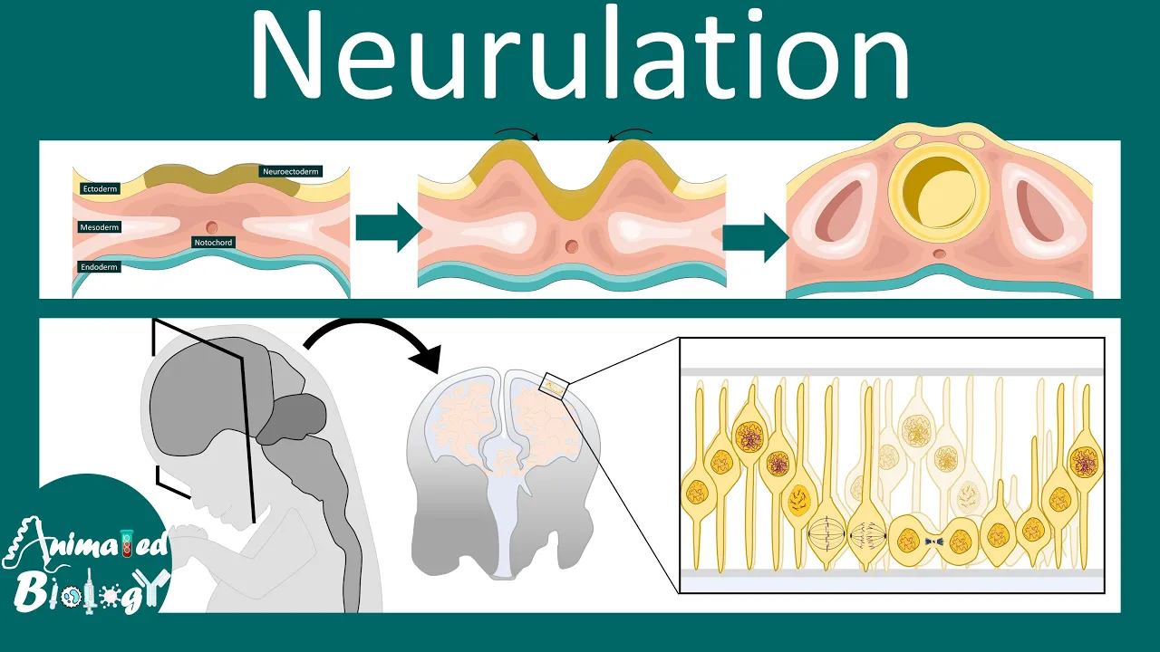
Neurulation: The subsequent embryonic stage where the ectoderm forms the neural tube, laying the foundation for the central nervous system.
Organogenesis and Germ Layer Derivatives: Exploring how the ectoderm, mesoderm, and endoderm differentiate into specific tissues, organs, and systems.
Embryonic Induction and Signaling: Investigating the molecular signaling pathways (such as Wnt, BMP, and Nodal) that coordinate gastrulation and pattern the body axes.
Developmental Pathology: Studying clinical abnormalities and birth defects that arise from errors during gastrulation, such as caudal regression syndrome or teratomas.
205.2K views1.9Klikes24:21Original Release: 2013-09-23
[Music] Hello friends, we are talking about developmental biology and we have seen the introductory part about developmental biology and why we study developmental biology and then we have seen uh the sequential step of embryogenesis which is drawn here. Uh that means the fusion of egg with sperm then production of zygote then the division of the zygote. Now cell division of the zygote which which you can call the as cleavage will give rise to mass of cells layered in different uh or oriented or placed in different planes to make a three-dimensional cellular structure which is called a blast stage. Then these cells will start to mature and as they mature they get the signal of producing different layers during the gastula gastulation step. Now this is the most important step above all because at this stage those cells that we can derive from this blastula will be destined and committed to produce a special type of cells. For example during this blastula events the most important event is the production of different layer of cells. Now those different layer of cells will generate different type of organs and different type of organ system inside the organism's body. For example, during this gastrolation, it will develop into three different layers. One is the outer layer which is called an ectoderm and innermost layer is called the endoderm and the layer which is placed middle of ecto and endoderm is called the mesoderm. So these three layers are generated. Now from those three different layer of cells different type of tissues generated during the development or during the embryoenesis.
Now a part we have to talk about this whole picture is called the embryogenesis. Embryioenesis means it is a production and maturation of a fully functional embryo from one cell which was previously zygote. Okay. Now we'll be studying this gastula phase in detail in this particular video. So the process of the production of cells and production of these different cell layers during the embryogenesis at this stage will be called gastrulation.
Okay. The phase is called gastrula and the production of this type of cells from a chunk of one one type of cells which are totally potent in nature and they commit into a particular special type of cell via the process is called the gastrolation.
Okay. Now in this gastulation the most important event I've told before that the production of uh or arrangement of the cells in different layers outer layer, inner layer and middle layer.
So for this particular thing to happen there are again sequential events because remember in developmental biology everything happens in sequential events. Okay. And timing is very much important. And we have seen in previous video that in this gastulation at the very beginning of gastulation at the early stage of gastula the cells are not yet committed to a particular the production of a particular cell type or organ type after the gastula is going gastric phase is going on at the later stage of gastrolation the cells are committed to to do their job. Okay. So now here let us talk about gastrolation.
So we'll first draw uh the structure of uh or or from the blast the late blast then we come to the gastula. Now actually the step is drawn here is not particularly blast because blast stage is also important because it provides uh the the structural integrity of the cell. It provides uh the 3D structure or a structure like a ball a hollow ball which is in turn converted into gastrol.
So simply what we'll be seeing is an hollow ball but all the cells that are present there is destined to produce every type of cell inside our body but once it is converted into gastula it produces three different layers that is the difference during this process.
Okay.
Okay. So let us consider this. So let's consider this as a blastula. Now during this blastula stage what we can have? We are having layers of cells. layers of cells arranged in different regions.
Okay, for example, we need to draw this like that.
Say this blue colored region is a type of cells. So the cell differentiation begins from the blast phase. It will be completed after the end of gastrolation.
Okay. And gastrolation means most of the part of this embryogenesis is done. So from the blast to face we are having this. So what we are having we're having a layer of cell. Now this is a blue layer of cell. So I I'm just drawing it like that. So so that you can find this as a blue layer. So it's a layer of cell.
Then we what we are having we are having this red region of cells.
Okay. And then finally we're having this black part.
Okay. Okay.
So we have something like that. Okay.
Now picture uh look at the picture and the color representation is very important in this case. Now this blue colored region cells are arranged. So it's an hollow ball. Why I'm telling it's a hollow ball? Because all these shaded regions are solid mass of cells.
All of these shaded regions are solid mass of cells and rest part this this blank region is blank. Nothing is there.
So it's a hole actually it's a vacule like it is called blastoal.
Okay. And the whole thing is called a blastula and blastocyst. And this hole is called blastoil.
Yes, this is called a blastoil. Okay, and less rest of the part is there. And the rest of the part means this blue colored lining of cells. Remember I've told you that the differentiation stage begins from the blastula and then it complete completed in this gastula. So this blue colored things blue color cells here are ectomal cells.
red colored region is messodermal cells and this black colored thing is endodermal cells. So we are having layers of cell in this particular stage. So from the beginning in the blasto stage we are having these different type of cells ectoermal, endodermal and messodermal cells. Okay. But they are arranged just in a small amount like that. But during the gastulation what they does is that they start to arrange in such a way which is uh giving rise to the organism. Okay. So these are simply mass of cells. Now the cells are arranged in in a region and make a hollow blastoil at the middle.
Okay. So this is a structure but the cells will arrange in such a way but they are differentiated in ectomal, endoderm, misodermal type of cells but they are not yet layered. Okay. But after this gastrolation what we can see the cells will be layered in such a way that in this whole uh whole ball like structure outermost will be the ectoderm middle one is the endodm meodorm and the inner one is the endoderm. But in this case we haven't find these things just cells give rise to different type but yet they are not arranged in layers.
Okay. So in the grulation we'll be seeing the arrangement of these cells in layers. Okay. Let us consider now. Now this is called a blastoyst and it is having blastoil and blasto stage. Now what will happen? This cell will start to modify. How? Because the cell as you can see here in this endodmal region there are some new arrangement of cells.
Those cells are called bottle cells.
Okay. Or bottleneck cells. So this bottle cells here. So let me draw this here like that. So this is a joint where bottle cells start to so some bottle cells I don't know whether you're able to see it or not. This is this green color cells are the bottle cells. Now the bottle cells are really important because they are called the guide cells.
Okay, the chunk of cells these are some bottle cells not one many bottle cells.
Now why they are called bottle cells?
Because they are act so they arranged in this like this orientation. So the epical region of the bottle cells are narrower but the bessel region of the bottle cells are broader. So that's why they looking like a bottle uh like a bottleneck. and uh bottom part is uh spreading. So it is called a bottle cells. Now these bottle cells are called guide cells because these cells guide other cells during this gastulation.
Because what we know in this gastula the most interesting event is the movement of cells and lining of the cells one after another. This is sheetlike arrangement and movement of cells will be required.
Now what we'll be seeing in the future events the bottle cells will guide other type of cells like the cells of ectodermoderm and endoderm to move at this directions.
This direction it's a anticlockwise direction we'll be seeing it.
Okay. So we'll be seeing this anticlockwise direction of the cells and then they will arrange it in layers. So our actual goal is to produce something like that. Let me draw this. So, so actual goal is a production of this thing endo ectoderm in the outside, misoderm in the middle and endoderm at the last. Okay. So, this will be our arrangement. At the end, we will end up with something like that. So, after the gasolation, we'll find something like that. And at the middle we will having the hole which will make the gut. Right?
Now in this case it will be converted into this kind. How? Let us consider here. Okay. So here the bottle cell will guide other cells to move. Now as the bottle cells are formed it will guide other cells to move in this direction.
The cells will start moving and as they are moving they start to make a small hole-like structure. And this joint point of this bottle cells and the ectoderm and the mesodermal cells it is forming a lip like structure just like a lip this opening right a small opening like structure it is called the dorsal lip. So at as they start to move so let me draw it again here like this. So as they start to move the movement will look like this.
So as all the cells are spreading and remember so not only the cells are spreading so if we need to spread the cells we must have access to development of more cells. So cell division is going on in a rapid speed during this stage and they are moving. So say here the cells are moving and let us consider this like this way.
Okay. And here are the bottle cells.
Okay. something like that. Okay. And say okay.
Okay.
So this will be the structure after some time. Okay.
Movement of cells is going on. So you can see the movement of cells are going on in this way. And sorry one mistake is there.
So say here it is and okay so this is a movement and uh those bottle cells are placed there. So as they start migrating the cell starts migrating in this direction anticlockwise direction and what we start to form a structure like that. So it will form a lip here. Now this region the formation of lip is called dorsal lip.
Okay.
And as the cells start migrating what they're producing here they're producing region which was previously blastoil which is a large region but now this blastoil is constricted because other cells are start migrating and filling the uh space. So it will lead up uh it will lead up to the formation of a light a small region of blastoil it is called this particular region is called so there must be this part you must draw this.
Okay. So this part is called then arenton. So the blastoil is constricted due to the movement and uh movement of cells onto this region and what they produce is called arenton. This is again another hole another type of vacule but this is now called arenton and it is also called primitive gut because this is the first type. So it's a very earlier type of gut of the organism.
It's called primitive gut or or also called arenton right. So let me write arenton or it is also called primitive gut. Okay. So you need to learn this terminologies because lot of questions come in CSN providing this terminologies. Okay. So the movement of cells is going on. So from this part what we can see so these cell lines are moving cell layers. So these messodermal cell layers are moving here endodermal moving and the ectoderm is also moving.
So this can eventually give us the idea is that so this is the endodermal. Now the idea is giving us that this endodmal region is going to be inside and rest of the regions meodorm will be the middle of the ectomal endoderm. So actually from this part we can see that if the cells continues to move at this direction it will form a structure like this right and at the middle is the gut or arenteran here. Okay, this is called dorsal leap and as they are moving and this this site is expanding and it is it will form a blastoal and a blastol like structure what is called a blastopppore because this will form a porous uh region it will be called a blastoore from this region those blastoore region will become anus of that particular organism. Now here we are discussing about the development and gamtogenesis uh not gamtoenesis sorry embryogenesis of frog xenopus. So write it down the example is given for frog xenopus. So they are moving. Now after the stage what will happen here what we'll we end we can see the movement of the cells. So after the movement after few gener time or uh after a few days the cells at this position again give rise to another type of bottle cells. So bottle cells previously formed at this particular location. Now it will form the junction between the messoderm and ectoderm of another region because we are having the messodermal ectomal access in two different points. Bottleneck generates at the region of meodormal ectodermal region. It generates first here. Now it will generate here. So from here we'll be seeing that bottleneck bottle cells will generate. So another set of bottle cells will generate. Okay. So let me take here bottle cells will generate here again.
Now as bottle cells are generate as we all know bottle cells are guiding the the ability of bottle cell is to guide the cells to guide the cells to form layers. Right? So here they again start to guide this type of cells to go and form layer like structure. Okay.
Okay. So what we we can see they start to form again a layer like structure. So as they are making invaginations like that and start to form the layer like structure. So they are moving here in this direction and as they're moving in in this direction. So eventually they they're making the space for endoderm really shorter. Endoderm comes at the at the inside and rest of the regions will be outside. So what will it look like?
It looks like something like this.
So again, say this is the beginning point. Here it was for the beginning. So try to follow this because it's very important. Now as they are moving in this direction. So they are moving here in this direction and also the meodmal region is also moving. So they end up with something formation like this. And the endodermal segment are constricted. So they will be here like that. Okay. So at the end what we can form something like this.
Okay. So at the end we will form something like this because this bottle cells again guide the cells to go at this. So what will happen again this lining of the cells happen and form something like that. Okay. So what we end up with here we end up with three different layers. Outermost layer which is a blue color layer which is ectoerm.
Middle layer red color region which is the messoderm and the inner black color region or endoderm. Okay. So after certain time this will fuse with one another and form something like that.
Now in this particular place the regions ectoderm and this is a misoderm and again the endoderm.
So remember in case of the blastola or the late blastola where we are having the blastoyst and blastoil and the formation of a hollow ball we are also having the different layers not layers different type of cells ectodermal endodmal meodermal but the important thing is that in those case we don't have the alignment we don't have the arrangement of cells in this layer structure but after the gastrolation we end up with the layering of the cells which is the most important part during the gastulation the layering of the cells arrangement of the cells and what it forms it forms two different regions.
Okay, it it forms a region and where we end up with the blasto pore and this will become the anus of the organism and during the second bottle cells formation it forms another lip. We call them vententral lip.
Now formation of this dorsal lip and ventral lip are also important because this dorsal lip and ventral lip will dictate whether which part is going to be the dorsal region and which part is going to be the vententral region of that organism. Okay. So end up with this kind of structure. Now this ectodermodorm and endoderm the different layer of cells are having different functionalities. What are the functionalities? They these type of cells will give rise to specific type of organ systems in the body of organisms.
For example, the ectoermal cells will give rise to the skin cells and also sensory organs.
Skin cells and also sensory organs.
Mesodermal cells which is in the red color here. They will give rise to here. This messodermal cell will give rise to uh sorry this meodermal cell will give rise to uh reproductive system, bone system, okay and also blood circulation system.
But the endodermal region cells will generate and this misoderm also uh produce a reproductive system. The endodmal cells will produce digestive system and respiratory system.
Okay. So the type of cells. So these cells this ectoermal, messodermal and endodermal cells are specialized cells.
they will generate specific type of organ systems in the organism's body.
Now there's differentiation made in the less blastular phase but the commission commitment of the cells is seen in the late gastric phase and the arrangement after the arrangement. So we can find this the the the commitment of this type of cells to produce the specific type of organ system or cells of organ system only after the late glastula phase when the arrangement of the cells happen in these three lines. Okay. So this three layer arrangement of the cells during the gastrolation is very important for the commitment of those cells to produce specific type of organ systems. Then when they produce specific type of organ system combining them we can have what is called an organism here in this case xenopus xenopus frog or it is a normal xenopus fro frog. Okay. Now you need any problem during in this different stage any problem during the phases of this gastulation will lead to the malfunctioning or abnormal embryo and this abnormal embryo will develop into an abnormal uh adult xenopus frog. Okay. Now this is very important developmental biology again I'm telling you it's a sequential steps and timing is very very important.
Okay. Now the stages of this gastrolation can be easily visualized and utilizing dyes because if you utilize if you stain those cells with different dye we can track the movement of the dye during time or during this development with time and you can find something this okay so these are the sequential steps of gastrolation and I hope this will help you to understand.
Thank you.
Up Next

26:01
Tissue Flow Modularity in Avian Gastrulation | Serra Lab Talk
@bppbseminar134
267 views•2024-09-16

50:12
Bacterial Communication: Quorum Sensing and Biofilm Formation Explained
@JHUAAP
12.6K views•2011-06-28

14:34
RNA Interference Mechanism: siRNA and shRNA Pathways Explained
@shomusbiologyofficial
379.7K views•2015-02-21

7:08
Bacteriophages: Earth's Deadliest Killers and Future Antibiotics
@kurzgesagt
34.6M views•2018-05-13
Related Study Plans & Knowledge Roadmaps
Structured learning paths in Biology
biology
RNAi: Biogenesis, RISC & Gene Silencing
01Molecular Biology Foundations: DNA to Protein
02Introduction to RNA Interference (RNAi)
03Molecular Biogenesis of miRNA and siRNA
+ 2 more learning modules
10h5 modules
Explore Roadmap
biology
Mycorrhizae: Signaling, Biophysics & Ecology
01Foundations of Mycorrhizal Symbiosis
02Molecular Signaling and Root Colonization
03Biophysics and Bioenergetics of Nutrient Exchange
+ 1 more learning modules
11h4 modules
Explore Roadmap
biology
Circadian Clocks: TTFL, SCN & Periphery
01Foundations of Molecular Biology & Gene Expression
02Introduction to Chronobiology and Actograms
03The Core TTFL: Molecular Architecture of the Clock
+ 3 more learning modules
14h6 modules
Explore Roadmap
biology
Population Genetics: Math, Drift & Selection
01Genetic & Probability Foundations
02The Hardy-Weinberg Principle and Baseline Models
03Mathematical Modeling of Natural Selection
+ 3 more learning modules
18h6 modules
Explore Roadmap