A chemical synapse is a junction between neurons that enables communication without physical connection; when an action potential reaches the presynaptic terminal, voltage-gated calcium channels open, triggering synaptic vesicles to release neurotransmitters (like acetylcholine) via exocytosis, which then diffuse across the synaptic cleft and bind to ligand-gated sodium channels on the postsynaptic membrane, causing depolarization and allowing the action potential to continue propagating along the neuron.
Chemical Synapse Animation: Neurotransmitter Release Explained
Added:Deep Dive
Basic anatomy of a neuron, specifically the roles of the axon, dendrites, and the axon terminal.

Neuron Structure & It's Function || Dendrite, Cell body, Axon, Axon Hillock & Axon terminal ||
796
A neuron is the basic structural and functional unit of the nervous system, consisting of three main parts: the cell body (containing the nucleus and organelles that control metabolism), dendrites (highly branched projections that receive nerve impulses from other neurons), and the axon (a long, unbranched fiber that transmits nerve impulses away from the cell body to muscles or glands); the axon may be surrounded by a myelin sheath (which acts as an electrical insulator and enables faster impulse conduction through nodes of Ranvier) and terminates at axon terminals that release neurotransmitters to transmit signals to other neurons or tissues.
Click to watch →

GENERALIDADES NEUROANATOMIA - 2025 EN VIVO
532
The neuron is the anatomical and physiological unit of the nervous system. It consists of: (1) the cell body or soma containing the nucleus; (2) the axon originating from the axon hillock; (3) axon terminals (botones terminales) that secrete neurotransmitters like acetylcholine, glutamate, serotonin, and dopamine; and (4) dendrites that receive signals. The axon transmits electrical impulses and terminates in axon terminals containing vesicles with neurotransmitters.
Click to watch →

الخلية العصبية| الدرس الثاني| الوحدة الأولى| احياء
4.5K
Neurons have specialized structures for signal transmission. Dendrites are short, branched cytoplasmic projections that receive signals from other neurons and transmit them to the cell body. The axon is a long cylindrical extension that carries signals away from the cell body to other cells. The axon branches into terminal branches ending in synaptic terminals (synaptic knobs), which are swollen structures where nerve impulses are transmitted to adjacent cells. The direction of signal transmission is always from dendrites to the cell body, through the axon, and finally to synaptic terminals.
Click to watch →

Parts of a Neuron
308
Neurons have three main parts: the cell body (soma), dendrites, and axon. The cell body contains the nucleus surrounded by cytoplasm with organelles like lysosomes, mitochondria, and Golgi complex. Dendrites are tree-like projections (from Greek 'dendron') that receive nerve impulses through receptor sites on their plasma membrane. Neuroglia cells (astrocytes, oligodendrocytes) provide support and nourishment. The myelin sheath is an insulating layer made of fat cells, manufactured by oligodendrocytes in the CNS and Schwann cells in the PNS. The axon carries impulses away from the cell body. The axon terminal is the swollen end portion, and the synaptic knob transmits impulses to the next neuron. The cell body contains free ribosomes and Nissl bodies (clusters of rough endoplasmic reticulum) for protein synthesis supporting neuron growth and regeneration. The cytoskeleton includes neurofibrils (bundles of intermediate filaments) providing structural support and determining cell shape, and microtubules (cylindrical structures) that move materials between the cell body and axon terminal. Lipofuscin is a yellowish-brown pigment formed from accumulated lysosomes in aging neurons, not harmful to the cell. A ganglion is a collection of neuron cell bodies outside the CNS. Nerve fibers are any neuronal processes emerging from the cell body, including dendrites (multiple) and the axon (single). The axon hillock is a cone-shaped elevation where the axon joins the cell body. The initial segment is the part of the axon close to the hillock. The trigger zone (between initial segment and hillock) is where nerve impulses originate. The axon contains mitochondria, microtubules, and neurofibrils but lacks rough endoplasmic reticulum, so protein synthesis does not occur there. The axon cytoplasm is called axoplasm, and its membrane is the axolemma. Axon collaterals are side branches arising from the main axon at right angles. Axon terminals (synaptic knobs) are fine processes at the ends of axons and collaterals. The synapse is the communication site between neurons or between a neuron and effector cell. The axon terminal swells into a synaptic knob containing membrane-bound vesicles with neurotransmitters. When a nerve impulse reaches the terminal, these vesicles release neurotransmitters into the synaptic gap. The neurotransmitters bind to receptor sites on the postsynaptic cell (dendrite), propagating the nerve impulse to the next cell.
Click to watch →
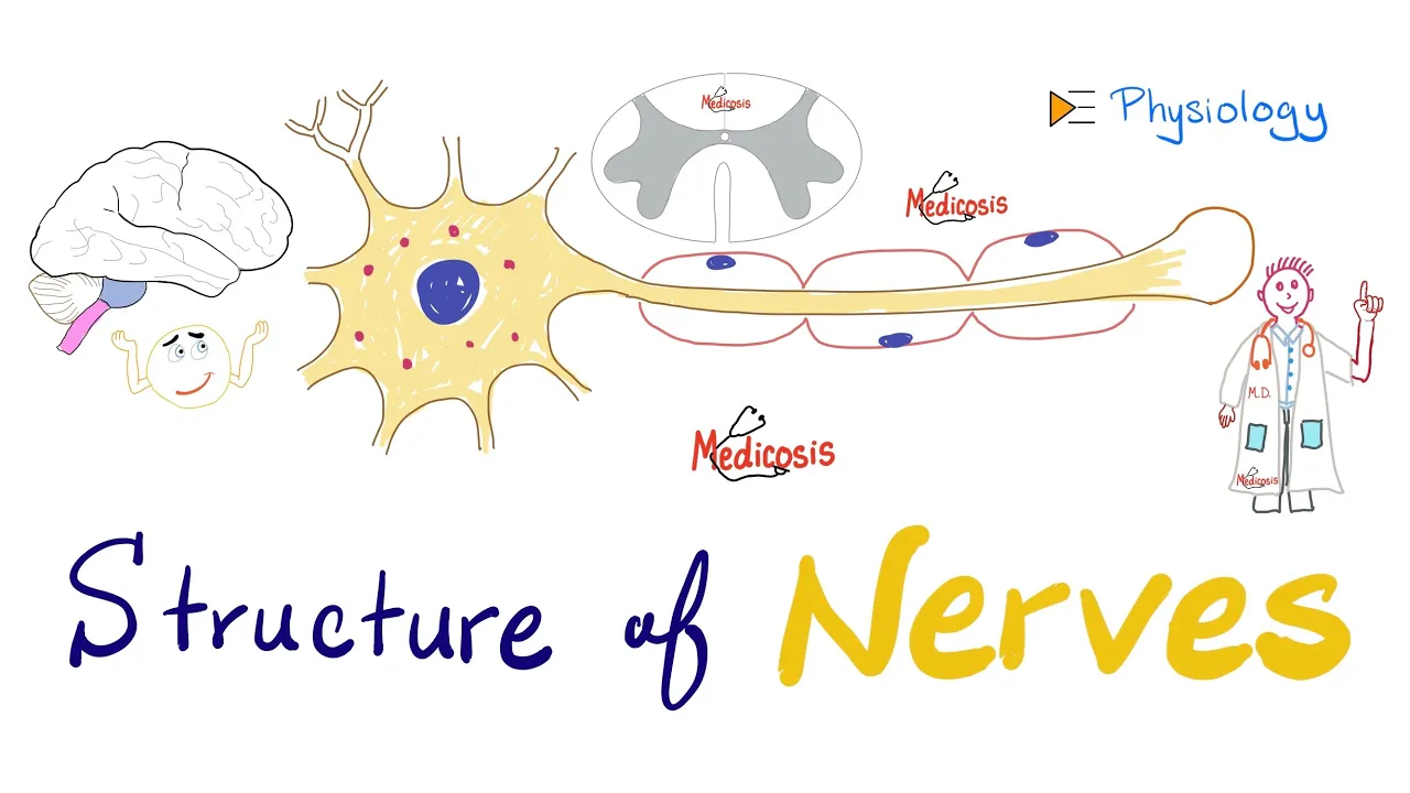
Structure of Neurons - Nerve Physiology Quick Review - Antomy and Physiology
34.2K
A neuron consists of four main structural components: dendrites, soma (cell body), axon, and axon terminus. Dendrites are branched extensions that increase surface area for receiving synaptic inputs from other neurons. The soma contains the nucleus with nucleolus and cytoplasm, serving as the metabolic center of the neuron. The axon is a long fiber that transmits nerve impulses away from the soma in one direction. The axon terminus (synaptic knob) contains vesicles storing neurotransmitters such as acetylcholine, norepinephrine, or dopamine, which are released into the synaptic cleft during signal transmission.
Click to watch →
Deep Dive
The generation and propagation of an action potential, including depolarization and repolarization phases.

🛑 أقوى مراجعة لوحدة الإتصال العصبي 🦞 !!
77.8K
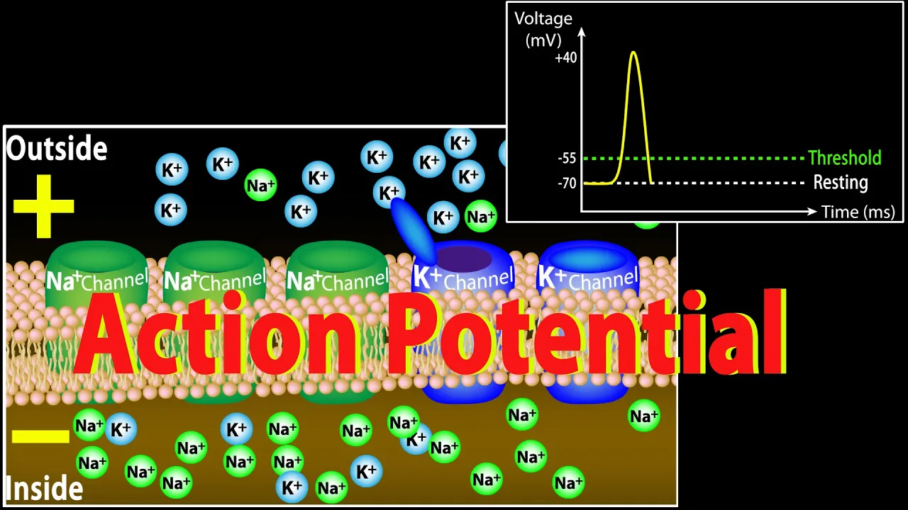
An action potential is a rapid, transient change in membrane potential that propagates along neurons. It consists of five phases: (1) Resting potential at approximately -70 mV, (2) Depolarization when voltage-gated sodium channels open and sodium rushes in, (3) Repolarization when sodium channels close and voltage-gated potassium channels open allowing potassium to exit, (4) Hyperpolarization when potassium channels close slowly, causing the membrane to become more negative than resting potential, and (5) Return to resting potential as the sodium-potassium pump restores ion distribution. The action potential is generated when a stimulus depolarizes the membrane to threshold, triggering the sequential opening and closing of voltage-gated ion channels.
Click to watch →
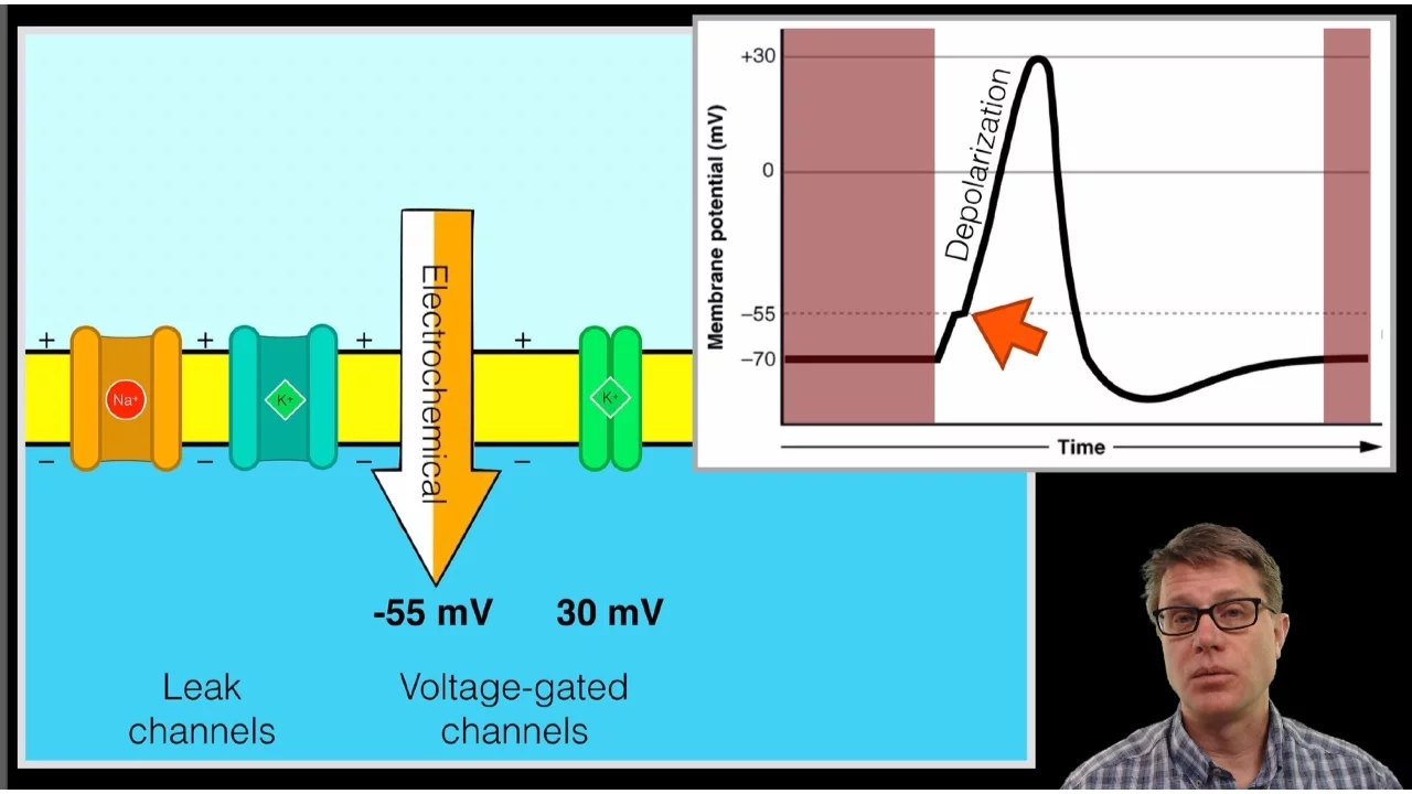
The Action Potential
1.5M
An action potential consists of two main phases. During depolarization, voltage-gated sodium channels open at threshold (-55 mV), causing rapid sodium influx that dramatically increases the membrane potential. During repolarization, sodium channels inactivate and voltage-gated potassium channels open at around -30 mV, allowing potassium efflux that brings the membrane potential back toward resting levels.
Click to watch →
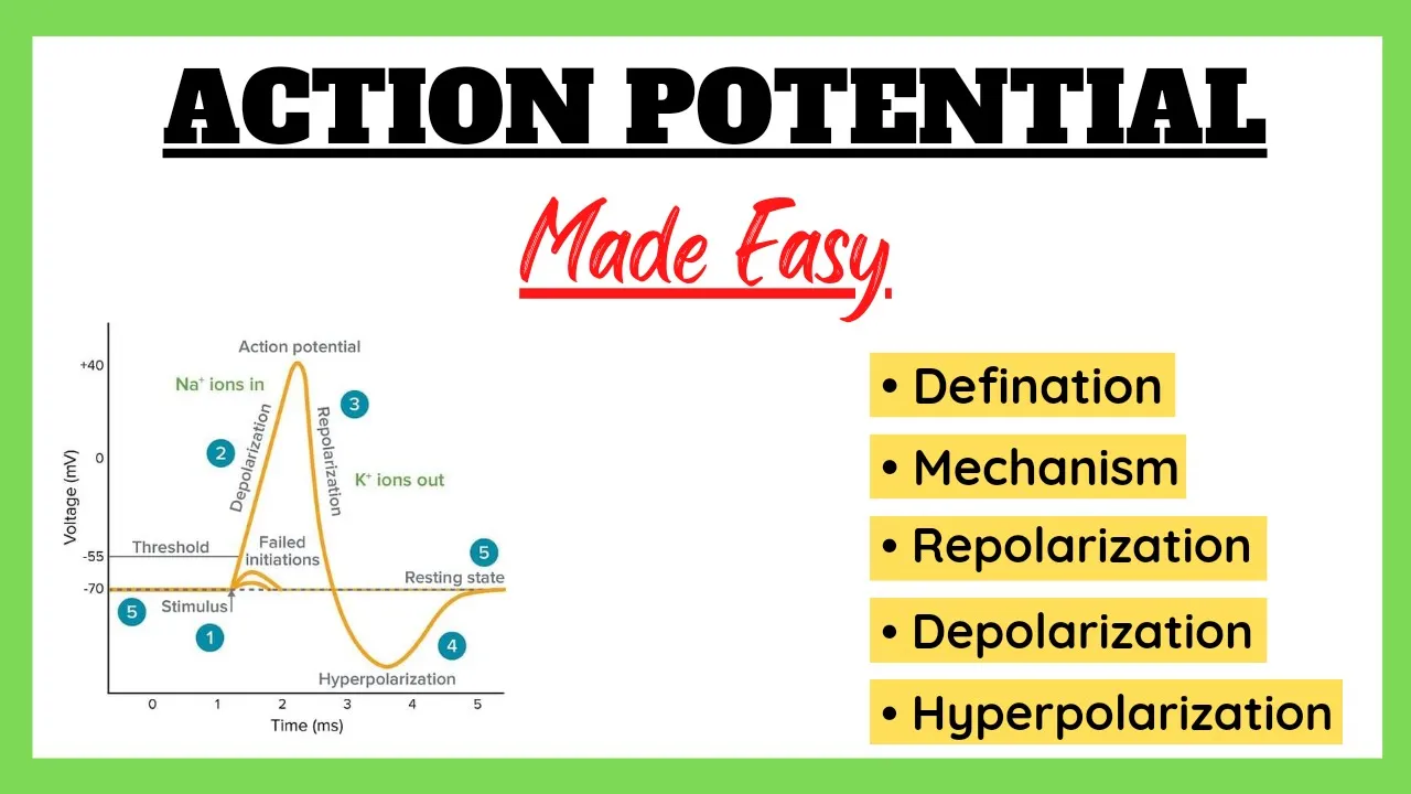
Action Potential, Depolarization, Repolarization, Refractory Period, Physiology Made Easy
199.7K
An action potential is a wave of depolarization and repolarization caused by voltage-gated sodium and potassium channels opening and closing in sequence: when a neuron reaches threshold (-55 mV), sodium channels open causing rapid depolarization (inside becomes positive), followed by potassium channels opening causing repolarization (restoring negative charge), and finally hyperpolarization occurs before the refractory period returns the neuron to resting potential (-70 mV).
Click to watch →
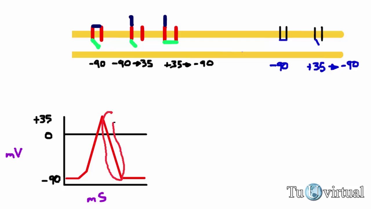
Potencial de acción - reposo, despolarización y repolarización de una fibra nerviosa
292.5K
An action potential is a rapid change in membrane potential that propagates along nerve fibers, consisting of three phases: (1) Resting phase where the membrane is polarized at approximately -90 mV, (2) Depolarization phase where sodium ions enter the cell through voltage-gated sodium channels, causing the membrane potential to become less negative and eventually positive (reaching +35 mV), and (3) Repolarization phase where potassium ions exit the cell through voltage-gated potassium channels, returning the membrane potential back to its resting negative value. The sodium channels have two gates: an activation gate that opens during depolarization and an inactivation gate that closes to prevent further sodium entry, while potassium channels open during repolarization to allow potassium efflux and restore the negative internal charge.
Click to watch →
Action Potential in Neurons: Phases and Mechanisms
1.6M
An action potential is a brief reversal of electric polarity across the cell membrane that occurs when excitatory signals cause membrane voltage to reach the threshold (-55mV), triggering voltage-gated sodium channels to open and allowing sodium influx that depolarizes the membrane; this is followed by potassium efflux that repolarizes and hyperpolarizes the membrane, with the refractory period preventing immediate re-firing and ensuring unidirectional propagation along the axon.
Click to watch →
Deep Dive
The concept of electrochemical gradients and the function of voltage-gated ion channels.

5-2 التواصل العصبي ج2 آلية جهد القعل
3K
Voltage-gated ion channels open or close in response to electrical potential changes, while leak channels remain open continuously allowing limited ion movement. During resting potential, voltage-gated channels remain closed. The electrochemical gradient combines concentration gradients and electrical gradients, driving ion movement. For sodium ions, both gradients favor movement into the cell. This combined gradient is fundamental to understanding how action potentials are generated, as it determines the direction and driving force for ion movement across the membrane.
Click to watch →

cours de cytologie suite 2 ( la perméabilité membranaire Plasmique)
4K
Ion channels facilitate the passage of ions across the plasma membrane according to electrochemical gradients, combining concentration and electrical forces. Voltage-gated ion channels open in response to membrane depolarization, allowing ions like calcium, potassium, sodium, and chloride to pass through. Ligand-gated ion channels open when specific molecules (neurotransmitters, hormones) bind to receptors on the channel, causing conformational changes that open the pore. These mechanisms enable rapid electrical signaling in cells and are fundamental to neuronal communication and muscle contraction.
Click to watch →

Clase de repaso - UBAXXI y CBC
11.3K
This segment covers ion channel function and electrochemical gradients. Ion channels facilitate passive transport of ions based on both concentration gradients and electrical gradients (membrane potential). For example, sodium ions move into cells because they are positively charged and attracted to the negatively charged interior, in addition to their concentration gradient. The action of channels depends on the existence of these electrochemical gradients, which drive ion movement without energy expenditure.
Click to watch →
Mechanism of Drug Action: Ion Channels and Drugs Action in Hindi Detailed explanation #pharmacology
242
Mechanosensitive ion channels open in response to mechanical touch or friction, enabling the sensation of touch. Voltage-gated ion channels are activated by electrochemical gradients across the membrane. The extracellular fluid has higher sodium concentration than intracellular fluid, creating an electrochemical gradient. When membrane potential changes, sensors on the channels detect this and activate the channel. Voltage-gated sodium channels exist in three states: closed but capable of opening, open (activated), and closed but not capable of opening. The resting membrane potential is -70 to -90 mV, and action potentials reach -50 to +30 mV.
Click to watch →

CELL BIOLOGY-8: CSIR/ GATE/ IIT-JAM/ ICMR/ SET/ DR. ASHISHH GUPTA: BIOMAGNET
870
Voltage-gated ion channels are transmembrane proteins whose conformational state depends on the difference in ionic charge across the membrane (membrane potential). Changes in membrane potential serve as the stimulus for opening and closing these channels. Key ion concentration differences across the plasma membrane are essential for cellular function: Sodium (Na+) has high extracellular concentration (~150 mM) and low intracellular concentration (~10 mM); Potassium (K+) has low extracellular concentration and high intracellular concentration; Chloride (Cl-) has high extracellular concentration and low intracellular concentration; Calcium (Ca2+) has high extracellular concentration and low intracellular concentration. These gradients are maintained by active transport mechanisms and are essential for generating action potentials in neurons and cardiac cells. Specific inhibitors block voltage-gated ion channels: Tetrodotoxin (TTX) and Saxitoxin (STX) block sodium voltage-gated channels; Local anesthetics (procaine, lidocaine, xylocaine) block sodium channels to prevent action potential generation in specific areas; Tetraethylammonium (TEA) blocks potassium voltage-gated channels.
Click to watch →
Deep Dive
Fundamental cellular processes, particularly exocytosis and receptor-ligand binding mechanisms.

Mod-01 Lec-30 Introduction to Receptor - Ligand Binding
3.9K
Receptor-ligand binding is a fundamental biochemical process where specific proteins called receptors on cell surfaces or organelles bind to specific ligands (such as neurotransmitters, hormones, or cytokines) following a lock-and-key mechanism, inducing conformational changes that initiate intracellular signaling cascades; this process is essential for most biochemical pathways and plays a critical role in diseases like familial hypercholesterolemia, where genetic defects impair proper receptor-ligand interactions.
Click to watch →

المحاضرة الرابعة | (Cytology 4(Function of cell membrane -rER & sERــ Histology |
7.2K
Exocytosis is the reverse of endocytosis, expelling materials from the cell to the extracellular space. Receptor-mediated endocytosis provides selective uptake of specific substances like growth hormones. The process involves receptor binding, membrane invagination forming coated pits, vesicle formation, and receptor recycling. Growth hormone receptor deficiency causes dwarfism even with normal hormone levels, demonstrating receptor importance in cellular signaling.
Click to watch →

Fisiologia 2.0: Aula 3 - Fisiologia celular - processos fagocitários
16.7K
Cells communicate with their environment through specialized transport mechanisms. Small molecules use simple diffusion (if lipid-soluble) or transport proteins (if hydrophilic). Large molecules require endocytosis for internalization and exocytosis for externalization. Endocytosis includes phagocytosis (solid particles) and pinocytosis (fluid), occurring in clathrin-coated regions. Receptor-mediated endocytosis involves ligand binding, GRK phosphorylation, beta-arrestin recruitment, and clathrin vesicle formation. Exocytosis is calcium-dependent, using SNARE proteins (synaptotagmin, synaptobrevin, SNAP-25, syntaxin) for vesicle fusion. Calcium binding intensifies SNARE interactions, forming a fusion pore that expands until complete membrane fusion releases contents extracellularly.
Click to watch →

This Is Your Body On Cannabis
10.3M
Receptors function using a lock-and-key mechanism where the receptor acts as the lock and the binding molecule acts as the key. When a cannabinoid binds to its corresponding receptor, it initiates or modulates cellular responses. This binding triggers chemical reactions within the cell, leading to changes in cellular behavior.
Click to watch →
Cell Biology || Transport across Plasma Membrane || Active Transport || Zoology || B. Sc. || M.Sc.
715
Receptor-mediated endocytosis involves specific extracellular macromolecules binding to specific receptors on the plasma membrane. Clathrin proteins coat the vesicle forming a coated pit. After receptor-ligand binding, the vesicle forms, undergoes uncoating, and receptors are recycled. Exocytosis is the process by which large materials are expelled outside the cell through membrane-bound vesicles that fuse with the plasma membrane and release contents.
Click to watch →
Prerequisite Knowledge
- Concept 01Basic anatomy of a neuron, specifically the roles of the axon, dendrites, and the axon terminal.
- Concept 02The generation and propagation of an action potential, including depolarization and repolarization phases.
- Concept 03The concept of electrochemical gradients and the function of voltage-gated ion channels.
- Concept 04Fundamental cellular processes, particularly exocytosis and receptor-ligand binding mechanisms.
Subsequent Learning
- Step 01The difference between excitatory and inhibitory postsynaptic potentials (EPSPs and IPSPs) and how summation occurs.
- Step 02The distinct classes of neurotransmitters (e.g., amino acids, monoamines) and their specific receptors (ionotropic vs. metabotropic).
- Step 03Mechanisms of neurotransmitter inactivation and clearance, such as enzymatic breakdown and reuptake transporters.
- Step 04The pharmacological effects of drugs, toxins, and medications (such as SSRIs or neuromuscular blockers) on synaptic transmission.
Synapse Basics
Key Structures
Signal Trigger
Vesicle Release
NT Diffusion
Channel Binding
Signal Continue
Synapse Basics
0:05Playing Section
- 1
Chemical synapse is a junction between neurons.
- 2
Enables communication without physical connection.
The 'Kiss-and-Run' Vesicle Fusion Model
While standard animations typically depict neurotransmitter release through classical "full-collapse fusion"—where synaptic vesicles merge completely with the presynaptic membrane—an alternative model known as "kiss-and-run" exists. In this model, vesicles form a transient, narrow pore with the presynaptic membrane to release neurotransmitters, and then quickly pinch back off to be reused without ever fully integrating into the membrane. This mechanism allows for much faster recycling of synaptic vesicles, which is crucial during high-frequency neuronal activity. Although historically debated, evidence suggests that both full fusion and "kiss-and-run" coexist, challenging the singular, simplified view of vesicular exocytosis often shown in traditional educational animations.
Deep Dive
The difference between excitatory and inhibitory postsynaptic potentials (EPSPs and IPSPs) and how summation occurs.

EPSPs and IPSPs
128
Neurons integrate multiple inputs through graded membrane potentials that sum together to determine whether an action potential fires; excitatory postsynaptic potentials (EPSPs) depolarize the neuron by bringing voltage closer to threshold, while inhibitory postsynaptic potentials (IPSPs) hyperpolarize it by moving voltage further from threshold, and this integration occurs through spatial summation (inputs from different synapses) and temporal summation (inputs from the same synapse over time).
Click to watch →

Neuro - Aula 1
10.4K
Excitatory postsynaptic potentials (EPSPs) are caused by sodium influx through ligand-gated channels, depolarizing the neuron. Inhibitory postsynaptic potentials (IPSPs) are caused by chloride influx or potassium efflux, hyperpolarizing the neuron. The summation of EPSPs and IPSPs at the axon hillock determines whether an action potential will be generated.
Click to watch →
Action Potentials, Refractory Period, and Summation
5.1K
Since individual synaptic inputs are often small and insufficient to reach threshold alone, neurons use summation to accumulate signals. Excitatory postsynaptic potentials (EPSPs) raise membrane potential while inhibitory postsynaptic potentials (IPSPs) decrease it. There are two types of summation: (1) Temporal summation - occurs when a single presynaptic neuron fires rapidly, sending EPSPs in quick succession; each EPSP partially decays before the next arrives, allowing them to add up and potentially reach threshold. (2) Spatial summation - occurs when multiple presynaptic neurons fire simultaneously, with their EPSPs adding together at the same time. Both mechanisms allow the neuron to integrate multiple inputs and determine whether threshold is reached. IPSPs can counteract EPSPs if they occur simultaneously, preventing action potential generation.
Click to watch →
General Biology - Nervous System Part I
1.1K
Synaptic potentials are classified by effect: excitatory postsynaptic potentials (EPSPs) depolarize the postsynaptic neuron, making it more likely to fire, while inhibitory postsynaptic potentials (IPSPs) hyperpolarize the postsynaptic neuron, making it less likely to fire. Both can summate temporally (from repeated stimulation of the same synapse) or spatially (from simultaneous stimulation of different synapses).
Click to watch →
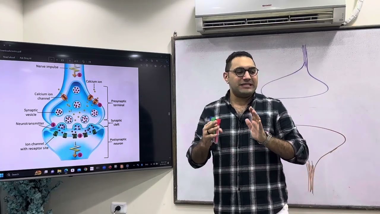
CNS Physiology -3 د/الشريف
33.7K
EPSPs are characterized by: (1) Depolarization of the postsynaptic membrane, (2) Movement toward the threshold, (3) Short duration (milliseconds), (4) Graded magnitude (can vary in size), and (5) Local spread (decay with distance). EPSPs have amplitudes of about 0.5-1.0 millivolts, much smaller than the threshold change needed to trigger an action potential. Multiple EPSPs must summate to reach threshold. IPSPs are hyperpolarizing changes that move the membrane potential away from threshold, caused by opening of ion channels allowing chloride to enter or potassium to leave the cell. Hyperpolarization occurs through two main mechanisms: efflux of potassium ions from the cell and influx of chloride ions into the cell. Both mechanisms make the inside of the cell more negative, moving the membrane potential away from the threshold.
Click to watch →
Deep Dive
The distinct classes of neurotransmitters (e.g., amino acids, monoamines) and their specific receptors (ionotropic vs. metabotropic).

Types of neurotransmitter receptors | Nervous system physiology | NCLEX-RN | Khan Academy
473.4K
Neurotransmitter receptors are classified into two major classes with fundamentally different mechanisms. Ionotropic receptors are ligand-gated ion channels that open directly when neurotransmitters bind, allowing rapid ion flow that causes immediate graded potentials—either excitatory (sodium/calcium influx causing depolarization) or inhibitory (chloride influx or potassium efflux causing hyperpolarization). Metabotropic receptors, in contrast, do not form ion channels; instead, they activate intracellular second messenger systems that can modify ion channel activity, alter protein function, or change gene expression. While metabotropic responses are slower than ionotropic effects, they offer greater signal amplification and can produce longer-lasting changes to neuronal behavior, either at rest or during activity.
Click to watch →
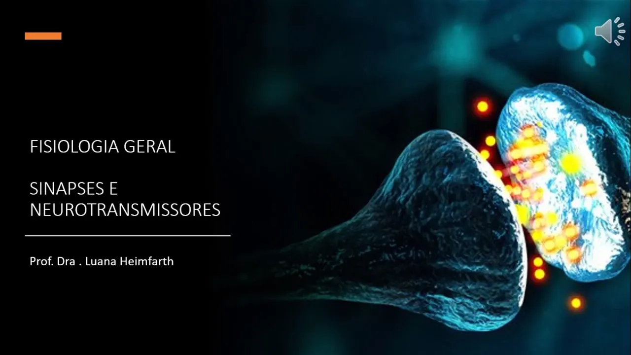
MÓDULO 1 - Aula 3: Sinapses e neurotransmissores
57.6K
Neurotransmitters are classified into three categories: (1) Neurotransmitters - small molecules with rapid, short-term effects (acetylcholine, dopamine, serotonin, glutamate), (2) Neuromodulators - larger molecules with slower, longer-lasting effects, and (3) Neuropeptides - secreted into the bloodstream as hormones. Receptors are divided into: (1) Ionotropic receptors - ligand-gated ion channels that open directly when neurotransmitter binds, fast-acting and local, (2) Metabotropic receptors - G-protein coupled receptors that activate second messenger systems, slower-acting and can modulate other ion channels, activate enzymes, and affect gene transcription.
Click to watch →

Neurotransmitters of the human body
250.3K
Neurotransmitters are chemical messengers that transmit information across the body, categorized into monoamines (serotonin, dopamine, norepinephrine), catecholamines (dopamine, norepinephrine), amino acids (glutamate, GABA, glycine), and acetylcholine; each neurotransmitter has specific locations, functions, and receptor types (ionotropic or metabotropic) that determine whether they excite or inhibit neural activity, with clinical applications including Alzheimer's disease treatment (acetylcholine), depression and sleep regulation (serotonin), Parkinson's disease and schizophrenia (dopamine), ADHD and anxiety (norepinephrine), ALS (glutamate), anxiety and drug abuse rehabilitation (GABA), and spasticity (glycine).
Click to watch →

CNS: Biochemistry 1: neurotransmitters and degenerative disorders
106
Neurotransmitter receptors are classified into ionotropic (ligand-gated ion channels causing rapid effects) and metabotropic (G-protein coupled receptors triggering slower intracellular signaling cascades). The receptor type determines whether a neurotransmitter produces excitatory or inhibitory effects. Amino acid neurotransmitters are the most abundant in the nervous system. Glutamate is the major excitatory neurotransmitter, synthesized from glutamine and released upon depolarization. Glycine is synthesized from serine and acts as an inhibitory neurotransmitter. Both are stored in synaptic vesicles and released through the same mechanism. After release, they are reuptaken by specific transporters for reuse or taken up by astrocytes for recycling through the glutamate-GABA-glutamine cycle.
Click to watch →
Técnica para aprender de sinapsis neuronal, canales iónicos y metabotrópicos fácil y sencillo
2.8K
Neurotransmitter receptors are classified into two types: ionotropic (ligand-gated ion channels) and metabotropic (G-protein coupled receptors). Ionotropic receptors directly open ion channels when neurotransmitters bind, causing rapid membrane potential changes. Metabotropic receptors activate intracellular signaling cascades: neurotransmitter binding activates G-proteins, which activate adenylyl cyclase to produce cAMP (second messenger), which activates protein kinase A to phosphorylate target proteins. This multi-step cascade allows for signal amplification and complex modulation of cellular responses.
Click to watch →
Deep Dive
Mechanisms of neurotransmitter inactivation and clearance, such as enzymatic breakdown and reuptake transporters.

Sinapses Químicas: Fisiologia (aula básica)
15.5K
Two main mechanisms inactivate neurotransmitters: enzymatic degradation and reuptake. For classical neurotransmitters like acetylcholine, enzymes in the synaptic cleft (e.g., acetylcholinesterase) break down the neurotransmitter, with fragments recycled for resynthesis. For neurotransmitters like dopamine, transporters in the presynaptic membrane reuptake the neurotransmitter for recycling. Cocaine inhibits dopamine reuptake, increasing synaptic dopamine concentration.
Click to watch →

Chapter 3: Neurotransmitters
318
Neurotransmitters must be removed from the synaptic cleft because if they remain, they will continue to activate receptors indefinitely, preventing new signals from being transmitted. There are four primary mechanisms for neurotransmitter clearance: (1) Diffusion - neurotransmitters simply diffuse away from the synaptic cleft following concentration gradients; (2) Enzymatic degradation - enzymes in the synaptic cleft actively break down neurotransmitters; (3) Reuptake - specific membrane transporters bring neurotransmitters back into the presynaptic neuron for reuse; (4) Astrocyte uptake - neighboring astrocytes take up certain neurotransmitters, some of which can be stored for later re-export to axon terminals. These mechanisms ensure rapid termination of neurotransmitter action and proper timing of neural communication.
Click to watch →
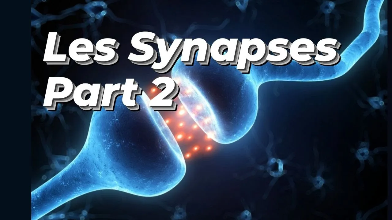
SYNAPSE PART 2 ( NEURO-NEURONALE )
8.1K
After neurotransmitter release, three main mechanisms remove them from the synaptic cleft: diffusion into extracellular space, enzymatic degradation, and reuptake by presynaptic membrane or glial cells (astrocytes, oligodendrocytes, microglia). Catecholamines (dopamine, norepinephrine, epinephrine) undergo intracellular degradation after reuptake. These mechanisms ensure proper synaptic function, prevent continuous stimulation, and maintain signal fidelity. The balance between release and clearance determines synaptic efficacy and contributes to neurological disorders when disrupted.
Click to watch →

Umbrella Metaphor
336
After neurotransmitter release, excess molecules must be cleared to prevent continuous stimulation. Three primary mechanisms accomplish this: (1) Reuptake pumps actively transport neurotransmitter back into the presynaptic neuron; SSRIs (selective serotonin reuptake inhibitors) are antidepressants that block this process; (2) Enzymatic breakdown uses enzymes to break down neurotransmitters, as seen with acetylcholinesterase in neuromuscular junctions; (3) Diffusion allows neurotransmitters to diffuse away from the synaptic cleft, where they may be taken up by astrocytes. Proper clearance ensures each receptor is activated only once per neurotransmitter release event.
Click to watch →

De synaps deel 1 - De werking van synapsen
25.2K
Neurotransmitter clearance occurs through three mechanisms: reuptake by membrane pumps, enzymatic breakdown, and diffusion. Proper clearance is essential for normal function; failure causes pathological conditions. Many drugs and toxins target these clearance mechanisms by blocking reuptake or enzymatic breakdown, causing neurotransmitters to remain active longer. This prolongs their effects on postsynaptic neurons, either enhancing or inhibiting neuronal activity depending on the neurotransmitter type.
Click to watch →
Deep Dive
The pharmacological effects of drugs, toxins, and medications (such as SSRIs or neuromuscular blockers) on synaptic transmission.

194 Physiology 204 CNS module written questions for 2nd year medical students 2026
110
Various substances affect synaptic transmission: caffeine, theobromine, and theophylline increase transmission by blocking adenosine receptors; strychnine blocks inhibitory glycine receptors; anesthetics and sedatives decrease transmission by enhancing inhibitory effects. Botulinum toxin blocks acetylcholine release at the neuromuscular junction, causing flaccid paralysis used therapeutically for wrinkles and overactive bladder. Tetanus toxin blocks inhibitory neurotransmitter release in the spinal cord, causing uncontrolled muscle spasms. These examples demonstrate how synaptic mechanisms can be pharmacologically modulated for therapeutic purposes.
Click to watch →
Efeitos de Drogas nas Sinapses
4.3K
Drugs affect synaptic transmission by acting as agonists or antagonists on neurotransmitter receptors, altering the production, storage, release, or degradation of neurotransmitters such as serotonin, dopamine, and GABA; depressants like alcohol and opioids reduce CNS activity, stimulants like caffeine increase alertness through adenosine receptor blockade, and hallucinogens like LSD alter perception by acting as partial agonists on serotonin receptors.
Click to watch →
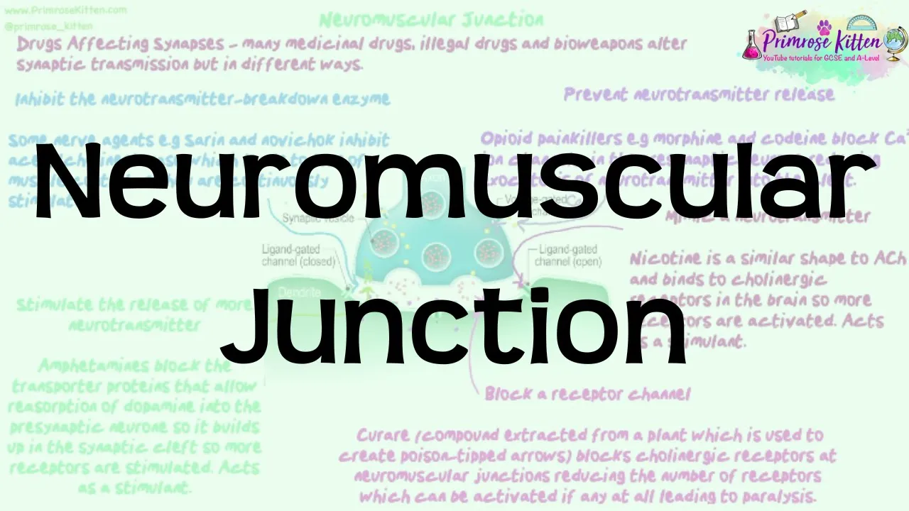
Neuromuscular Junction | Revision for Biology A-Level and IB
742
Various drugs and toxins affect synaptic transmission through five primary mechanisms: (1) Opioids block presynaptic calcium channels, preventing neurotransmitter release; (2) Nicotine mimics acetylcholine to overstimulate receptors; (3) Curare blocks postsynaptic receptors, causing paralysis; (4) Amphetamines block neurotransmitter reuptake, increasing synaptic concentration; (5) Nerve agents inhibit acetylcholinesterase, causing acetylcholine accumulation. Each mechanism produces distinct effects: opioids block pain signals, nicotine enhances stimulation, curare causes paralysis, amphetamines produce euphoria, and nerve agents cause fatal overstimulation. Understanding these mechanisms allows prediction of drug effects based on their site of action.
Click to watch →

USMLE QUIZ, Synaptic Mechanisms & Drug Effects
161
Drugs that affect the nervous system primarily work by targeting three key stages of synaptic transmission: altering neurotransmitter release (such as botulinum toxin blocking release or amphetamines stimulating excessive release), mimicking or blocking receptors (acting as agonists or antagonists), and interfering with reuptake or degradation (like SSRIs blocking serotonin reuptake to prolong its action in the synaptic cleft).
Click to watch →

Lecture - Neuro Physiology: (Basic Function of Synapses and Neuromuscular Transmission)
3.7K
Since synaptic transmission is a chemical process, it can be affected by drugs and toxins. Receptor antagonists inhibit the action of neurotransmitters at receptors—for example, curare blocks acetylcholine at the neuromuscular junction, preventing action potentials in muscles and potentially causing paralysis. Receptor agonists mimic the action of neurotransmitters—for example, morphine activates opiate receptors in the brain to cause pain relief.
Click to watch →
Synapse Basics
Key Structures
Signal Trigger
Vesicle Release
NT Diffusion
Channel Binding
Signal Continue
Synapse Basics
0:05- 1
Chemical synapse is a junction between neurons.
- 2
Enables communication without physical connection.
The 'Kiss-and-Run' Vesicle Fusion Model
While standard animations typically depict neurotransmitter release through classical "full-collapse fusion"—where synaptic vesicles merge completely with the presynaptic membrane—an alternative model known as "kiss-and-run" exists. In this model, vesicles form a transient, narrow pore with the presynaptic membrane to release neurotransmitters, and then quickly pinch back off to be reused without ever fully integrating into the membrane. This mechanism allows for much faster recycling of synaptic vesicles, which is crucial during high-frequency neuronal activity. Although historically debated, evidence suggests that both full fusion and "kiss-and-run" coexist, challenging the singular, simplified view of vesicular exocytosis often shown in traditional educational animations.
Basic anatomy of a neuron, specifically the roles of the axon, dendrites, and the axon terminal.
The generation and propagation of an action potential, including depolarization and repolarization phases.
The concept of electrochemical gradients and the function of voltage-gated ion channels.
Fundamental cellular processes, particularly exocytosis and receptor-ligand binding mechanisms.
The difference between excitatory and inhibitory postsynaptic potentials (EPSPs and IPSPs) and how summation occurs.
The distinct classes of neurotransmitters (e.g., amino acids, monoamines) and their specific receptors (ionotropic vs. metabotropic).
Mechanisms of neurotransmitter inactivation and clearance, such as enzymatic breakdown and reuptake transporters.
The pharmacological effects of drugs, toxins, and medications (such as SSRIs or neuromuscular blockers) on synaptic transmission.
668.2K views7.2Klikes1:13@retwedtOriginal Release: 2013-12-17
a chemical synapse is a special Junction between two neurons that allows them to communicate without being physically connected a synapse has several structures the Press synaptic neuron ends in a small bulb called the pre synaptic terminal the post synaptic terminal membrane is directly adjacent separated by a small space called a synaptic CFT an action potential causes voltage iated calcium ion channels to open in the pre synaptic terminal the influx of calcium ions prompts synaptic vesicles to release neurotransmitters via exocytosis this neuron is releasing acetylcholine the neurotransmitter quickly diffuses across a synaptic CL two acetylcholine molecules bind with one Liang gated sodium Channel at the post synaptic membrane opening the channel when enough sodium ion channels open the post synaptic cell depolarizes and the action potential continues along the neuron [Music]
Up Next

11:57
Sleep Cycle & EEG Waves: Non-REM vs REM Mnemonics
@dranshumantripathi3599
71.7K views•2018-03-18

21:55
Deciphering Neural Circuits Controlling Anorexia in Mice | Richard Palmiter
@AllenInstitute
2.3K views•2011-10-10

4:10
Vagus Nerve (CN X): Anatomy, Nuclei & Functions Explained
@Alilamedicalmedia
305.2K views•2022-10-31

13:03
How Exercise Benefits Your Brain: Science Explained
@TED
11.4M views•2018-03-21
Related Study Plans & Knowledge Roadmaps
Structured learning paths in Neuroscience
neuroscience
Appetite Regulation: Brain Circuits & Hormones
01Foundations of Endocrinology and Brain Anatomy
02Introduction to Energy Homeostasis
03Peripheral Signals: Leptin, Ghrelin, and Insulin
+ 2 more learning modules
14h5 modules
Explore Roadmap
neuroscience
Neuroeconomics: Value, Risk & Brain Mechanisms
01Foundations of Neuroscience and Brain Anatomy
02Introduction to Neuroeconomics and Subjective Value
03Dopaminergic Prediction Errors & Reinforcement Learning
+ 3 more learning modules
18h6 modules
Explore Roadmap
neuroscience
Spatial Navigation: Cells, Grids & Brain Maps
01Foundations of Brain Anatomy and Memory
02Place Cells and Head-Direction Cells
03Grid Cells: The Brain's Coordinate System
+ 2 more learning modules
12h5 modules
Explore Roadmap
neuroscience
BCIs: Neural Decoding & Prosthetic Control
01Neuroscience Foundations: How the Brain Signals Intent
02Signal Acquisition: Recording the Brain's Activity
03Neural Decoding: Translating Brain Waves into Data
+ 2 more learning modules
25h5 modules
Explore Roadmap