























This video presents a comprehensive approach to coma management, emphasizing that neurological emergencies require immediate treatment before definitive diagnosis. The lecture covers early management principles including ABC stabilization, toxic/metabolic screening, and rapid neurological assessment. It details the neuroanatomy of alertness through the ascending reticular activating system, explaining how lesions in four key locations (dorsolateral pons, paramedian midbrain, diencephalon, and bihemispheric) cause coma. The three main herniation syndromes—central, uncal, and tonsillar—are explained with their characteristic signs and progression. Intracranial pressure management strategies include head elevation, hyperventilation, sedation, osmotherapy, and surgical interventions. The differential diagnosis spans vascular pathologies (stroke, hemorrhage, venous sinus thrombosis), infectious causes (meningitis, encephalitis, abscess), traumatic injuries (hemorrhages, diffuse axonal injury), neoplastic lesions, toxic-metabolic conditions (Wernicke's encephalopathy, osmotic demyelination, hypoglycemia), immune-mediated disorders (autoimmune encephalitides, paraneoplastic syndromes), and hereditary conditions. Status epilepticus management follows a tiered protocol from benzodiazepines to general anesthesia. Outcomes range from locked-in state to brain death, with prognosis based on age, baseline function, coma duration, and underlying cause. Brain death declaration requires irreversible coma cause, exclusion of confounders, clinical brainstem death criteria, and apnea testing.
Approach to Coma: Diagnosis & Management | Neurology Guide
Added:Hello neurophiles.
Dr. Rybinnik here.
For this topic, I enlisted the help of a friend.
[Announcer voice] So give a warm welcome, to the critical care neurologist extraordinaire, the chief encephalopathist, Dr. Ram "Think Outside the Box" Gowda [Normal voice] Take it away, Ram.
Hello, friends.
I'm Ram Gowda, and I'm one of the attending neurointensivists here at Rutgers Robert Wood Johnson Medical school.
So, today's talk is about coma.
So first, we are going to review early management of patients with acute confusional state Next we'll discuss the neuroanatomy of alertness, the coma exam and the three main herniation syndromes We are going to learn how to address intracranial pressure crises and how to manage cerebral edema Next, we'll go over some of the choice pathologies that can cause coma And finally we will conclude with the discussion of outcomes for patients in a coma, and review the steps for brain death declaration.
So, let’s start with a case: We have a 55 year old woman, previously healthy She developed 3 days of diffuse headaches, and was found by her family on the floor of her kitchen, lethargic and vomiting.
So they called EMS.
Initially, the patient was able to converse, but quickly progressed to unresponsiveness, and had to be intubated by EMS after their arrival.
There wasn't any shaking, no incontinance.
There wasn't any arrhythmia or significant trauma noted.
On the initial evaluation in the ED: Vital signs are normal.
obtunded, grimacing to sternal rub.
She has some withdrawal to pain on the right side, but not on the left.
The pupils are responsive to light but uneven (the right pupil larger than left pupil).
The right eye also seems to be slightly deviated outward.
She has a cough, and gag reflexes.
But there is also extensor posturing on the left side.
The patient was already intubated in the field by EMS for airway protection.
and she is currently hemodynamically stable in the ED.
What is our initial approach to the management of the acute confusional state?
This is clearly a neurological emergency!
Treat first and ask questions later!
That’s a little hyperbolic, since you’re obviously still continuing your assessment.
But the guiding principle here is that you shouldn’t wait for a definitive diagnosis before initiating treatment for dangerous diseases.
So let’s review the general approach to a patient in acute confusional state.
History is still key.
Doesn't matter if the patient's comatose.
You might need to make some phone calls since the patient is usually unable to help you here.
But you still have to gather history.
The time last known at baseline is extremely important when you're considering eligibility for any kind of stroke interventions.
And the description of the actual event, past medical history, medications, toxic exposures, any previous episodes... All of these can give a clue to pathology.
[Dr. Cameron] "Trauma is out of the picture.
It could still be infection."
[Dr. Foreman] "Or neurological..." [Dr. House] "Or genetic!
"We need a better history."
If the symptom onset is within 24 hours, you should really consider activating a stroke code especially if you see focal neurologic deficits.
You really don’t want to miss a treatable ischemic lesion.
Next, ABCs.
This patient is no different than any other emergent patient.
Make sure that airway, breathing, and circulation are all stabilized.
If they are are unstable, your first priority is intubation and hemodynamic support.
Luckily, the EMS already took care of that in our case.
After that, let's work on IV access, cardiac monitoring, any respiratory support, such as supplemental oxygen, send labs, and if it's a trauma patient, immobilize the c-spine.
So why did we send all those labs?
Well, toxic/metabolic causes for the acute confusional states are far more common than other causes.
So first things first, let's fix any electrolyte issues (especially sodium and calcium), consider sepsis, vitamin deficiencies (especially thiamine), and any kind of poisoning.
If the cause of encephalopathy is known (let's say a person was found next to a bottle of benzodiazepines) then we can address it at this point.
So thiamine plus glucose for suspected Wernicke’s encephalopathy.
Flumazenil for benzodiazepine.
Naloxone for opioid overdose.
I'm sure you've read a ton about the opioid crisis, and these people are showing up in your ED, so think about it.
But don't forget, opiods aren't the only thing that they're taking.
All of these have been covered in the “Toxidromes” lecture, so refer to that for more information.
Next, do a rapid neurological evaluation.
In the real world, of course, you’re doing this in parallel with getting your IV access, hooking up monitors, sending labs.
All of these things are happening at the same time.
So what’s the goal of the neuro exam?
Really it's to look for localizing signs, any focal findings that point to a focal causative lesion in the brain as opposed to a diffuse disease process.
And you can’t spell “neuro exam” without neuro-anatomy, so let's take it back a bit to some basics.
Shown here is a sagittal cut through the midline where you can see all the important components of the ascending reticular activating system that keeps you awake.
So we are talking about the pons, midbrain, thalamus/hypothalamus, and cerebral cortices.
It follows, that there are essentially four kinds of lesions that cause coma: Dorsolateral pons, paramedian midbrain, diencephalic, and bi-hemispheric.
So for example, if the straight sinus is thrombosed, the diencephalon (the thalamus) will become ischemic, causing coma.
Or if the basilar artery is occluded, the pons and midbrain can be damaged.
Coma again.
Generalized tonic clonic seizure?
Bilateral cortices are inactivated.
Coma.
You get the point.
By the way, since the reticular activating system is located dorsally in the pons, ventral pontine lesions, which can still be devastating do not cause coma.
What's the name of the syndrome in which patients have severe motor disability and appear to be unresponsive, but actually are cognitively intact?
That’s right.
Locked-in syndrome.
Damage to the corticospinal and corticobulbar pathways results in total paralysis save eye movements.
So to cause disordered consciousness, a lesion must be in one of four locations.
So how do you narrow it down?
[Spock] "An ancestor of mine maintained that if you eliminate the impossible, whatever remains, however improbable must be truth."
So this is where brainstem reflexes come in.
You know how eyes are window to the soul?
Well, pupils are the window to the midbrain.
Remember the parasympathetic response to a light shining in the eye?
Cranial nerve II in, and cranial nerve III out with a brief stop in the midbrain pretectal nuclei.
So, if your comatose patient has a blown pupil, then chances are the ipsilateral midbrain is affected.
Next is the vestibulo-ocular reflex.
This is elicited by the oculocephalic maneuver (you might call it the "dolls-eye maneuver") or ice water injections into an ear, (also called cold calorics).
This uses the vestibular nuclei, which are in the medulla to activate horizontal eye movements from the pons.
Before we get further though, if you learned the mnemonic COWS, (cold opposite, warm same) forget it!
Put it out of your brain immediately!
It doesn't help you at all.
You might remember that vestibular nuclei control the pontine paramedian reticular formation in a contralateral fashion, just like the frontal lobe.
So, if you infuse ice water into the right ear, that inhibits the signal to the right cranial nerve VIII and vestibular nucleus.
The left vestibular nucleus is now hyperactive relative to the right.
So it activates the right PPRF, pushing the eyes towards the right the side of the ear where ice water was injected.
In a conscious person, the eyes will deviate towards the ear in which cold water was injected, but then the frontal eye fields will cause a corrective saccade back to midline.
In a comatose patient, there won’t be a corrective saccade, since the frontal eye fields won’t be active.
So the eyes will remain deviated towards the ear in which cold water was injected.
The pons can be additionally tested via the corneal reflex.
The corneal touch is sensed with cranial nerve V, and the eyes respond with a blink via cranial verve VII.
Nuclei for both of those are in the pons.
Cough and gag reflexes are activated through the nucleus ambiguus and the nucleus of the solitary tract.
Both of those are in the medulla.
Why in the world are we testing the medulla?
We just said that the ascending reticular activating system starts in the pons, above the medulla.
Well, you're going to learn that mass lesions can cause damage in a progressive fashion, and absent medullary reflexes can suggest extensive destruction.
So by the time lesions reach the medulla, the patient is typically intubated, as pictured here.
So, we can use the suction cannula in the endotracheal tube to test the cough reflex.
Finally, we are moving from brainstem reflexes to see motor responses.
We will resort to painful stimuli to try to elicit meaningful responses from unconscious patients.
There is a lot of ways you can do this: Sternal rub, applying pressure to the supraorbital ridge, nailbed pressure, or pressure over the temporomandibular joint.
It's the only way to get a decent exam.
Just don't do it without explaining why you're doing it to any family at bedside.
So it's one thing to observe a response, but how do you describe it when you're presenting a case?
Well, normal people are awake, oriented and following commands.
One step below that is disorientation or aphasia.
Drowsy patients are lethargic, but arousable to stimulation, like voice or light touch.
Obtunded patients need more vigorous stimulation to arouse them, and stuporous patients only localize to deep pain – basically reach out to push away the painful stimulus.
Drowsy, obtunded, and stuporous patients all exhibit meaningful responses.
Once meaningful responses are absent, a patient is described as comatose and either withdraws reflexively, exhibits abnormal posturing, or doesn't respond to pain at all.
Really, only "comatose" here has a specific meaning.
The other terms can be pretty vague and often inconsistent between different providers.
I think it's more important for you to describe the actual responses from a patient to the stimulus you are applying.
Let’s see how this plays out in a hypothetical situation.
Say a patient develops a mass lesion – hematoma, tumor, cerebral edema, etc. Here we've got a subdural hematoma.
That mass lesion will exert mass effect on the brain, causing different parts to herniate.
The medial frontal lobe can herniate under the falx cerebri (which separates the two hemispheres).
Diencephalon can herniate centrally downward into the midbrain.
Medial temporal lobe, the uncus, can herniate onto the midbrain.
And finally, the cerebellar tonsils can herniate through the foramen magnum.
So, what signs do you expect to see in central herniation?
Let’s start with breathing.
What drives breathing?
It's really to blow off CO2, more than taking in oxygen.
So, the hypothalamus is our best chemoreceptor.
But when hypothalamus is damaged, we have to rely on the slower secondary chemoreceptors in the fourth ventricle to sense the rising CO2.
So, patients with hypothalamic damage hold their breath, until CO2 rises enough to trigger ventilation.
They then breathe really fast and hyperventilate to blow off that CO2.
But then when the CO2 declines they lose that drive again, and go back to apnea.
That bouncing back between hyperventilation and periods of apnea is Cheyne-Stokes breathing.
You can see it in other diseases, but it's also characteristic of central herniation.
So here is me Chayne-Stoking... [Dr. Gowda hyperventilates, then holds his breath repeatedly] Ok that's enough.
I'm getting lightheaded.
Hypothalamus is also where sympathetics originate.
What do the sympathetics do to the pupils?
They dilate them.
So if the sympathetics are damaged, the pupils will be small but still reactive.
These are so-called "diencephalic pupils."
Unfortunately, many toxic metabolic derangements can also result in the same pupil responses, so this isn't specific.
Aside from the pupils, brainstem reflexes will be normal in central herniation.
And what about the motor response?
Patients may become paratonic.
This means that they feel like they are resisting your exam.
Or they can have flaccid or flexor (decorticate) posturing with upgoing plantars when there is sufficient damage to corticospinal tracts above the red nucleus.
Central herniation is symmetric, but uncal herniation is asymmetric.
Breathing can be normal, or your can see Cheyne Stokes respirations.
The earliest and most subtle sign of uncal herniation is the dilation of the ipsilateral pupil.
Midbrain damage progresses to involve the cranial nerve III nucleus, and the ipsilateral eye develops medial rectus weakness.
So on this image, the left eye with the dilated pupil does not adduct with vestibulo-ocular reflex testing.
So you've got the left eye is down and out with a blown pupil.
Got it?
Motor responses are similar to central herniation, except abnormalities are contralateral.
So in this scenario, left uncal herniation causes a left blown pupil, and flexor posturing on the right.
Makes sense?
So why did we spend so much time identifying central and uncal herniation syndromes?
These are the herniation stages where you can intervene with reasonable success.
If you miss this window, herniation can progress to the more serious tonsillar phase.
Breathing becomes apneustic (with long respiratory pauses) or ataxic (which is very irregular).
The pupils become fixed and unresponsive.
The brainstem reflexes disappear.
And motor responses progress from extensor (decerebrate) posturing (meaning a lesion below red nucleus), to quadriplegia or even clonus.
So now that we understand some of the herniation syndromes and the associated neurologic signs, let's get back to our case.
Our patient has minimal withdrawal to pain on the right side.
The right pupil is large than left, and the right eye is down and out.
Lower brainstem reflexes are present, but there is extensor posturing on the left side.
So, is our patient herniation?
If so, what type of herniation?
I hope you said "uncal herniation," cause that's absolutely right.
So what do we do next?
The presence of a herniation syndrome usually suggests there is an intracranial pressure crisis.
Let’s learn how to manage that.
By the way, you may have noticed that we haven’t even done any imaging or considered the cause of patient’s herniation.
Remember, it's a neurologic emergency!
It's called a crisis for a reason.
We're treating first.
We'll ask questions later.
The container illustrated here represents the human skull.
I know, it's a box, but work with me.
The four major compartments are the brain, the blood in the arteries (which is really driven by the brain's metabolism), the blood in the veins, and the cerebrospinal fluid.
These boxes are not drawn to scale.
The brain takes up most of the space.
The Monro-Kellie doctrine supposes that the sum of those four compartments is always equal.
After all, your skull can’t suddenly get bigger or smaller.
Let's add a mass lesion.
So now, if there is more volume in the skull, then the pressure has to go up.
Initially, venous blood and cerebrospinal fluid are pushed out of the skull to compensate for the new mass.
But eventually, as the mass effect worsens, you've exhausted these mechanisms of compensation, and the brain gets squished.
[Astronaut Jim Lovell] "Houston, we have a problem."
So let’s help our patient’s brain, and lower the ICP.
First, we're going to raise the head of bed greater than 30 degrees, and we'll to turn the head to midposition to improve venous return.
These might seem like small things, but I will tell you it has a huge effect on a patient's ICP.
You'd be surprised how much the ICP can go up when one of the neck veins is compressed when the head is turned to one side.
Our patient is already intubated, so we start hyperventilating her.
Within minutes, CO2 is blown off, and the low CO2 causes vasoconstriction in the cerebral vessels.
Less blood in the arteries means less volume in the brain, which mean less intracranial pressure.
This effect is rapid but short-lived, so we've got to think of other things to do as well.
We also should be careful of rebound hypercarbia when we stop hyperventilating the patient.
If the patient is coughing and fighting the ventilator or otherwise agitated, the patient is doing a lot of Valsalva and that's increasing ICP as well.
Sedation can achieve a calm motionless state, which can help with venous return.
But also, it can reduce cerebral metabolism and that will reduce the need for cerebral blood flow reducing the amount of blood in the arteries, and reducing the volume inside the skull.
Next, we can address cerebral edema with either osmotherapy (Mannitol or hypertonic saline) or glucocorticoids.
So, let's talk about edema.
Fluid movement into and out of brain parenchyma is controlled by the neurovascular unit.
I'm telling you that the neurovascular unit is like the fundamental particle of our world of neurocritical care.
You probably know it by another name – the blood brain barrier.
Here it is.
There are astrocyte foot processes that are wrapped around the capillary endothelial cells and they are all connected by by tight junctions.
This is a very secure barrier.
But, what happens when this barrier breaks down?
If these tight junctions give way, the water is coming out of the capillaries through the not-so-tight junctions and into the brain parenchyma, and that causes vasogenic cerebral edema.
The blood brain barrier is disrupted when infections, also tumors cause inflammation.
So this vasogenic cerebral edema is really responsive to anti-inflammatory agents like glucocorticoids.
How is this situation different?
Here everything is swollen.
The fluid regulation is disturbed, and the excess fluid is inside the cells not in the interstitium.
This classically happens with ischemia where neurons and astrocytes can no longer drive their sodium potassium pumps to maintain their electrochemical gradients.
Water rushes into the cell.
In this situation, we need to dehydrate these cells.
And we turn to the superheroes of the neuro ICU: Mannitol and hypertonic saline.
In an emergency situation like ours when you don’t necessarily know which type of edema you're dealing with, osmotherapy is always your first choice.
The Monroe-Kelly doctrine tells us that all these compartments have to add up to a fixed volume.
Well, let's think outside the box.
Crack open the skull.
For some of these pathologies, surgical intervention may be appropriate.
We are really talking about two procedures: One is a craniotomy – removal of a piece of skull to decompress the brain under pressure and access a mass lesion for resection.
Like you want tot take out a tumor, or take out a hematoma, and decompress the brain that way.
And an external ventricular drain, which as the name suggests diverts CSF externally from a ventricle.
So here, we are taking CSF out, and reducing that compartment.
An EVD can be both a pressure monitor and a treatment, especially in cases of obstructive hydrocephalus.
When all of these interventions fail, you really have to resort to some desperate measures.
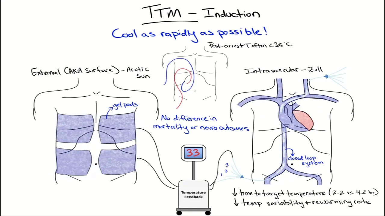
The two options remaining are therapeutic hypothermia and/or induced coma with pentobarbital.
These both greatly reduce cerebral metabolism and thus the need for cerebral blood flow.
But, they have a lot of other problems and you're not going to see them outside of an ICU.
I'll tell you though, if my back is against the wall, and I've got nothing else to turn to I'm going to "throw the kitchen sink" at my patients, and that includes these therapies.
Let's get back to our patient.
We elevated the head of bed.
We hyperventilated her.
And we gave her hyperosmolar therapy.
After that, we started seeing signs that the herniation was improving.
Her anisocoria improved, and now she's got more meaningful motor responses on the right side.
[Sighs] Whew… I am breathing a sigh or relief now, cause we have a moment to start thinking about what else we can do for our patient.
What is her pathology?
Let's talk about the differential diagnosis of coma.
The list of disorders is long enough to fill a whole book.
In fact, there is a book – Plum and Posner’s Diagnosis of Stupor and Coma, which is a real classic of neurology.
[Marvin Gaye's "Let's Get it On" plays] In this section, I’ll give you a sampling of some of the common and testable conditions that cause coma.
It is long list.
So now is probably a good time to pause this talk and take a coffee break.
Are you ready?
Caffeinated?
Let's move to the next section.
Here are the usual suspects causing a severe encephalopathy including coma.
We've got vascular, infectious, hereditary, traumatic, neoplastic, toxic-metabolic, and immune-mediated.
Prion disease is really hard to categorize.
You know, it behaves like an infection, but it can be hereditary too.
I'm shoving it into the hereditary box for now.
First up, vascular.
Vascular pathologies include ischemic and hemorrhagic strokes, and also hemodynamic issues.
Basilar artery occlusion, massive hemispheric and cerebellar infarction, and venous sinus thrombosis are the usual ischemic suspects.
Thalamic hemorrhages, large hemispheric hematomas with mass effect and subarachnoid hemorrhages can all cause coma.
Posterior reversible encephalopathy syndrome even has encephalopathy in the name, so you know it's important.
Global anoxic injury, such as in the setting of cardiac arrest, rounds out the list.
Here you are looking at the axial head CT slice.
By convention when we look at head imaging, the patient's left is on the right hand side of the screen.
It's as if we are looking from the patient's feet up towards the head of the bed.
This slice is at the level of the pons.
You see the bright spot?
That's the basilar artery, and it's hyperdense.
That's concerning for a thrombus.
So after seeing that bright spot, we got a CT angiogram of the head.
Here the two vertebral arteries are labeled.
Look at the tracing though.
This is where the basilar artery supposed to be but isn’t.
The clinical presentation of basilar artery thrombosis can be pretty variable, but it usually involves acute weakness, brainstem signs, eye movement abnormalities, ataxia, dysarthria/dysphagia.
Oh, and I forgot... Coma.
You remember why were are doing this talk?
If you want more information on this, watch the stroke talk.
It's definitely a diagnosis you don't want to ever miss.
Now you try reading this image.
I’ll give you a minute.
You probably said that these are non-contrast head CT axial slices – one through the midbrain, and the other at the basal ganglia level.
And you probably noticed that all the sulci are hyperdense.
The sulci are supposed to be filled with CSF and that should be hypodense on CT.
So, hyperdensity means blood in the subarachnoid space.
So, this is a classic high-grade subarachnoid hemorrhage.
There is also some intraventricular hemorrhage marked by the white arrows.
So tell me, how is this patient going to present clinically?
That's right.
Worst headache of life and meningeal signs.
In the emergency room though, every patient with a headache is having the worst headache of their life.
After all, that's why they are in the ED.
So we're often leaning more on imaging and LPs to rule out this diagnosis.
Here is another example of a coma producing lesion.
Imagine a patient with a history of hypertension who develops sudden gait instability, nausea, vomiting, and might even have fallen to the ground followed by rapidly progressive coma.
Again, two axial head CT slices, one through the pons, and the other at the level of the temporal horns of lateral ventricles.
You can see a large right cerebellar hematoma with some intraventricular extension of blood to the 4th ventricle.
When blood enters the CSF space, it clots, plugs up the flow on CSF and causes obstructive hydrocephalus.
One of the early radiographic signs of obstructive hydrocephalus is prominent temporal horns of the lateral ventricles, as pictured on the right.
Normally temporal horns should be slit-like.
These are dilated.
That's a clue that this patient has hydrocephalus.
Now look at this axial head CT slice through the basal ganglia.
What is abnormal?
That's right.
The basal ganglia and the cortical ribbon appear to be symmetrically hypodense.
Imagine a brain suffers a global anoxic ischemic injury, such as during cardiac arrest or drowning.
Which areas of the brain are most sensitive to the lack of oxygen?
Which areas do you expect to be damaged?
[Doctor from Star Trek Voyager] "I'm a doctor, not a database."
Well, obviously the parts that require the most energy to run.
That includes the basal ganglia, the hippocampus, layer 4 of the cortex (especially visual cortex), and the Purkinje cells of the cerebellum.
These are the big energy hogs of the brain, and those are the ones that are most vulnerable to any lack of oxygen.
This is the pattern of injury that you see on this CT (except you don’t see the cerebellum).
If you do the more sensitive diffusion weighted MR sequence, you can see diffuse cortical ischemia even better.
See how the cortex is bright on this MRI?
That tells us it's ischemic.
Now, this patient presented with hypertensive crisis (systolic in the 220s), worsening headache, bilateral vision loss, seizures, and lethargy.
You probably remember the old Voltaire quote, that the Holy Roman empire This is posterior reversible encephalopathy syndrome.
You know the old Voltaire quote that the Holy Roman Empire was neither holy, Roman, nor an empire?
Of course you do, everyone loves French philosophers.
Anyway, same thing here.
It’s not always posterior or reversible.
In the setting of highly elevated blood pressure, it can be part of hypertensive emergency.
It’s probably the same entity as eclampsia in the obstetric patient.
Hydrostatic pressure and/or increased permeability of the blood brain barrier are the cause.
Certain immunosuppressants, like tacrolimus and cyclosporine given to renal transplant patients can also cause this disease.
And you can see that right here.
These are axial FLAIR MR images (FLAIR is T2 with CSF digitally subtracted to better highlight abnormalities).
The slice on the left is through the basal ganglia, and the slice on the right is higher up in the brain.
On both of these slices, you can see extensive hyperintensities that correspond to vasogenic edema in the white matter predominantly in the occipital and parietal lobes.
Hence, posterior encephalopathy syndrome.
This is a good example of vasogenic edema that's not caused by an infection or a tumor.
We usually treat it by lowering the blood pressure and/or removing the offending agent.
Now, let’s take a look at a few infections.
Typical infectious causes of coma include: Acute meningitis and encephalitis, (that can be be bacterial, viral or fungal) brain abscess, and progressive multifocal encephalopathy due to JC virus infection Here is a T1 MRI axial slice with contrast.
What’s abnormal?
The meninges are abnormally enhancing.
Well, this is a nice example of acute bacterial meningitis.
The patient here typically develops headache, fever, stiff neck, and alteration of mental status, along with seizures.
A small number of cases (especially children) develop severe vasogenic edema, and elevated intracranial pressure, which is translated to the optic nerve, causing papilledema.
Meningitis can be caused by a variety of organisms including bacteria, fungi, and viruses.
The most common bugs are streptococcus pneumoniae, Neisseria meningitidis, and good old enteroviruses.
Add aspergillus and listeria to this list in immunosuppressed patients.
Invasion of meninges is usually done via the bloodstream, so don’t forget to check blood cultures to identify the organism.
Meningitis can also develop if there is communication between the meninges and the surface (such as a CSF fistula).
Think of patients with open head injury or just post cranial surgery.
In our Neuro ICU, this is one of these things we always look out for.
Lumber puncture is the key to diagnosis.
But there is one thing that I want you to take away from this: You should not delay starting empiric antibiotics for the LP.
Typically, cover the patient with vancomycin and ceftriaxone adding ampicillin in immunosuppressed patients.
You should also consider adding dexamethasone This can really help with severe cerebral edema.
Bacterial, fungi and parasites all form localized infections.
But viruses are usually widespread and bilateral and cause coma more often.
Here is an example of encephalitis that's caused by Herpes Simplex Type 1 virus.
This is by far the most common viral encephalitis.
This patient developed progressive personality changes, memory difficulties, and seizures two weeks prior to presentation.
Here are axial T2 images that both show hyperintensity in the frontal and temporal lobes as well as the insula.
These are the areas where the infection usually begins, and where you expect lesions to cause the symptoms I just described.
The CSF usually shows elevated protein with lymphocytic pleocytosis, and HSV PCR is diagnostic.
EEG can also be helpful in showing epileptiform activity coming from the damaged temporal lobes.
As with bacterial meningitis, early treatment is key.
So don't delay starting acyclovir for the LP.
If an infection can't be eradicated by the body, it's walled off in an abscess.
Just like you show all of your junk in the closet and don't want to look at it anymore.
That's what the body does.
And you can get abscesses in the brain just like anywhere else.
Abscess symptoms are really the same as any other mass lesion (like a tumor or hematoma), except they progress more rapidly with headaches and seizures.
You don't always get fever, so you really try to should elicit a history of known infection source (like infected heart valve or dental abscess, or immunosuppression).
Here is an imaging example.
This first image is an MRI.
It's a T1 sequence with contrast axial slice through the basal ganglia.
You can see a medical student favorite here – ring enhancing lesion.
There is nothing particularly magical or pathognomic about ring enhancement.
It's just a descriptor.
It just indicates that there is an area of blood brain barrier disruption surrounding the mass.
Many disorders can have the same appearance, so we need to review other MRI sequences.
In the middle is a T2 Axial image.
The abscess is confined to the temporal lobe but look at the massive amount of subcortical vasogenic cerebral edema.
Abscesses are highly irritating to the brain and often generate more edema than tumors of the same size.
You can imagine all that junk that's walled off in your closet just starts spilling out.
And finally, on the right is a DWI sequence, and you see lightbulb-bright diffusion restriction.
Diffusion weighted imaging highlights areas of viscous fluid filled with cellular debris, bacteria and inflammatory cells.
You might remember from the stroke talk that diffusion restriction is really our gold standard to diagnose stroke.
But not all things that restrict diffusion are strokes.
Here we have a mass lesion that's not really obeying any vascular territory, and is accompanied by a huge amount of vasogenic edema.
How do you treat it?
Start antibiotics, and just like with a pocket of pus anywhere else in your body, drain it surgically.
Traumatic injury is next.
There is usually no mystery about the diagnosis here.
Post traumatic coma is commonly caused by extra axial hemorrhages (such as epidural or subdural hemorrhages), but also hemorrhagic brain contusions, diffuse axonal injury, brain edema, and fat embolism with long bone fractures.
Let’s take a look at some examples.
Here is a non-contrast CT axial slice through the basal ganglia.
What are the abnormalities?
There is a lens shaped hyperdensity outside of the brain in the epidural space.
[Little girl] "Sounds like a subdural hematoma to me."
[Dr. Hfuhruhurr] "Oh, it does, does it?"
"Well it's not your job to diagnose."
[Little girl] "But I thought..." [Dr. Hfuhruhurr] "You thought!
You thought!"
"Just go!"
"Three years of nursery school, and you think you now it all."
"But you're still wet behind the ears."
"It's not a subdural hematoma.
It's epidural!"
"Ha!"
Why am I saying it's outside the brain and in the epidural space?
Well, you probably remember that epidural space respects the suture lines, and this lens shape doesn't pass beyond them.
This bleed was likely caused by a tear of the middle meningeal artery, which was damaged when the temporal bone fracture crossed the middle meningeal groove.
On the right, the CT image has been adjusted to highlight the bone.
You can see there is a fracture in the skull overlying the hematoma.
Classically, these patients start with a headaches post trauma and progress rapidly to coma over hours because of the rising mass effect of the expanding hematoma.
Only one fifth actually have the ‘‘classic’’ history though of this traumatic loss of consciousness, followed by a lucid interval.
Take a look at this non-contrast head CT axial slice through the midbrain.
This patient was taking Rivaroxaban, which is a direct factor Xa inhibitor, for the treatment of deep venous thrombosis.
She slipped and fell in the kitchen, hitting her head.
She was comatose on arrival.
Your first clue of something being wrong is the absence of any fluid-filled hypodense CSF spaces around the midbrain.
Usually there is a nice moat around the midbrain.
Here, there is no space at all.
It's squished up right against the rest of the brain.
Look around for a reason.
Do you see those two crescent-shaped hyperdensities outside of the temporal lobes that are compressing the brain?
These are clearly spreading out along the brain, crossing the suture lines.
So it's not an epidural hematoma.
It's a subdural hematoma.
This is a classic example of an acute subdural hematoma, that was probably worsened by anticoagulant use in this patient.
Finally, diffuse axonal injury is an important mechanism of traumatic coma, especially in patients who don't have an overt large traumatic lesion.
Diffuse axonal injury is a type of traumatic brain injury where acceleration and deceleration leads to shearing forces on the white matter tracts of the brain, causing damage to the axons.
The first three images are axial FLAIR MRI sequences ascending from the level of the basal ganglia.
You see multifocal white matter hyperintensities, which by themselves are generally not specific.
Except here you see them in the corpus callosum, and also scattered in the periventricular and frontal areas.
Combined with a history of a high-speed motor vehicle accident, these hyperintensities are very suggestive of diffuse axonal injury.
To really make the diagnosis, though, we need to see blood.
The last two images are Gradient Recalled Echo (GRE), axial cut.
GRE is a specific sequence that's tuned to pick up hemosiderin, which is to say blood.
And here, you see several areas of susceptibility (dark spots) that correspond to microhemorrhages in the white matter.
Next up, tumors.
Neoplastic lesions causing coma are generally either massive intracerebral tumors (primary gliomas or metastasis), leptomeningeal disease (lymphomas), dural-based tumors with mass effect (meningiomas) or pituitary tumors.
That last group can cause altered mental status not by mass effect, but it's effect on the endocrine system.
Here is Axial and sagittal T1 sequences both with contrast, as well as axial T2 sequence on the right.
You can see the abnormality from across the room There is a large, ugly, enhancing bulky mass involving the corpus callosum and surrounded by cerebral edema, that bright hyperintensity you see on the T2 sequence.
This patient has glioblastoma multiforme - the highest grade of primary CNS glioma.
Gliomas typically invade the substance of the brain and then create mass lesions.
They have bulk!
They push everything out of the way.
As they get bigger, they outgrow their blood supply and develop areas of necrosis or hemorrhage inside.
Now here is a similar MRI.
We are looking at the Axial T2.
Do you see the vast amount of vasogenic edema?
I say vasogenic because the cortical ribbon does not seem to be affected.
The hyperintensity respects the boundaries of the white and grey matter.
This edema even seems to be involving the corpus callosum.
So what do you think this tumor is?
Another glioblastoma?
[Prof. Van Helsing] "Yes!
And no."
[Dr. Harker] "Then what are you saying?!"
[Prof. Van Helsing] "I'm saying 'no,' but I'm leaning towards 'yes.'"
[Dr. Seward] "Then you're saying 'yes?'"
[Prof. Van Helsing] "No!"
[Dr. Seward] "Then it's 'no?'"
[Prof. Van Helsing] "Not necessarily."
[Dr. Harker] "You sound dubious."
[Prof. Van Helsing] "No, I'm positive."
[Dr. Harker] "Of what?"
[Prof. Van Helsing] "My theory!"
[Dr. Harker] "And that would be?"
[Prof. Van Helsing] "The theory of 'yes,' or 'no.'"
Here is a T1 Axial and Coronal slices with contrast to help you.
What do you see?
This mass is outside the brain.
This mass is based in the dura, and I say that because it has an enhancing dural tail – the region of the dura adjacent to the mass that's also enhancing.
So what is the most common dural based mass?
A meningioma.
Meningiomas typically grow over the convexities, along the falx, or along the base of the skull at the sphenoid wing.
Because these are typically slow growing tumors and usually don't create this much mass effect acutely, coma is an unusual presentation.
One exception would be a meningioma that hemorrhages within itself.
More likely meningiomas present with focal deficits by compressing the brain structures, and also with seizures.
The treatment here is as expected – corticosteroids to treat the edema and surgery to remove the tumor.
Finally, let’s take a look at an example of encephalopathy caused by extensive neoplastic invasion of leptomeninges and also the subarachnoid space.
You are looking at an axial and sagittal T1 sequence here, both with contrast.
The first image is through the pons, with occipital lobes showing up posteriorly.
The second image is just to the left of midline, and you can see the cerebellum and the occipital horn of lateral ventricle.
The brainstem would be more medial, so you do not see it yet.
Look at the patchy enhancement that perfectly follows the sulci.
This is enhancement of the subarachnoid space and pia.
Subarachnoid space surrounds the entire CNS.
Cranial nerves travel through this subarachnoid space.
So, the hallmark of subarachnoid tumors is multilevel disfunction – cranial or spinal nerves, spinal cord, brainstem, or the hemispheres.
So why do you get coma?
Well, a number of reasons: There can be hydrocephalus from obstruction of spinal fluid pathways, direct invasion of the brain through the pial vessels, status epilepticus, or overactive immune response to tumor.
Biopsy is the gold standard here for diagnosis.
CSF cytology via lumbar puncture does not have a lot of sensitivity.
You can do multiple high-volume lumbar punctures for CSF cytology and still not find neoplastic cells.
In this case, this patient was diagnosed with meningeal lymphoma.
If you are rotating through the emergency department, and you see a comatose patient, toxic metabolic causes are probably going to be first on your mind.
You could be thinking of toxidromes – drug overdoses or drug-drug interactions (for example serotonin syndrome from a combination of selective serotonin receptor blockers and monoamine oxidase inhibitors).
You could be thinking side effects of poisons, like alcohol.
You could be thinking – derangements of glucose.
There is a reason why we check fingerstick glucose in any unresponsive patient.
You could also look at electrolyte disturbances (hyponatremia, hypercalcemia, and acidosis).
Renal failure (uremia).
Hepatic failure, causing hyperammonemia And endocrine disturbances, including the adrenal glands, pituitary gland and thyroid gland.
Obviously, that’s a long list.
Let’s look at some examples.
This patient has chronic malignancy, who then developed confusion and balance issues, and presented with a rapidly progressing coma with a characteristic finding of ophthalmoplegia.
That means the eyes don't move.
You are all experts in neuroimaging by now.
So what is abnormal on these coronal FLAIR images?
Did you see the hyperintensities in the frontal cortex, medial thalami, and the mamillary bodies?
What’s the diagnosis?
Yep, this is severe thiamine deficiency otherwise known as Wernicke’s encephalopathy.
All these neuronal areas are deprived of thiamine to the same extent, but certain cell groups – the neurons surrounding the third ventricle, the cerebral aqueduct and the 4th ventricle – are pathologically more sensitive to this deficiency and show the greatest amount of damage.
Remember that the diencephalon is part of the reticular activating system.
So, hence damage to the thalamus causes coma.
The oculomotor nuclei are periaqueductal (they are right next to the cerebral aqueduct).
Hence you get ophthalmoplegia.
You probably already remember this: Giving glucose without thiamine can precipitate excitotoxicity in this disease, and worsen the outcome.
Who get’s Wernicke’s?
Well obviously you think alcoholics, but really anyone who is malnourished.
That includes cancer patients like this one.
What's the treatment?
High-dose thiamine.
You give it intravenously, high-dose for several days.
The nystagmus, and oculomotor paralysis can be reversed by thiamine.
But often times the damage to thalamus and mamillary body can't.
This can cause striking memory failure and confabulation when patients awaken.
That confabulation makes up the Korsakoff part of Wernicke-Korsakoff syndrome.
Autonomic insufficiency in this disease is very common – think orthostatic hypotension and even shock.
So these patients are usually monitored in the intensive care units.
Ok.
By now, you know the drill.
Axial T2 and FLAIR cuts, through the lower and upper pons, with bilateral temporal lobes visible as well.
What do you see?
Hyperintensity in the white matter of the central pons, and also in the medial temporal lobes.
What's the diagnosis?
This is central pontine and extra pontine myelinolysis.
I put this in just to remind you that lysis of myelin does not have to be limited to the pons.
That's why the name of this disease is now osmotic demyelination syndrome.
Usually these are patients that have sufferred chronic hyponatremia and got rapidly overcorrected in the hospital.
The majority of these patients become locked-in, but a quarter of them can also develop impaired consciousness.
Quick flashback to the start of this talk.
Remember that damage to the ventral pons can cause locked-in syndrome, but the reticular activating system is located in the dorsal medial pons.
So depending on where the osmotic damage is, it can cause either of these symptoms.
Final example - hypoglycemic coma.
This diabetic patient accidentally overdosed on insulin.
There are few things more dangerous to the brain than profound, prolonged hypoglycemia.
The longer it lasts the more likely it will produce irreversible neuronal damage.
Glucose is brain’s fuel, and each 100 grams of brain uses 5.5mg of glucose per minute.
Basically, the brain consumes almost all of the glucose that the liver produces.
This Axial T2 image through basal ganglia shows extensive damage to the areas of the brain that need glucose the most – the cortical ribbon and the basal ganglia.
And you are going to say, well this looks exactly like the image of patient with hypoxic ischemic encephalopathy.” And you would be absolutely right.
The same parts of the brain that need oxygen also need glucose.
Finally, let’s wrap up the pathological differential with a discussion of immune-mediated disorders.
When it comes to immune-mediated comas, we are talking about: Autoimmune encephalitides, like acute disseminated encephalomyelitis (ADEM), Bickerstaff encephalitis, neurosarcoidosis, and lupus cerebritis.
Vasculitides, like Behcet’s, or primary angiitis of the central nervous system are also under this category.
And paraneoplastic disorders, like anti-NMDA receptor encephalitis and limbic encephalitis, where underlying malignancy triggers autoimmune damage to the CNS.
A lot of "itises," and I'm tired from saying all of them, but let's move to the cases.
At this point, I won’t call out the imaging technique or brain structures.
You are on your own now.
Time to channel your inner neuroradiologist.
Imagine you are in a very dark room, and describe what you are looking at.
I will give you one clue.
This is a 6 year old, previously healthy young boy who had a mild upper respiratory tract infection two weeks ago, and now comes to the emergency department with 2 days of fever, headache, slurred speech, gait instability, excessive sleeping and disorientation, and increased muscle tone throughout.
Did you say that this is a T2 FLAIR axial image with diffuse white matter hyperintensities?
Yeah, I knew you would.
This is a classic case of ADEM.
I'm going to say ADEM since it's a lot easier to pronounce than acute demyelinating encephalomyelitis.
Patients are usually younger, and develop multiple neurological symptoms including cognitive and behavioral changes (that's the encephalitis) over a period of several days, about 1 to 2 weeks after a febrile illness.
ADEM is an example of a “parainfectious encephalomyelitis.” The “itis” here is presumably caused by a deranged, aggressive immune response to a virus or vaccination.
What causes this?
We don't know.
Usually we throw in phrases like “molecular mimicry,” but just imagine me waving my hands a lot right now.
The CSF may show mild lymphocytic pleocytosis (usually less than 500 cells), and elevated protein.
But is also may be normal.
This illness is monophasic.
It usually resolves with some steroids and immunosuppressants, and time.
It rarely recurs.
So imagine you are an ED doc who is now seeing this child.
What is the diagnostic challenge?
We really have to differentiate between “itises” caused by active viral infection, requiring antiviral treatment, and autoimmune parainfectious response, requiring immunosuppression.
One clue is that patients with viral encephalitis tend to be more severely ill with higher fevers for longer periods of time, and autoimmune stuff tends to start when viral illness has passed.
Next case.
Here is a 25 year old woman who presented with 1 week history of progressive headaches, vertigo, audio-visual hallucinations, and progressed to developing coma, seizures and myoclonus.
Here are three FLAIR slices from the same brain.
They show temporal, insular and frontal hyperintensities suggesting an encephalitis.
MRI is not very specific in this disease, since temporal abnormalities can look like HSV infection (though without enhancement), cortical abnormalities can look like stroke (but without restriction of diffusion on DWI), and the white matter abnormalities can look like ADEM.
It's clearly a big mimic.
You get the picture.
History is much more important here, and thankfully there is a diagnostic testing.
This is an example of NMDA receptor antibody encephalitis.
Think of this cause of coma in a young woman with initially viral-like prodrome who then goes on to develop psychiatric symptoms and eventually progresses to coma, seizures, hyperkinetic movement disorders with abnormal involuntary jerky movement and autonomic storming (bouts of hypertension, tachycardia, and diaphoresis).
The NMDA receptor mediates glutamate, which excites neurons.
So you can see all these symptoms are caused by overexcitation of neurons in the brain.
Diagnosis can sometimes be difficult, because after all, you're getting young patients who are having their first bout of psychosis.
It all happens at about the same age.
Beware of patients who don't have any personal or family history of psychiatric illness, who suddenly develop psychosis, and any abnormal neurologic findings.
I can tell you, I've taken care of teenagers who initially were diagnosed with schizophrenia and spent days in a psychiatric unit before developing seizures and other findings that clearly pointed to a different type of disease.
This paraneoplastic syndrome is often associated with ovarian teratoma or carcinoma.
It's important to stick with treatment with these patients.
Their treatments are often very prolonged.
Although after sometimes weeks or months of immune therapy, these patients can go back to normal life - school and work.
Well, after that long detour, have you forgotten about our case?
Let's get back to it.
When we last left her she was exhibiting signs of uncal herniation, which then reversed after our management.
What do we do next?
The top three disorders that cause sudden coma and need to be emergently addressed are vascular (basilar artery occlusion, hemorrhage, straight sinus thrombosis), status epilepticus, or a mass lesion (such as a neoplasm).
Our patient had mass effect, so it stands to reason that hemorrhage and mass would be at the top of our early differential.
Let’s look for that structural lesion.
What the best initial test?
That's right, a non-contrast head CT.
If we were worried about a basilar artery occlusion or other vascular issue, we would get vascular imaging with a CT angiogram of the head and neck with contrast.
That's less likely in this case.
Here is our patient’s head CT at different axial cuts in the brain.
The right image is the customary view through the basal ganglia, and the left image is above that.
Try to read this scan.
I’ll give you a minute.
You can probably make out the heterogeneous right frontal hyperdensity surrounded by hypodensity.
There is also a significant amount of mass effect.
See how the right hemisphere is pushing over into the left side.
That's called midline shift.
Our patient underwent an emergency craniotomy, decompression and evacuation of the lesion, which turned out to be hemorrhagic tumor (glioblastoma multiforme in this case).
Here you can see her post-op head CT.
The skull has been removed on the right, there is mild amount of air in the cavity where Here you can see her post-operative head CT.
The skull has been removed on the right, so there is a small amount of air (black) in the cavity where the tumor used to be.
The mass effect has improved, and there is a small amount of hemorrhage spilled over into the lateral ventricles.
Post-surgery, our patient alertness has improved to the point where we were able to extubated her.
She is now arousable to voice.
She is following commands.
There is persistent left sided weakness, but she has some movement on that side.
The next day though, she was once again difficult to arouse and her left sided weakness became worse.
Well that’s puzzling.
What's going on here?
Her vitals and labs are normal, and there are no sedatives confounding the picture.
We repeated the head CT, and that was stable.
Take my word for it.
So the mass effect has been dealt with, and yet our patient’s deficits are out of proportion to the scan.
Why?
[Prof. Van Helsing] "How does your modern science explain zat?"
"Can you explain zat?"
"Can you explain zat?"
[Dr. Weber] "I can't explain 'zat.'"
[Dr. Seward] "I can't explain 'zat.'"
[Prof. Van Helsing] "No one can explain zat!"
How do we figure this out, and what's the next best step in the management?
Well, neurologically injured brains are prone to seizures.
Up to one third of neurologically critically ill patients develop status epilepticus.
So, we should consider treating empirically.
Start antiepileptics.
Then, let's make the diagnosis.
Start with a spot 30 minute EEG and then progress to 24 hour continuous EEG to assess for seizures.
Unfortunately, our hunch was right.
The patient had right fronto-temporal epileptiform activity, probably coming from the brain surrounding the tumor cavity, and two electrographic seizures were captured during a 30 minute recording.
What do we do next?
This is a progression of status epilepticus management.
On the left side of the screen, the patient has just started seizing, and we are just starting our initial therapies.
As we move towards the right, we are going through higher tiers of therapies and interventions.
We’ve already secured the airway, and the ABCs are stable.
The first line medications are short acting benzodiazepines like lorazepam or diazepam, and that would typically abort seizures early.
But if these seizures continue, we move on to second line medications like fosphenytoin, valproic acid, phenobarbital, or Levetiracetam.
Now, I advocate moving directly to general anesthesia, if your first line medications fail, since it takes time to start with second line medications.
The sooner we stop the status epilepticus, the more secondary brain damage we will avoid.
In our case, the patient is already intubated and will need sedation anyway.
So we start midazolam or propofol, which are short-acting and titratable anesthetics.
We then titrate the anesthesia to stopping the seizures on the EEG.
This basically acts as a reset for the injured brain.
In cases where status epilepticus persists for more than 30 minutes despite therapy, at that point it is classified as malignant.
As you have guessed, malignant anything is not good for the brain.
So just like in intracranial pressure crisis, we may proceed to higher levels of therapy, including inducing pentobarbital coma.
And it goes without saying, ICU level care and continuous EEG monitoring are a must for a patient in status.
So we treated our patient with midazolam and levetiracetam, and the seizures stopped.
Sadly, not all is well that ends well.
Her ICU course was prolonged and very complicated.
At some point, she suffered a brief cardiac arrest.
CPR was started and return of spontaneous circulation was achieved after 15 minutes.
But even 72 hours later, her neurological exam remains poor.
She has no brainstem reflexes and no meaningful responses.
Obviously this is not what we wanted.
The family is understandably devastated.
They want to know what is going to happen to her.
That brings us to the last part of our talk – outcomes.
Imagine a graph of cognitive function on the x-axis and motor function on the y-axis.
So the best possible outcomes are in the top right corner, and the worst possible outcomes are in the bottom left.
Full cognitive and motor recovery is what we all hope for, but few achieve after a coma.
Locked-in state belongs in this first column.
This is a state of preserved cognition, but severely limited motor function.
Locked-in patients are not comatose, and they can still move their eyes.
Look for vertical eye movements and blinks.
That's how you can communicate.
The next column is moderate to severe cognitive or motor disability.
The important distinction here is that patient still has reliable functional communication.
There are following commands and making at least some of their wishes known.
On the left side of the dotted line, reliable functional communication is absent, and these patients are said to be in a minimally conscious state.
Basically this is someone with a severe alteration of consciousness.
They spend the majority of their time unresponsive, but they can show meaningful awareness of self or environment by attending to loved ones (by looking at them).
They may attempt purposeful movements, and so on.
Next step down is persistent vegetative state.
Patients have sleep wake cycles, but show no meaningful awareness or interaction.
“Persistent” refers to passage of time, and can be diagnosed at least one month after brain injury.
Of course, for patients in this state, outcomes for meaningful neurological recovery are generally poor.
However, because a subset of patients in the persistent vegetative state do end up recovering higher levels of consciousness, this term has fallen out of favor, and we prefer to call it unresponsive wakefulness.
Finally, the most dismal outcome is death.
Brain death in this case.
That's absence of brain activity including brainstem function.
We will talk about the criteria for diagnosing brain death in a moment.
There is a lot we don't understand about the injured brain, and making a prognosis can be very difficult Age, baseline function and the patient’s neurological examination are arguably the best prognostic factors.
So we would be very optimistic about meaningful long term neurological recovery for a young healthy woman who suffers a coma because of severe traumatic brain injury after a motor vehicle accident, but she is now awake and following some commands.
The cause of coma is also important.
The best prognostication data we have is for cardiac arrest and for traumatic brain injury patients.
Although some data exists for outcomes post autoimmune encephalitis, such as NMDA receptor encephalitis.
I mentioned that it's important to be aggressive with treatment for these patients, because outcomes can be quite good, even after months of treatment.
Neuro imaging can be helpful.
Earlier in this talk, we saw examples of diffuse anoxic and hypoglycemic injury on MRI, portending bad outcomes.
But, ultimately, these are just pictures.
Never make a prognosis based on a scan alone.
In a comatose patient without meaningful neurological responses, EEG with absent normal brain reactivity may serve to confirm poor outcome, but it doesn't always reliably predict the extent of future recovery.
Serum biomarkers, like neuron specific enolase have been used as another confirmation of poor outcome after cardiac arrest.
The use of these biomarkers though is still being studied.
Other important predictors of poor outcomes are the duration of coma, and other comorbid conditions.
Obviously, someone who has been in a coma for 2 months has a poorer outcome, than someone who's only had a coma for several days.
And a person who is in a coma due to metastatic cancer has a poorer outcome than someone with traumatic brain injury but otherwise healthy.
In general, our tests are better able to tell us the patients who do poorly, than predict the patients who are going to recover.
The important point here is that there are no shortcuts to prognostication.
Every case is unique.
In our case, you may remember that things are pretty dire.
Brainstem reflexes are absent in our patient, and she does not have any response to pain.
We are really worried that she may be brain dead.
So, how do we go about declaring this?
Brain death is death.
But, it's not as easy to understand as cardiopulmonary death.
You can feel the heartbeat.
But it's a lot more difficult to figure out if the brain is still working.
So, let's go through the process by which we to establish brain death.
First, we need to establish that there is irreversible cause of coma.
Neuroimaging can be very helpful here.
Cardiac arrest, basilar artery thrombosis, large hemorrhage – all of those qualify.
If you don't know the cause of coma, you CANNOT establish brain death.
Next, exclude confounding factors.
There can't be any CNS depressant drugs.
The patient can't be paralyzed chemically.
The patient can't be hypothermic, or very hypotensive.
And the patient can't have any major metabolic derangements.
All of these things can cloud the neurologic exam and give an impression of a brain dead patient.
Next, you need clinical evidence of loss of brainstem function.
The pupils must be fixed and unreactive to light.
There must be no oculocephalic, and oculovestibular reflexes.
No corneals, no cough or gag reflexes.
There can be no meaningful motor responses present.
Although reflexes, like upgoing plantar or triple flexion, are allowed.
And the patient must not exhibit any spontaneous breathing.
The patient must be completely dependent on a ventilator for breaths.
The final element of brain death testing is to establish that there is no spontaneous respiratory drive off of the ventillator.
That's called the apnea test.
That is done by pre-oxygenating the patient and establishing a ventilatory rate to normalize the CO2 as much as possible.
How do we know it's normal?
We take an arterial blood gas.
Next, we disconnect the ventilator for 8-10 minutes.
We allow the CO2 to rise above 60 mmHg, or at least a 20 mmHg increase over the baseline.
Both of these should induce respiration in someone with intact respiratory center in the medulla.
If the patient becomes unstable during the apnea test (the patient becomes hypotensive or desaturates), we have to abort the test and reconnect the ventilator.
In this case, we may rely on ancillary tests to help complete the picture of brain death.
These tests ARE NOT confirmatory tests.
You CANNOT do these in absence of meeting the other criteria for brain death.
Ancillary tests in general look for brain death in one of two ways: Either looking for absent cerebral blood flow, or absent electrical brain activity.
Examples of ancillary tests include: Nuclear SPECT imaging, transcranial dopplers, a cerebral angiogram, or electroencephalogram (EEG).
Thanks Ram.
Congrats, you made it!
Now, let’s briefly discuss the entire algorithm.
Say you have a patient with an acute confusional state – this could be disorientation, aphasia or lethargy.
History is key.
A good beginning would be to find out the time last known at baseline, details of event onset, comorbid conditions, medications and toxic exposures, and previous such episodes Next.
Is the patient presenting within or outside of a 24 hour window from last known at baseline?
Within 24 hours?
Consider activating a stroke code, especially when you see focal neurological deficits.
Vascular pathologies are a common and treatable cause of acute coma especially with focal deficits.
Once you activate a stroke code, you go down a rapid stroke assessment pathway, which I reviewed in the stroke talk.
Next, initial stabilization.
We are talking about airway, breathing, circulation - the ABCs.
A common resuscitation process would include a large bore IV, or preferably two, a cardiac monitor, oxygen via face mask, and labs, including basic panel, complete blood count, fingerstick glucose, liver function tests with ammonia level, thyroid function tests and drug levels were appropriate.
When sepsis is suspected, blood cultures and lactate are also checked.
And in trauma patients, we should immobilize the c-spine.
If ABCs are unstable, the patient would need intubation and hemodynamic support.
Once ABCs are stable, in an acutely altered patient without focal neurological deficits, toxic and metabolic etiologies are by far the most common cause.
So we consider issues like electrolyte disturbances (especially sodium and calcium), sepsis, vitamin deficiencies (especially thiamine), and poisonings and toxidromes.
If the cause of encephalopathy is known, then we can address it at this point.
We can use toxin-absorbing activated charcoal and induce vomiting within 2 hours of ingestion.
If thiamine deficiency is suspected, you need to treat with IV thiamine.
For significant benzodiazepine overdose, there is flumazenil, but consider that carefully because it can induce withdrawal seizures.
For opiod overdose, there is naloxone, which can be given intravenously or sprayed into the nose.
Next, we need to move on to a rapid neurological evaluation.
We are looking for localizing or focal findings to help identify a structural lesion.
But also we check for meningeal signs and observe for abnormal involuntary movements, such as myoclonus.
That last one is to help with pathological differential.
These movements can suggest seizure, serotonin syndrome, or NMDA receptor encephalitis.
Our coma exam essentially has 4 components: Respirations, pupils, brainstem reflexes, and motor responses.
Next.
Are there signs of elevated intracranial pressure?
Yes?
The proceed with emergent ICP management: Head position optimization, hyperventilation, sedation where appropriate, cerebral edema management, surgical intervention, and so on.
Once the intracranial pressure crisis has been addressed, we need to consider this short but dangerous differential, especially when a patient has focal neurological deficits or your examination is suboptimal.
Vascular lesions (such as basilar artery occlusion, venous sinus thrombosis and hemorrhage), mass lesion, (which by the way can be neoplastic or infectious, like a brain abscess), and status epilepticus.
First things first, assess for a structural lesion.
We start with a stat non-contrast head CT.
We should also get an emergent vascular study, such as the CTA of head and neck with contrast in patients with concern for a vascular pathology, which is basically everyone within 24 hours of abrupt onset of symptoms especially when they present with focal deficits.
In fact, CTAs are an integral part of our stroke code protocol.
If we identify a hemorrhage, then we treat according to the hemorrhage protocol outlined in your stroke talk.
For hemorrhages and mass lesions with mass effect, we can consider urgent surgical intervention.
Basilar artery thrombosis may be treated with thrombolytics and thrombectomy when appropriate.
If at this point, there is no obvious structural lesion, we can consider an urgent MRI of the brain with and without contrast to look for entities like completed stroke without and occluded vessel, encephalitis changes, and PRES.
But MRI takes considerable effort, especially in a thrashing disoriented or intubated patient, so it’s usually delayed until later in the algorithm.
Say there is no mass lesion or basilar artery thrombosis.
At this point, we should consider cerebral venous sinus thrombosis.
Thrombosis of straight sinus for example.
To diagnose that we will likely check a rapid CT Venogram with contrast.
We can also use an MR venogram, but as I mentioned, it’s more difficult to get in the ED.
If thrombosis is confirmed, we consider anticoagulation.
Imagine all of the imaging is negative.
Now let’s assess for status epilepticus.
We can consider treated empirically according to the status epilepticus protocol, while getting an EEG.
Start with a spot 30 minute EEG, and progress to 24 hour EEG monitoring as needed.
Now, let’s say all of our workup has been unrevealing thus far.
Well, now we need to cast a wider net.
This is where we consider: Vascular and traumatic pathologies, like PRES, anoxic ischemic injury, diffuse axonal injury, and fat embolism.
Infectious and inflammatory pathologies, like meningitis, herpes encephalitis, brain abscess, ADEM, vasculitis, and paraneoplastic encephalitis, like NMDA receptor encephalitis or limbic encephalitis.
Toxic metabolic pathologies are giant category and include diseases like osmotic demyelination syndrome, carbon monoxide poisoning, hepatic and uremic encephalopathies, pancretopathies (think diabetic keto acidosis and hyperosmolar non-ketotic coma), thyroidopathies, adrenal disorders, panhypopituitarism, and drug intoxication, like serotonin syndrome and neuroleptic malignant syndrome.
Obviously this is not an exhaustive list.
Hereditary category includes disorders like MELAS (mitochondrial encephalopathy with lactic acidosis and stroke like events) - say that three times fast - and prior disease.
As we discussed before, prion disease probably belongs in its own category, but we lumped it here with hereditary disorders just to simplify things.
We didn’t discuss the hereditary category much in this talk, because these disorders are usually rare in the adult patient.
Finally, don’t forget neoplastic pathologies, like leptomeningeal carcinomatosis, gliomas, metastasis, and pituitary neoplasms.
If you suspect infection, consider treating empirically at this point.
And finally, now is the time for lumbar puncture and the first or even repeat MRI.
You may want to call for reinforcements – neurology consult, infectious disease consult, oncology consult.
You know, the House MD team.
And repeating certain labs may not be a bad idea to help with diagnosis since disease processes do progress.
That's all!
Thank you for your kind attention, and we'll talk again soon.
Up Next
Hyperkalemia Pathophysiology, ECG Changes & Treatment Explained
@Alilamedicalmedia
611K views•2017-03-13

Graphic Medicine: Comics for Collaborative Healthcare Communication
@nationalpatientadvocate
189 views•2023-12-04

Neuroanatomy: Central and Peripheral Nervous System Divisions Explained
@AKLECTURES
136.2K views•2014-09-20

Stages of Labor and Vaginal Birth | Childbirth Animation
@nucleusmedicalmedia
52.1M views•2017-08-18
Related Study Plans & Knowledge Roadmaps
Structured learning paths in Medicine