This video provides an animated overview of DNA and genome structure, designed to help students understand fundamental concepts of genetics and cell division at the beginning or end of a biology unit.
DNA Replication and Genome Overview Animated Educational Guide
Added:Deep Dive
Basic eukaryotic and prokaryotic cell structure, particularly the function of the nucleus as the cellular control center.

TYT Biyoloji FULL TEKRAR | MSÜ Son Tekrar
394.4K
Cells are classified as prokaryotic (bacteria, archaea) or eukaryotic (protists, plants, fungi, animals). Prokaryotic cells lack a nucleus and membrane-bound organelles. Eukaryotic cells have a nucleus and membrane-bound organelles. The nucleus is the control center containing genetic material. Organelles are classified as non-membrane-bound (ribosomes, centrosomes), single-membrane-bound (endoplasmic reticulum, Golgi, lysosomes, peroxisomes, vacuoles), or double-membrane-bound (mitochondria, plastids).
Click to watch →

Cell structure :Target 360 in Biology NCERT आयाम By Dr Rishabh Choubey Sir | Bio Guru
202.1K
The nucleus is the control center of the cell that contains the genetic material (DNA). It is surrounded by a double membrane called the nuclear envelope. The nucleus directs all cellular activities by controlling gene expression and regulating the synthesis of proteins and other molecules. The nucleus is essential for cell survival and function.
Click to watch →

Cell Structure and Function: Eukaryotic vs Prokaryotic Cells | Biology
33.6M
Cells are the smallest living units of organisms, sharing three common components: cell membrane, cytoplasm, and genetic material (DNA). Cells are broadly categorized into eukaryotic cells (found in plants and animals) which have a nucleus and membrane-enclosed organelles, and prokaryotic cells (such as bacteria) which lack a nucleus and membrane-bound organelles. Key organelles include the nucleus (control center containing DNA), ribosomes (protein synthesis), endoplasmic reticulum (protein transport), Golgi apparatus (protein modification), vacuoles (storage), lysosomes (cellular digestion), mitochondria (energy production via cellular respiration), and chloroplasts (photosynthesis in plant cells). Both plant and animal cells contain mitochondria, while only plant cells have chloroplasts.
Click to watch →

8th Science | Chapter 1 | The Living World | Lecture 1 | Maharashtra Board | JR Tutorials |
83.6K
All living organisms are composed of cells, which are microscopic structures visible only under a microscope. Inside each cell, there is a nucleus surrounded by a nuclear membrane that contains genetic material. Prokaryotic cells lack a well-developed nucleus and nuclear membrane, with no distinct organelles. Examples include bacteria and blue-green algae in Kingdom Monera. Eukaryotic cells have a well-defined nucleus enclosed in a nuclear membrane and can be either unicellular (Kingdom Protista) or multicellular (Kingdom Fungi, Plantae, and Animalia). This fundamental distinction in cell structure is the basis for classifying all living organisms.
Click to watch →
Núcleo Celular | Biologia | Prof. Paulo Jubilut
812.4K
The cell nucleus is the control center of eukaryotic cells, discovered by Robert Brown in 1833, containing genetic material (DNA) organized as chromatin (DNA-protein complexes) and serving as the site for nucleic acid synthesis; it is surrounded by a double-membrane nuclear envelope with pores that regulate molecular transport, and contains the nucleolus where ribosome formation occurs.
Click to watch →
Deep Dive
The chemical structure of nucleotides, including the sugar-phosphate backbone and the four nitrogenous bases (adenine, thymine, cytosine, and guanine).

Molecular basis of inheritance lec03
391
The chemical structure of a nucleotide consists of three main parts: (1) A sugar molecule (ribose in RNA or deoxyribose in DNA), (2) A nitrogenous base (adenine, guanine, cytosine, thymine, or uracil), and (3) A phosphate group. The sugar and nitrogenous base form a nucleoside, and when a phosphate group is attached to the sugar, it becomes a nucleotide. The phosphate group connects nucleotides together to form the backbone of nucleic acid chains.
Click to watch →

The 4 Nucleotide Bases: Guanine, Cytosine, Adenine, and Thymine | What Are Purines and Pyrimidines
102.1K
DNA nucleotides consist of three components: a nitrogenous base (adenine, thymine, guanine, or cytosine), a deoxyribose sugar lacking an oxygen atom at carbon 2, and a phosphate group. The four bases classify into purines (adenine and guanine with two-ring structures) and pyrimidines (thymine and cytosine with one-ring structures). Nucleotides link via phosphodiester bonds formed between the 5' hydroxyl group of one sugar and the phosphate group of another, releasing water in a condensation reaction. This creates the sugar-phosphate backbone, where phosphate groups connect adjacent sugars through alternating phosphoester linkages, enabling long DNA strands to form.
Click to watch →

Sinh học phân tử P2
440
Nucleotides consist of three components: a five-carbon sugar (deoxyribose in DNA, ribose in RNA), a nitrogenous base (purines: adenine and guanine; pyrimidines: cytosine, thymine in DNA, uracil in RNA), and a phosphate group. Nucleosides lack the phosphate group. The phosphate group provides acidic properties. DNA and RNA differ in their sugar components and base composition (DNA has thymine, RNA has uracil). The sugar-phosphate backbone forms the structural framework, while nitrogenous bases project inward for base pairing.
Click to watch →

TOPIC # BIOMOLECULE (part-8)# (Nucleic acid)# chemistry by Dr.Mohd.Gaus(KGMU)
301
Nitrogenous bases are classified into purines (adenine and guanine) and pyrimidines (cytosine, thymine, and uracil). Purines have a double-ring structure, while pyrimidines have a single-ring structure. Adenine and guanine contain amino groups (NH2), while cytosine, thymine, and uracil contain carbonyl groups (C=O). Thymine is found only in DNA, while uracil is found only in RNA. A nucleoside consists of a nitrogenous base attached to a sugar molecule (deoxyribose in DNA, ribose in RNA) through a glycosidic bond at the 1' position. A nucleotide consists of three components: a nitrogenous base, a sugar molecule, and a phosphate group. The phosphate group is attached to the 5' carbon of the sugar, while the nitrogenous base is attached to the 1' carbon. Nucleotides are linked together through phosphodiester bonds to form the sugar-phosphate backbone.
Click to watch →

LF MUNI 2016 test chemie Mgr
2.5K
Nucleotides consist of: (1) A nitrogenous base (adenine, guanine, cytosine, thymine, uracil), (2) A pentose sugar (ribose in RNA, deoxyribose in DNA), (3) One or more phosphate groups. The base attaches to the sugar via a glycosidic bond. Adenosine = adenine + ribose. Adenine nucleotide = adenosine + phosphate(s).
Click to watch →
Deep Dive
An introductory concept of chromosomes and how they package genetic information.
Genetics and Heredity for A&P - Episode 79
177
Chromosomes are the microscopic structures that serve as packages of heredity. Inside the nucleus of almost every cell in the human body, chromosomes consist of long strands of DNA coiled and folded around proteins. These structures are packed tightly enough to fit into a microscopic space while carrying vast amounts of genetic information. The human genome contains 46 chromosomes arranged in 23 pairs, with each pair consisting of homologous chromosomes that are similar in size, shape, and gene content.
Click to watch →

Nhiễm sắc thể và bộ nhiễm sắc thể (phần 1) - KHTN 9 - OLM.VN
23.3K
Chromosomes are composed of DNA wrapped around histone proteins forming nucleosomes, which are organized through multiple levels of coiling. Heterochromatin regions have tightly packed nucleosomes and are transcriptionally inactive, while euchromatin regions have spaced nucleosomes and are transcriptionally active. Each chromosome contains two DNA molecules (one per chromatid), compressed thousands of times to fit within the nucleus. This packaging enables efficient storage and transmission of genetic information during cell division, making chromosomes the primary material basis for inheritance in eukaryotic cells.
Click to watch →
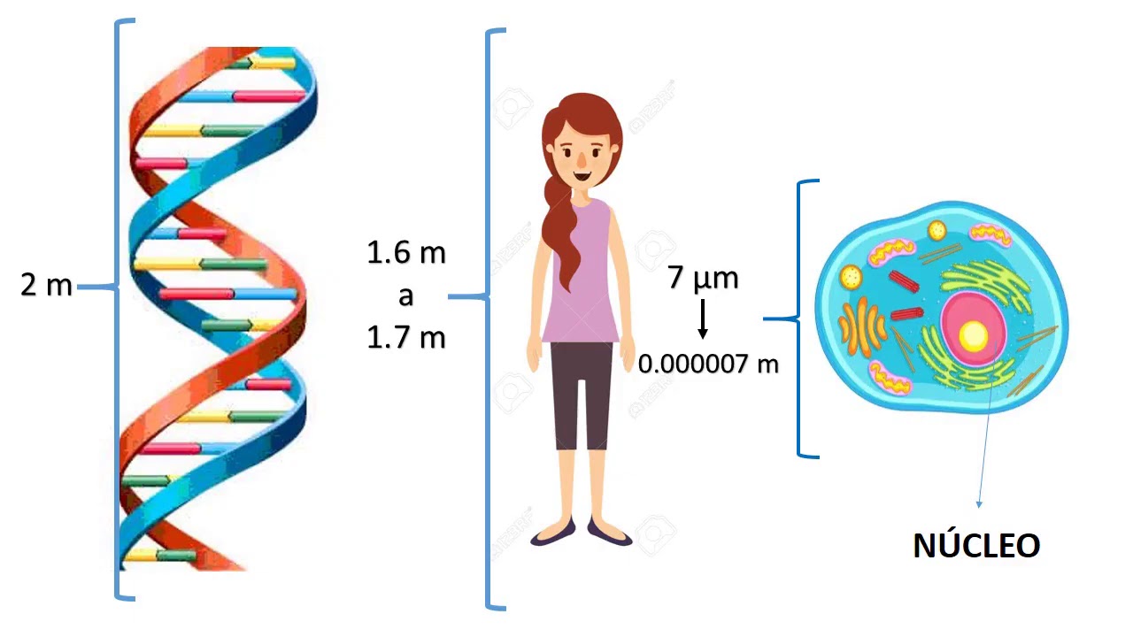
Conceptos Básicos de Genética PROPEDÉUTICO
125
DNA wraps around histone proteins to form nucleosomes, which package into fibers and then into chromosome arms called chromatids. Chromosomes are rod-shaped structures in the nucleus containing DNA and proteins, transmitting genetic material between cells. Human chromosomes arrange in 23 pairs called a karyotype. Somatic cells contain 46 chromosomes, while germ cells (ovule and sperm) contain 23 each. The 23rd pair determines sex: females develop when both parents contribute X chromosomes, males when one parent contributes X and the other Y. Chromosome numbers vary by species regardless of organism size, with whales having 44 chromosomes and butterflies up to 380.
Click to watch →

HSB Lecture 2a Genetics and Chromosomal Sex
131
Chromosomes are packets that organize and package DNA (genetic material). Humans have 46 chromosomes arranged in 23 pairs. Each chromosome contains genes, which are instructions for building different body parts. Chromosomes come in pairs (like left and right shoes), with two copies of each chromosome number (1-23).
Click to watch →
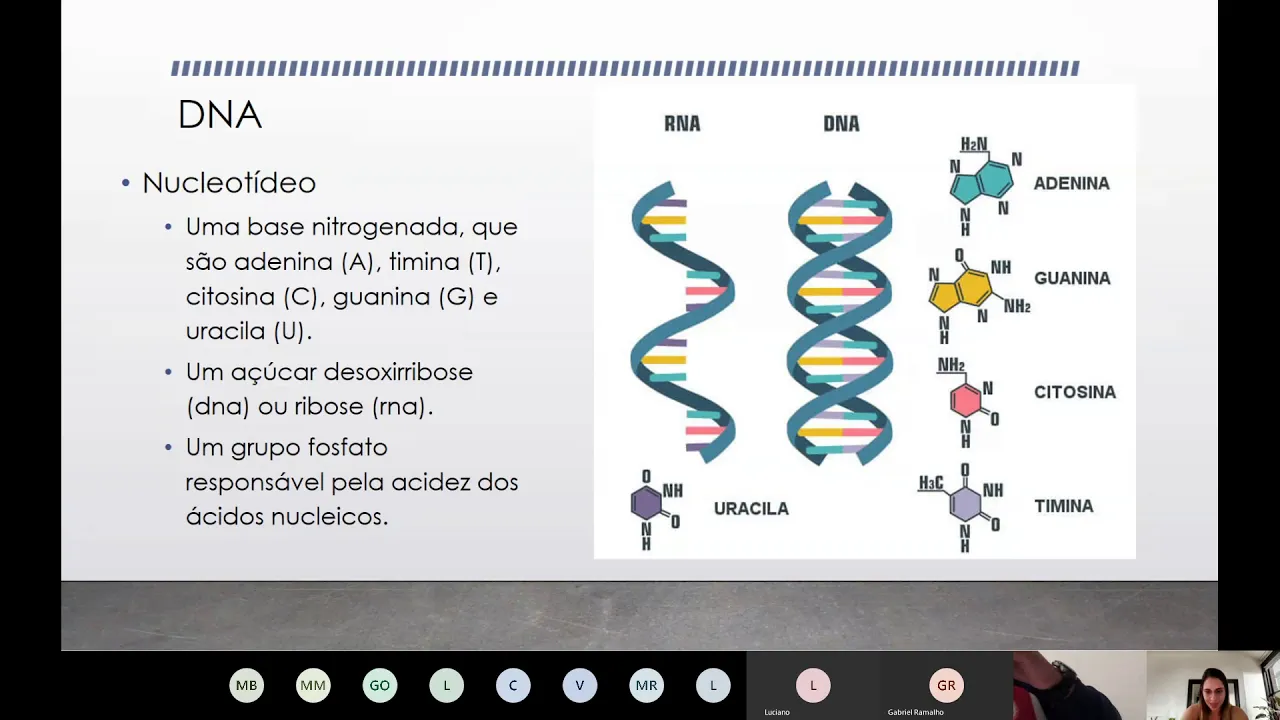
Aula ao vivo - Ciências Moleculares e Celulares
697
Chromosomes carry all genetic information for an organism, with each chromosome containing many genes determining physical and functional characteristics. Chromatin exists in two forms: euchromatin (active, loosely packed) and heterochromatin (inactive, tightly packed). This dynamic packaging allows cells to regulate gene expression. Chromosomes are homologous pairs, with females having XX and males having XY. The Y chromosome is smaller with fewer genes, contributing to sex differences. Alleles are different versions of genes at corresponding positions on homologous chromosomes.
Click to watch →
Deep Dive
The concept of the cell cycle (specifically interphase and mitosis) as a framework for cell division.

Cell cycle and Cell Division Concept of Interphase chapter no#07 Lecture no 01 By Dr.Mehtab Ali.
436
The cell cycle is the series of changes a cell undergoes from the beginning of its life until cell division is completed, consisting of interphase (the non-dividing phase where cells grow and prepare for division) and the mitotic phase (M phase) where actual cell division occurs. Interphase is divided into three stages: G1 phase (cell growth and preparation for DNA replication), S phase (DNA replication), and G2 phase (final preparation for mitosis). During interphase, cells do not divide but undergo growth, DNA replication, and organelle duplication to ensure each daughter cell receives a complete set of genetic material and cellular components.
Click to watch →

Unidad 2 Biología, Segundo Medio
1.9K
The cell cycle is a sequence of events leading to cell growth and division into daughter cells. Interphase consists of three stages: G1 (first growth stage where cell grows to full size and performs biochemical functions), S (synthesis phase where DNA replication occurs in the nucleus), and G2 (second growth stage where cell finishes growing and prepares for mitosis). Mitosis is the major division phase where duplicated DNA is separated into two new nuclei. Mitosis begins with prophase (chromosome condensation and spindle formation), followed by prometaphase (nuclear membrane breakdown and spindle fiber attachment to kinetochores), metaphase (chromosome alignment at the cell equator), anaphase (sister chromatid separation), and telophase (nuclear envelope reformation).
Click to watch →
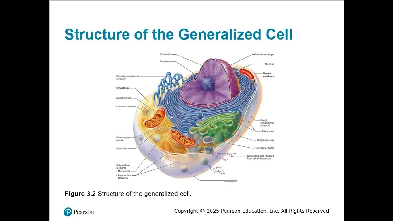
Chapter 03 Cells The Living Units Part 02
111
The cell cycle consists of interphase (cell growth and normal activities) and mitosis (cell division). Interphase is divided into G1 (growth and metabolism, centriole replication begins), S (DNA replication ensuring identical genetic copies), and G2 (preparation for division, centriole replication completes). Mitosis is divided into prophase (chromatin condenses into chromosomes, nuclear envelope breaks apart, mitotic spindle forms), metaphase (chromosomes align at cell equator), anaphase (sister chromatids separate and move to opposite poles), and telophase (chromosomes uncoil, new nuclear membranes form). Cytokinesis (cytoplasm division) begins in late anaphase and completes mitosis.
Click to watch →

BASIC CONCEPTS OF CELL CYCLE & CELL DIVISION || LT,TGT,PGT BIOLOGY || MUST WATCH VIDEO | BY ANJU MAM
157.5K
The cell cycle consists of two main phases: Interphase (non-dividing phase) and Mitotic Phase (dividing phase). Interphase is further divided into G1, S, and G2 phases. The mitotic phase includes mitosis (nuclear division) and cytokinesis (cytoplasmic division). Interphase accounts for approximately 95% of the cell cycle time, while the mitotic phase takes up less than 5%.
Click to watch →
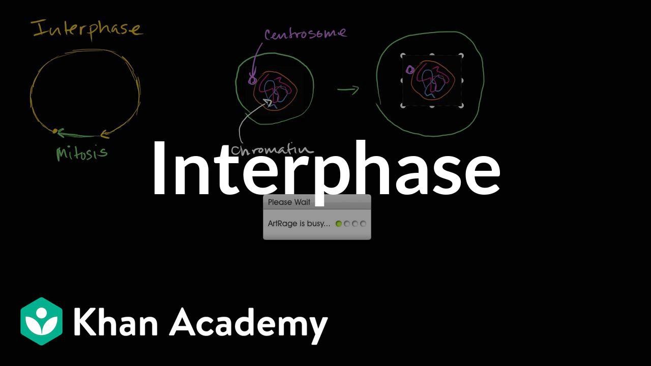
Interphase | Cells | MCAT | Khan Academy
924.4K
The cell cycle consists of two major phases: interphase and mitosis. Cells spend most of their life in interphase, performing normal cellular functions like growth, protein production, and metabolism. Mitosis is a shorter phase where the nucleus divides into two nuclei, enabling cell division. During interphase, chromosomes exist in their relaxed chromatin form, appearing diffuse under a light microscope. The cell grows, takes in nutrients, and prepares for DNA replication. This foundational understanding establishes how cells balance growth and division throughout their lifecycle.
Click to watch →
Prerequisite Knowledge
- Concept 01Basic eukaryotic and prokaryotic cell structure, particularly the function of the nucleus as the cellular control center.
- Concept 02The chemical structure of nucleotides, including the sugar-phosphate backbone and the four nitrogenous bases (adenine, thymine, cytosine, and guanine).
- Concept 03An introductory concept of chromosomes and how they package genetic information.
- Concept 04The concept of the cell cycle (specifically interphase and mitosis) as a framework for cell division.
Subsequent Learning
- Step 01The detailed molecular mechanisms of DNA replication, including the roles of specific enzymes like DNA polymerase, helicase, primase, and ligase on the leading and lagging strands.
- Step 02The processes of transcription and translation, which explain how the genetic code in the DNA is expressed as proteins.
- Step 03The causes and consequences of DNA mutations, and how cells use mismatch repair mechanisms to maintain genomic integrity.
- Step 04Biotechnological applications that utilize DNA replication principles, such as Polymerase Chain Reaction (PCR) and gene editing technologies like CRISPR.
Applause
Musical Flow
Melody Shift
Built Rhythm
Climax Point
Closing Notes
Applause
1:09Playing Section
- 1
Audience responds with applause.
- 2
Moment of recognition or appreciation.
- 3
Transition into musical content continues.
Epigenetics and Environmental Regulation
While traditional educational guides often present the genome as a static, deterministic blueprint where DNA sequence alone dictates organismal traits, modern genomics increasingly emphasizes epigenetics. This field reveals that chemical modifications to DNA and histones—influenced by environmental factors like diet, stress, and toxins—regulate gene expression without altering the underlying genetic sequence. Consequently, critics of strict genetic determinism argue that focusing solely on DNA replication and sequence oversimplifies how traits are inherited and expressed, advocating instead for a systems-biology approach that integrates environmental and epigenetic feedback loops.
Deep Dive
The detailed molecular mechanisms of DNA replication, including the roles of specific enzymes like DNA polymerase, helicase, primase, and ligase on the leading and lagging strands.
أسهل طريقة تفهم بيها تضاعف DNA في 8 دقائق بس🧬|جزء مهم من المحاضرة التالتة في فصل البيولوجيا الجزيئية
40.3K
DNA replication requires coordinated enzymatic action: Primase synthesizes RNA primers providing starting points since DNA polymerase cannot initiate synthesis de novo. DNA polymerase synthesizes new strands using parental templates and performs proofreading to correct errors. The leading strand is synthesized continuously 5' to 3' toward the fork, while the lagging strand is synthesized discontinuously in Okazaki fragments (100-1000 nucleotides) also 5' to 3' but away from the fork. DNA ligase then joins these fragments on the lagging strand. Different enzymes act with specific distributions: helicase on both strands, primase mainly on lagging, polymerase on both, proofreading on both, and ligase exclusively on lagging.
Click to watch →

DNA replication with enzymes /lec1
320
DNA replication is a semi-conservative process where the double helix unwinds to form a replication fork, with helicase breaking hydrogen bonds and topoisomerase II relieving supercoiling; DNA polymerase synthesizes new strands exclusively in the 5' to 3' direction, requiring primase to synthesize RNA primers that provide the necessary 3' hydroxyl group for initiation, resulting in continuous leading strand synthesis and discontinuous lagging strand synthesis through Okazaki fragments that are later joined by DNA ligase.
Click to watch →
7. Replication
85.3K
DNA replication requires coordinated action of multiple enzymes: helicase unwinds the double helix, single-strand binding proteins stabilize separated strands, primase synthesizes RNA primers, DNA polymerase extends primers to synthesize new strands, and ligase joins Okazaki fragments. The leading strand is synthesized continuously, while the lagging strand is made in discontinuous Okazaki fragments due to directional constraints of DNA polymerase.
Click to watch →

Cell Biology | DNA Replication 🧬
1.7M
DNA replication is a semi-conservative, bidirectional process occurring during the S phase of the cell cycle, where the double-stranded DNA unwinds at origins of replication (AT-rich regions), and each parental strand serves as a template for synthesizing a complementary daughter strand; the leading strand is synthesized continuously in the 5' to 3' direction, while the lagging strand is synthesized discontinuously in Okazaki fragments, requiring primase to lay down RNA primers and DNA polymerase III to synthesize DNA, followed by DNA polymerase I to remove primers and ligase to join fragments, with telomeres shortening over time due to incomplete replication at chromosome ends, though telomerase can counteract this in certain cells like stem cells.
Click to watch →
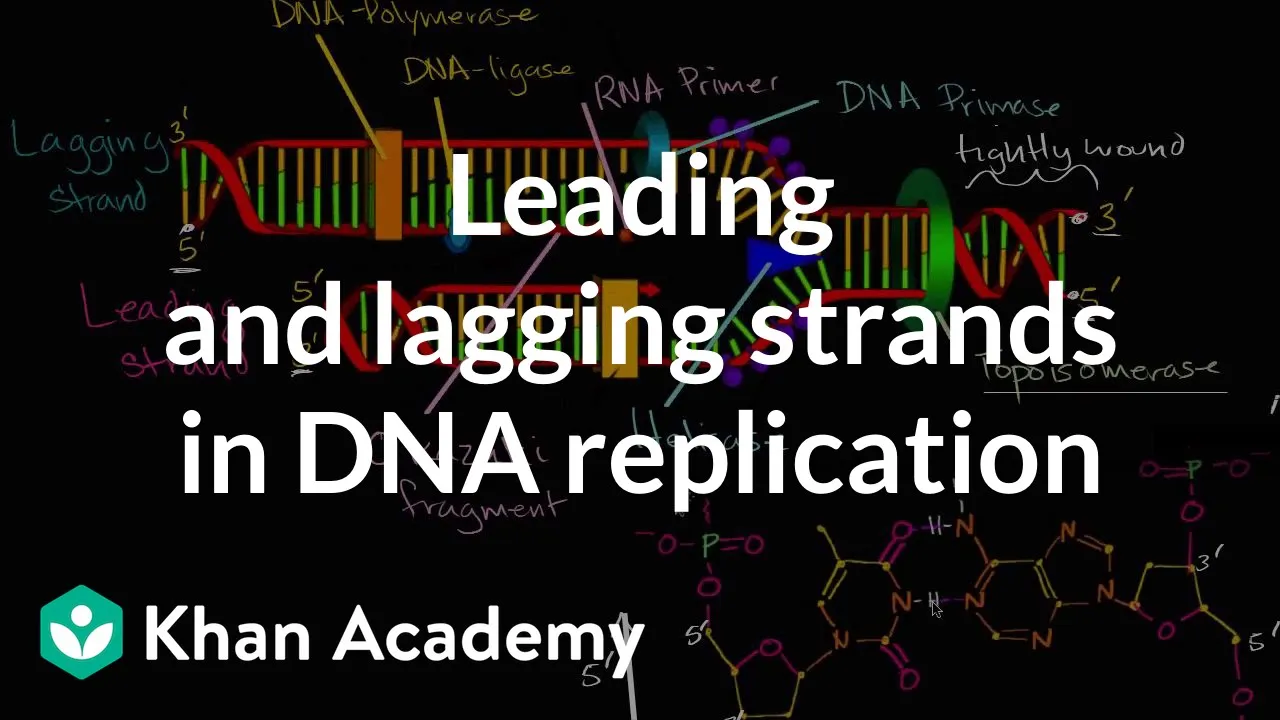
Leading and lagging strands in DNA replication | MCAT | Khan Academy
828.7K
DNA replication requires a coordinated team of enzymes working together. Topoisomerase first unwinds the tightly coiled DNA helix by temporarily breaking the phosphate backbone. Helicase then separates the two strands by breaking hydrogen bonds between complementary bases. DNA primase synthesizes RNA primers (typically 10 nucleotides long) that provide starting points for DNA polymerase. DNA polymerase extends these primers by adding complementary nucleotides at the 3' end, following the template strand in the 5' to 3' direction.
Click to watch →
Deep Dive
The processes of transcription and translation, which explain how the genetic code in the DNA is expressed as proteins.
April 6, 2026
135
Transcription is the process by which genetic information flows from DNA to RNA. Inside the nucleus, RNA polymerase unzips DNA and synthesizes complementary mRNA. The genetic code consists of nucleotide bases (A, U, C, G) that specify amino acid sequences. Translation occurs in the cytoplasm where mRNA is read in codons (groups of three nucleotides) to determine amino acid sequences. The genetic code chart is used to translate codons into corresponding amino acids, such as UAC for Tyrosine and CUG for Leucine.
Click to watch →
Agriculture Foundation Classes | Genetics & Plant Breeding #1 | Introduction | NABARD | IBPS-AFO
98.8K
Transcription is the process by which genetic information in DNA is copied to make RNA. Translation is the process by which RNA is used to synthesize proteins. These two processes are fundamental to gene expression - the flow of genetic information from DNA to RNA to protein. The course will cover the details of how these processes work in the body.
Click to watch →

Tgt biology classes || Tgt pgt biology previous years question papers || Upsessb latest news today
1.5K
Transcription is the process by which DNA is copied to RNA. Translation is the process by which RNA is used to synthesize proteins. These processes are fundamental to gene expression and the flow of genetic information from DNA to protein. The genetic code is nearly universal across all organisms.
Click to watch →

معــسكر الاحياء | اليوم الثاني - الحصة الثانية | تضاعف DNA وتصنيع البروتين - أ حسام عياش 2008
56.9K
Transcription copies genetic information from DNA to RNA in the nucleus. RNA polymerase binds to promoter sequences (TATA box, CAAT box) and synthesizes RNA in the 5' to 3' direction using one DNA strand as template. Primary transcripts undergo processing: 5' capping, polyadenylation, and splicing (removing introns, joining exons) to produce mature mRNA. Translation converts mRNA sequences into polypeptide chains at ribosomes. The genetic code uses 64 codons (3-nucleotide sequences) to specify 20 amino acids, with most amino acids having multiple codons (degeneracy). Three codons (UAA, UAG, UGA) serve as stop signals. Translation occurs in three stages: initiation (ribosome binds mRNA at start codon AUG), elongation (amino acids added one by one via peptide bonds), and termination (stop codons trigger release factors). Each amino acid addition requires 2 GTP molecules.
Click to watch →

مراجعة شرحيات الوراثة | المحاضرة الـ 4.
158.7K
Gene expression involves two main processes: transcription and translation. Transcription occurs in the nucleus where RNA polymerase binds to promoter sequences and synthesizes RNA from a DNA template, creating an RNA molecule complementary to the DNA template. Translation occurs in the cytoplasm at ribosomes where mRNA is decoded to synthesize proteins. Translation involves three stages: initiation (ribosome assembly at start codon AUG), elongation (polypeptide chain growth as amino acids are added via tRNA), and termination (chain release at stop codons UAA, UAG, UGA). The genetic code is nearly universal, with 64 codons specifying 20 amino acids plus stop signals.
Click to watch →
Deep Dive
The causes and consequences of DNA mutations, and how cells use mismatch repair mechanisms to maintain genomic integrity.

DNA Damage and Repair
23.8K
DNA is precious and fragile, requiring cells to evolve repair mechanisms against radiation, chemicals, and other hazards. The human body contains 10^14 cells, each sustaining ~100,000 DNA damage events daily. DNA damage occurs through intrinsic causes (replication errors, hydrolytic attack, reactive oxygen species) and extrinsic causes (radiation, mutagenic chemicals). Two main consequences exist: mutations introduced when replication errors escape correction, and structural alterations that stall transcription. The fidelity of DNA replication is 10^10, achieved through DNA polymerase proofreading (10^7) and mismatch repair (10^10). In E. coli, MutS recognizes mismatches, MutL recruits MutH, which nicks the unmethylated new strand. Hemimethylation (Dam methylase) distinguishes parental from new strands. Humans have homologous proteins but lack MutH, using PCNA for strand discrimination. Mutations in MSH2 cause familial colon cancer.
Click to watch →

MISMATCH REPAIR Mechanism || DNA damage &repair || Molecular biology
5.9K
DNA damage occurs due to radiation, chemicals, and mutations. Cells have multiple repair mechanisms including DNA proofreading and mismatch repair. Mismatch repair corrects errors that proofreading cannot fix. In normal DNA, A pairs with T (2 hydrogen bonds) and G pairs with C (3 hydrogen bonds). When incorrect base pairing occurs, the repair system identifies and corrects these mismatches to maintain genetic integrity.
Click to watch →
BIOL2416 Chapter 13 Gene Mutation and DNA Repair
9.3K
Cells employ multiple overlapping repair pathways to maintain genomic integrity: proofreading by DNA polymerase during replication; mismatch repair using methylation to distinguish old from new strands; direct repair enzymes restoring damaged nucleotides (e.g., O6-methylguanine methyltransferase); base excision repair removing damaged bases; nucleotide excision repair removing bulky lesions like pyrimidine dimers. These sophisticated mechanisms work together to correct errors arising from spontaneous mutations and environmental damage, ensuring faithful transmission of genetic information across cell divisions.
Click to watch →

Mismatch repair mechanism | DNA repair | Hindi | Notes | Part -4
19.2K
DNA damage refers to chemical and physical changes in DNA structure, including strand breaks and base alterations. DNA repair is essential because unrepaired damage can cause serious diseases and be inherited. Mismatch repair corrects errors during DNA replication by identifying mismatched base pairs. The process involves MutS recognizing mismatches, MutL coordinating repair, MutH performing strand discrimination on hemimethylated DNA, excision of the incorrect segment, DNA polymerase synthesizing the correct sequence, and DNA ligase sealing the backbone. This systematic repair mechanism maintains genomic stability and prevents mutations from being passed to daughter cells.
Click to watch →
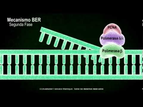
Mecanismo de reparación
39.5K
Cells employ multiple DNA repair mechanisms to maintain genomic integrity: Base Excision Repair (BER) corrects single-strand breaks and base modifications through glycosylases, AP endonucleases, and DNA polymerases; Nucleotide Excision Repair (NER) removes bulky DNA lesions like thymine dimers via XPC, TFIIH, and XPA proteins; Mismatch Repair (MMR) corrects replication errors using MSH proteins and exonucleases; Non-Homologous End Joining (NHEJ) repairs double-strand breaks by directly ligating broken ends; and Homologous Recombination (HR) uses sister chromatids as templates for accurate repair during S/G2 phases.
Click to watch →
Deep Dive
Biotechnological applications that utilize DNA replication principles, such as Polymerase Chain Reaction (PCR) and gene editing technologies like CRISPR.

الوراثة | منهج 2025 | المحاضرة 5 شرحيات مقدمة الوراثة
108.5K
DNA replication (التضاعف) allows genetic material to copy itself both naturally inside cells and artificially outside cells. PCR (Polymerase Chain Reaction) is a laboratory technique for amplifying DNA using DNA polymerase enzyme. The process involves repeated heating and cooling cycles that separate DNA strands and synthesize new complementary strands, exponentially increasing target DNA. This technique is essential for genetic analysis, forensics, and biotechnology applications requiring large amounts of specific DNA sequences.
Click to watch →

AULÃO ENEM 2018 | Biologia | Prof. Paulo Jubilut
358.1K
DNA replication is semi-conservative, meaning each new DNA molecule contains one original strand and one newly synthesized strand. The process begins during the S phase of interphase when the DNA double helix unwinds. Each separated strand serves as a template for building a complementary strand. PCR (Polymerase Chain Reaction) is a laboratory technique that amplifies specific DNA segments through repeated heating and cooling cycles, allowing exponential DNA production. This technique is fundamental in biotechnology for DNA analysis and cloning.
Click to watch →
NEET: Biotechnology - L5 | Live Daily 2.0 | Unacademy NEET | Seep Pahuja
31.9K
PCR (Polymerase Chain Reaction) amplifies specific DNA segments exponentially. The process involves thermal cycling: (1) Denaturation at high temperature separates double-stranded DNA into single strands, (2) Annealing allows specific primers to bind to complementary sequences, (3) Extension uses DNA polymerase to synthesize new strands. Each cycle doubles DNA molecules, producing billions of copies from a single template. PCR, invented by Kary Mullis (Nobel Prize 1993), revolutionized biotechnology by enabling rapid DNA amplification essential for genetic research, diagnostics (including COVID-19 testing), and molecular biology applications.
Click to watch →

UP बोर्ड 2026 Class 12 Biology महा REVISION🔥 CH 7 से CH 13 के Important Questions | 23 फरवरी पेपर✅
9.1K
Polymerase Chain Reaction (PCR), developed by Kary Mullis in 1985, amplifies specific DNA sequences through repeated heating and cooling cycles using heat-stable DNA polymerase from Thermus aquaticus. PCR involves denaturation, annealing, and extension steps. Biotechnology applications include producing insulin from genetically modified bacteria, creating Bt cotton resistant to cotton bollworms, and developing Golden Rice with beta-carotene for Vitamin A deficiency. RNA interference silences specific genes for pest resistance. Transgenic plants produce pharmaceuticals like heparin. GMOs offer benefits including shorter growth periods, pest resistance, and improved nutrition, though bioethics and bio-piracy concerns exist regarding genetic resource ownership.
Click to watch →

كلاميات الوراثة | المراجعة المركزة قبل الامتحان
258.5K
DNA can replicate in two ways: naturally within cells through cellular processes, and artificially through PCR (Polymerase Chain Reaction). PCR is an in vitro technique that uses the enzyme DNA polymerase to amplify specific DNA sequences. This capability allows scientists to produce millions of copies of a DNA segment from a small initial sample, which is essential for genetic analysis and biotechnology applications.
Click to watch →
Applause
Musical Flow
Melody Shift
Built Rhythm
Climax Point
Closing Notes
Applause
1:09- 1
Audience responds with applause.
- 2
Moment of recognition or appreciation.
- 3
Transition into musical content continues.
Epigenetics and Environmental Regulation
While traditional educational guides often present the genome as a static, deterministic blueprint where DNA sequence alone dictates organismal traits, modern genomics increasingly emphasizes epigenetics. This field reveals that chemical modifications to DNA and histones—influenced by environmental factors like diet, stress, and toxins—regulate gene expression without altering the underlying genetic sequence. Consequently, critics of strict genetic determinism argue that focusing solely on DNA replication and sequence oversimplifies how traits are inherited and expressed, advocating instead for a systems-biology approach that integrates environmental and epigenetic feedback loops.
Basic eukaryotic and prokaryotic cell structure, particularly the function of the nucleus as the cellular control center.
The chemical structure of nucleotides, including the sugar-phosphate backbone and the four nitrogenous bases (adenine, thymine, cytosine, and guanine).
An introductory concept of chromosomes and how they package genetic information.
The concept of the cell cycle (specifically interphase and mitosis) as a framework for cell division.
The detailed molecular mechanisms of DNA replication, including the roles of specific enzymes like DNA polymerase, helicase, primase, and ligase on the leading and lagging strands.
The processes of transcription and translation, which explain how the genetic code in the DNA is expressed as proteins.
The causes and consequences of DNA mutations, and how cells use mismatch repair mechanisms to maintain genomic integrity.
Biotechnological applications that utilize DNA replication principles, such as Polymerase Chain Reaction (PCR) and gene editing technologies like CRISPR.
1.5K views12likes5:03@0523HannahOriginal Release: 2019-02-04
[Music] [Music] [Music] [Applause] [Music] [Music] [Music] [Music] [Music] [Music] [Applause] [Music] [Music] you you
Up Next

8:11
Nanodrop 2000 Tutorial: Measuring DNA/RNA Concentration & Purity
@innovative_learner
32.1K views•2020-11-13

12:54
Triumph of Orthodoxy Icon: Byzantine Art & History Explained
@BenCallan
2.1K views•2024-08-06

9:49
FastAPI vs Flask vs Django: Choosing the Right Python Web Framework
@TechWithTim
302.5K views•2024-05-26

1:42
Game of Thrones Opening Credits: A Cinematic Analysis
@gameofthrones
46.3M views•2011-04-18
Related Study Plans & Knowledge Roadmaps
Structured learning paths in General & Interdisciplinary Studies
general-studies
Elevator Engineering: History, Physics & Cities
01From Hoists to Safety Brakes: The Dawn of Vertical Lift
02Hydraulic vs. Traction: The Engineering Physics of Elevators
03Modern Speed, Safety, and Algorithms
+ 2 more learning modules
12h5 modules
Explore Roadmap
general-studies
Rubber: Material Science, History & Geopolitics
01Origins of Natural Rubber & Mesoamerican History
02The Chemistry of Natural Rubber & Vulcanization
03The Geopolitics of the Rubber Boom & Colonization
+ 2 more learning modules
12h5 modules
Explore Roadmap
general-studies
Containerization: Evolution, Logistics & Trade
01Ancient Trade & Bulk Cargo: Amphorae to Barrels
02The Industrial Revolution & Cardboard Packaging
03The Containerization Revolution: Malcom McLean
+ 2 more learning modules
12h5 modules
Explore Roadmap
general-studies
Textile Tech: Evolution, Labor & Global Impact
01Pre-Industrial Textiles: Handloom & Cottage Industry
02Mechanization: The Spinning Jenny to the Power Loom
03The Factory System & Global Labor Transformation
+ 2 more learning modules
10h5 modules
Explore Roadmap