

























Forensic anthropologists estimate age, sex, and stature from skeletal remains using specific anatomical indicators: sex is determined primarily through the pelvis (sub-pubic angle, sciatic notch, birth canal) and skull (robusticity, brow ridge, chin shape, canine teeth), while age estimation relies on dental development patterns, epiphyseal fusion, osteon analysis, and symphyseal face changes; stature is calculated using long bone measurements applied to population-specific formulas. These methods form the foundation for identifying unknown decedents.
Forensic Anthropology: Estimating Age, Sex, and Stature
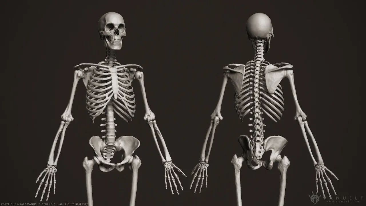
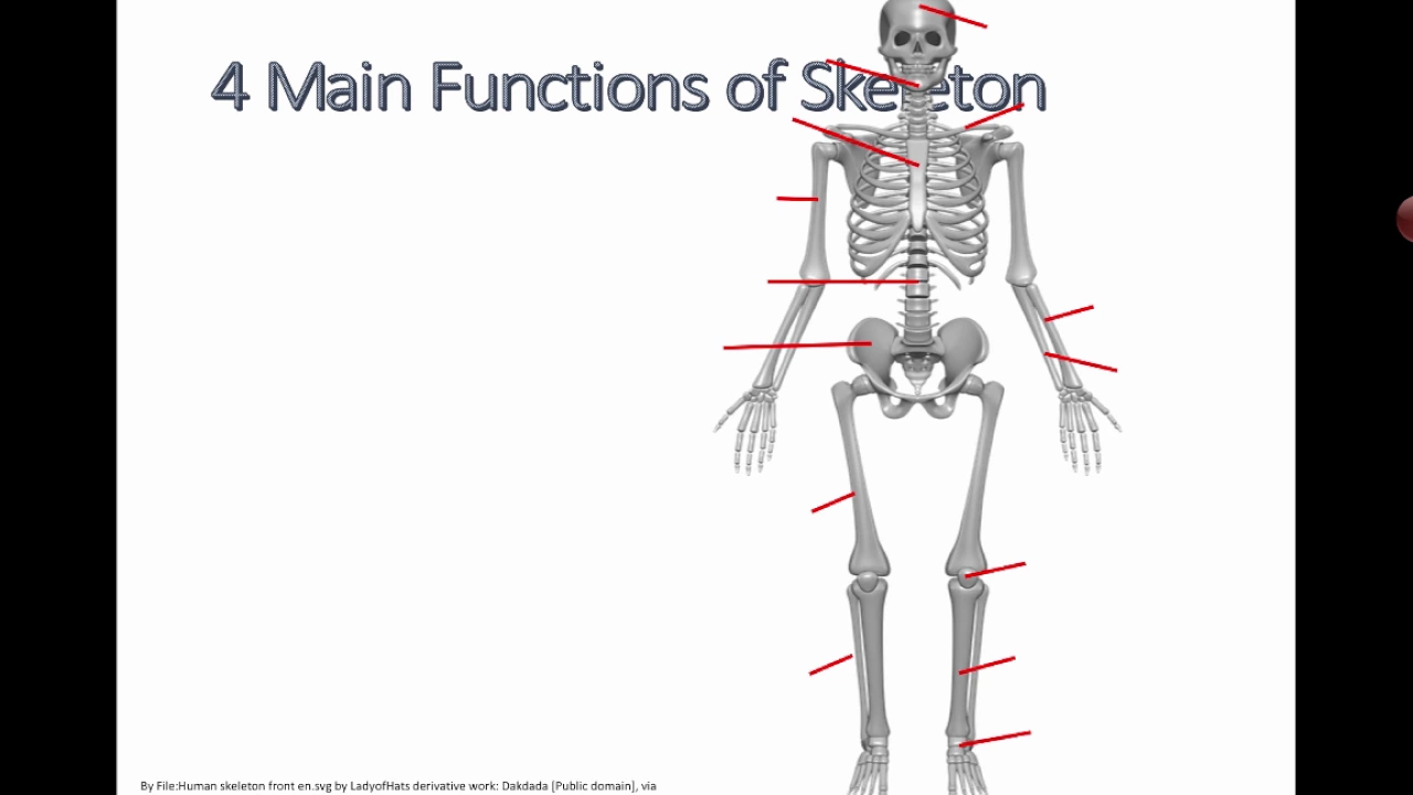
Added:Basic human osteology, including the names, locations, and anatomy of the major bones in the human skeleton.
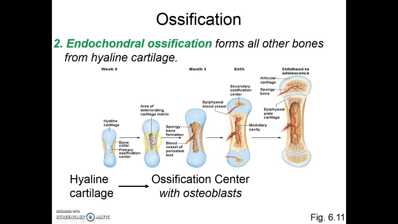
The concept of biological growth and development, specifically how bones ossify, grow, and fuse (epiphyseal fusion) from childhood to adulthood.
An understanding of sexual dimorphism, particularly how evolutionary and functional differences manifest in male and female skeletal anatomy.
Basic principles of scientific measurement (anthropometry) and the statistical variation inherent in human populations.
Estimating ancestral background (biogeographical ancestry) using craniometrics and macromorphoscopic traits to complete the biological profile.
Skeletal pathology and trauma analysis, distinguishing between antemortem, perimortem, and postmortem bone damage.
Forensic taphonomy, which explores how environmental factors, decomposition, and scavenging affect skeletal remains after death.
Advanced identification techniques, including forensic odontology (dental identification) and extracting DNA from skeletal remains.
2.2K views34likes39:56Original Release: 2020-10-01
hi folks welcome to the last unit unit three we're going to be talking it's all about identification how do we go about ideating a body now we've gone through the process of understanding how we find the bodies and how we understand the skeletal system and what we're looking at then we went into trauma and how to understand some of the traumas that we're going to look at now we're going to try to put a name on the deceit now we're looking at the individual as an individual and trying to figure out who they may have been the first step to doing that is determining their age their sex and their stature notice that we said sex not gender gender is a cultural construct it is not biologically determined gender is whatever the individual decides to choose sex on the other hand is fundamentally you are male or female there is very little ambiguity there is no admixture there are some cases but very few of somebody who is intersex being like the double x uh y chromosomal abnormalities or um incomplete eggs or hormone therapy these things will have an effect but they won't change it that much and to be honest it'll just add to the ambiguity but not really foil everything so the skull is the second most indicative anatomical feature of sex in the human body the pelvis is by far the best all of these ideas are based on something called sexual dimorphism dye means to morph means shape so sexual to shape-ism the understanding that if there are two sexes of a given species they will differ in shape and or size the most dramatic and the reason that i have an image of gorillas here are gorillas gorilla males are nearly twice the size of the average gorilla female sexual dimorphism follows basic principles that is in our species males are usually slightly larger not the dramatic thing that we see in gorillas we only see males being about on average 10 percent larger than our females however this isn't a guarantee is it we see larger women all the time we see women who are more robust than men sometimes depending on what they do but those things can actually help us determine what we're looking at they are still skeletally going to look female especially in their pelvis if they are female even if they're very robust or very large childbirth is what makes the female pelvis very unique it is geared toward that it's got a birth canal that males do not have so it tends to be wider and broader males on the other hand tend to have much more robusticity that is higher muscle content how male males muscles are usually uh more densely packed than females and they usually develop much quicker but there's more than that it is driven by hormones so at puberty males develop larger muscle origin and insertion points on the bone than do females this has nothing to do really with building that muscle up it has everything to do with hormones saying they will therefore this is an adaptation that happens so males tend to be what we call more robust than females but not always and we'll see how there's some abnormalities there too sexual dimorphism as i mentioned is hormonally controlled it's evident in adults not in children we cannot perceive the difference in prepubescent children between male and female it just does not show up it can vary within a population and between populations so you can have very large women or very small men in a given population or you can have the expectation is going to be more divergent in certain populations than in others there are certain populations where everybody tends to be smaller and certain populations where everybody tends to be larger so we'll see those uh differences happen but that has a lot to do with ancestry so you're going to see throughout this entire unit we cannot do one without knowing some of the other so we're going to talk about ancestry and we're going to talk about age and we're going to talk about other aspects and all of these things have to be taken into account before you can answer any of them so as we mentioned before the pelvis is the most clear anatomical portion of the body that tells us male from female females tend to have larger broader wider pelvis than do males for example as you see here in this image the sub-pubic angle what we're talking about here is this this angle here in males it would be much more acute it'll go more like that in females it's much broader a wider angle same thing over here in the sciatic notch we're going to see a much wider sciatic notch than what we see in males this is the front here we're facing this way is the front so this is toward the back side kind of in the hip area in fact this is the acetabulum where the femur meets up with the pelvis in order to make the hip so we're going to see a wider here in the front this is the front area here we're going to see the wider sciatic notch the sciatic notches are right back here and behind and we're going to see obviously a wider birth canal this includes this here is called the sacrum the sacrum will tend to not curve in as much on females as it does on males so on males everything is narrower the this area here the birth canal literally is narrower this angle is narrower the sciatic notch is narrower on males than it is on females here is a very male pelvis now you'll see the blatant difference here look at how narrow that sub-pubic angle is look at how narrow the greater sciatic notch is and if you look in this front image here look at how tiny this canal is there's no baby going to fit through that does that make sense males are designed for bipedal locomotion therefore everything's gotten very narrow females are designed for both by fetal locomotion and childbirth so you have this compromise going on the pelvic girdle is usually indicated by single bones as you noticed probably in that last picture we have the os coxa on the left the oscoxa on the right and the sacrum down the middle it's three fundamental bones we're often only given one or the other so the key here is to look at that sciatic notch which is here if it is generally three centimeters or narrower it is usually male okay over here we're looking at the sub-pubic angle and here we're going to take the angle instead of having them both coming together we get this greater angle like this now we only have one to go by so we've got to measure straight down this is called the pubic symphysis it's where the two halves of the pelvis come together in the front and you go straight down here now you're seeing i am now creating an angle here triangle then i can measure the width of this angle okay skulls are the second best way of determining male from female fundamentally i always say looking at a skull is very similar to looking at a black and white photograph of a human with all their hair shaved can you still tell the difference between male and female yes you can so what we're looking at here is overall size and that weird word robusticity that robustness of the skull the larger and more robust skulls tend to be the more male super orbital taurus also known as the sot are that's really your brow ridge the super orbital margin which is the margin in the upper eye orbit is the eye eye socket so super orbital taurus is the brow ridge super orbital margin is the margin right between the roof of the eye and curving around in the eye sock it's kind of hard to explain but i'll show you pictures in a minute the chin males tend to have a more squared off chin females tend to have a rounded or pointy chin teeth are usually larger in males particularly the canine teeth we'll see that in a minute the canines those are your vampire teeth those are the teeth over here in the corners of your mouth as you smile you can see them quite clearly muscle ridges are usually larger in males even in those areas where the muscle doesn't build up for example if you have big muscle ridges on your cheeks is that because you've been smiling a lot not really it is again going back to that hormonal thing when the androgynous hormones kick in during puberty that's when we see these muscle attachment points start to get larger these ridges the external occipital protuberance that's on the back side of the skull is often much more prominent in males don't worry i'm going to show you pictures of all these things the mandible that's your jaw bone is usually much more large and blunt it's smaller and sharper in females as we talked about with the chin also there's a little thing going on with the angle of the mandible which we'll talk about in a few minutes the mastoid process those are the processes that you can feel those little lumps that are right behind your earlobe if you feel your earlobe and go right behind it you'll feel a lump coming out of the base of your skull that connects up to a muscle that strap muscle actually helps you turn your head generally speaking those are larger in nails because again it's a muscle attachment point cranial bossing are usually much more prominent females again i'll show you what those are in a minute forehead males tend to have a more sloping frontal that is their forehead tends to slope back from the brow ridge more so than females females have to tend to have a much more upright kind of a a wider taller forehead here we are looking at a male and female jaw these these don't look that much different in this image and i apologize for it but there's an attempt basically speaking the male jaws tend to be wider they tend to have a more pronounced eversion going on right here this uh angle here is called the gonion the gonial angle and it tends to be everted it looks like somebody pinched and pulled outward on either side that's because that is again a muscle attachment point for the masseter muscle which helps you chew females tend to have as much a smaller less pronounced eversion here you can see this bumps out just a little bit but when we see real pictures it'll be much more clear the teeth tend to be smaller this angle and i'll show you it from the side tends to be greater in females and tends to be more well almost 90 degrees and nails this angle here and then female chins tend to be a little pointy and a little narrower from edge to edge really what we're doing is coming straight down from the canine teeth and then look at the width here whereas males coming straight down from the canine teeth is much wider and squared off usually sexual characteristics in the rest of the skull here's our super orbital taurus again now what we're looking at is that brow ridge this is that bump here the super orbital margin is right along here and it actually feeds into this taurus the toroid shape here is this big rounded almost like if you're making if you're working with clay and you make a little worm of clay that's kind of if you lay it across a male's brow that's sort of what you're looking at here so it tends to be this margin tends to be smoother and more rounded in males in females it tends to be much sharper and i'll show you some images of that the muscle ridges like up here down here and the external occipital protuberance here the mastoid process all these are muscle attachment points and they tend to be much more robust in males chin tends to be squared off that gonion angle you can see it's now bulging a little bit and you can see this angle this is the angle i was talking about in the last slide see this nearly 90 degrees in a male in a female it's going to be opened up a wider angle in females same images side by side from a male to female here you can see clearly the male skull which is the one on the left is a larger more robust skull you can see all those muscle attachments are a little bit larger a little bit more pronounced also you see how the male skull tends to sort of slope backwards the forehead from the brow ridge to the kind of middle of the skull whereas the females tends to be a much more vertical forehead nearly no brow ridge usually there's a little bit going on here but not much not compared to this bump out that we see in the males and we see that there's a lot of this smoothness going on in females the mastoid processes tend to be smaller and incorrectly drawn here this female jaw would usually tend to be a little bit more wider angle than what we see in the nails very tight angle we'll see all this when we see actual pictures in just a minute here we have a comparison between a male typical male and typical female skull now granted they cheated it a little bit by tipping the female skull down a little bit of what we would call frankfurt horizontal or the frankfurt plane but nonetheless that's done for a reason and i'll explain that in just a second first of all we're going to look at the male skull first of all notice down here these are those gonially versions that i'm talking about you see how these are bumped out almost like you grabbed this corner of this of the jaw from the back side and pulled it out to the side this is a muscle attachment point for the masseter muscle that goes from here up to here and helps with chewing up here we've got a much larger more ghost brow ridge we've got that sloping skull we've got these really blatant temporal lines which is another muscle attachment point we would have a rounded supraorbital margin in here we again have muscle attachment points that are relatively pronounced squared off chin and very large canine teeth here on the female side we have this rounded chin much smaller it's not as rugose all of these little muscle attachment points we see very little gonial eversion back here then we have a much sharper orbital margin nearly no brow ridge furthermore you see these little bumps here here and here those are those cranial bossing that i was talking about before it looks almost like if you had taken a clay pot and you used your thumb and pushed out from inside they're just little bumps okay this is actually a sexually selective aspect of the female skull we'll talk about that much later on maybe furthermore we'll see the same bossing on the back side of the skull as well it tends to show up in female skulls not in male skulls and it tends to be kind of prominent in some female foreheads so that's where you look for it but that's the best image i have of a really good cranial bossing okay females also females female females also tend to look very gracile this means the opposite of a robust it's from the same term as graceful same root word robusticity means big rugose whereas grass isle means slight and small so the idea here is if you look at these two skulls side by side you could pretty much clearly figure out okay the one on the left is most likely male the one on the right is probably female age estimation now we're getting into very difficult territory in juveniles almost the opposite of sex determination in juveniles this is very easy it becomes much harder the older people get we're going to see that the age estimations can go from within months with a newborn or a young child we can get it down to a matter of months as they get older we can get it into a matter of years in juveniles and young adults then we start slipping into five-year categories and then in old age like we're talking from 50 to about 80 to 90 100 years old plus we start going into decades because it's a lot less clear as to what we're working with i'll walk you through why those things are and how we determine age when we're looking at a skeleton estimating age of a skull forensic science needs to work with law enforcement so we have to give them a reasonable age grouping after dental eruption completes age is very difficult to estimate using only the skull but there's a lot of other things that we can use to determine age the skull is very very useful especially early on the skull can tell us a lot about the age of an individual the cranium of an infant is divided very clearly it tends to have more sutures the sutures have these holes in them which are called fontanelles that's where the sutures come together this is a growth area there's no bone here so if if the hole there is a fontanelle you know you're dealing with an infant skull okay moving on the neurocranium is much larger in relation to the face and mandible between ages 0 to 5. in other words their brain case is huge and they've got tiny little faces that's because that brain is growing very fast that's also the reason all these sutures are considered what we call open now after they get older we start seeing those sutures fusing together and going away this is where we start getting into the decades of difference it's tough to say studying a skull that's six to 21 years of age we have a lot that we can work on but the key is dental eruption it's the most accurate and the skull is usually about full grown but the face goes through a lot of changes so we'll talk about these things in piece by piece first of all let's look at the sutures at around age 20 the sutures are still what we would consider open now this is where the different plates of the skull are growing together and you can think of a suture that's these squiggly lines on the top of the skull that we're looking at you can think of these sutures as fingers that are kind of interlaced and they hold together that way we met when we were looking at we met these things in trauma when we were looking at explosive trauma that would pop these things apart as the individual gets much older like into their 70s 80s 90s we start to see these sutures completely obliterate here you can see over the age 70 person this suture this is called the sagittal suture and the lambda suture are almost completely obliterated they're going away these bones have now fused together these sutures will will obliterate at different rates and we'll talk a little bit more about that later on newborns with teeth are the easiest to determine first of all they have no teeth their two sets of teeth buds are still within the jaw in what we call the crypts so you can actually help to determine whether you're looking at an infant jaw or a non-infant jaw by shaking it because those little teeth will rattle on the inside um ages one to three primary teeth these are the deciduous also known as milk or baby teeth these are all coming out now by the age of three they've got them all in by the age of four five and six they start to lose those teeth and replace them with the permanent teeth all of the adult teeth the permanent teeth will be in by age 10.
now these dates and times have changed a little bit over recent years it's mostly because of prenatal and natal healthcare that is the babies are getting a lot of nutrition and they're growing very well very quickly so we're seeing these tooth eruptions moving forward just a little bit i'll give you a really good example as we get into young adult the jaw starts to get larger and thicker the jaw shows a complete set of all the permanent teeth that is 32 teeth the third molar is apparent and it used to be called the wisdom tooth also known as the 21 year molar not anymore nowadays most of you that are watching this probably got your third molar or your wisdom teeth probably came out closer to age 17 16. you can see this is a slightly older slide it's only a few years old but by now it's already changed old age now we start to see tooth erosion and tooth loss although we know people can lose all their teeth at a very young age it happens it's more rare but it happens we also tend to see more and more to reconstruction especially these days so you'll see a lot of crowns a lot of fillings those kinds of things start to happen more and more in old age to the eruption schedule now do not write this down because it has changed notice here we've got the central incisor on the lower mandible this is i'm looking over here on the left hand chart central incisor down here these are the baby teeth on the mandible all right the lower jaw central size used to show up around six to ten months not anymore you guys now it's showing up at three to six months most babies will have those two lower teeth and quickly followed by the two central upper incisor teeth those are your bugs bunny teeth these are the ones that come in first these are usually showing up eight to 12 months not anymore you guys now we're seeing them in six to eight months fairly regularly so everything is sped up again that's because of this neonatal uh healthcare and and nutrition uh then we go into the adult teeth the adult teeth eruption is again a little bit more common a little bit more scheduled normally we see the lower and upper third molar usually popping out right about 17 to 21 years of age oftentimes though it's happening a little bit earlier now so it's going to start to speed up again as healthcare and everything else gets better post cranial age estimation this is the rest of the body what we look at here are epiphyseal meaning at the end of the ephesium meaning the end of the long bones the sutures are starting to close so what we're looking at is these growth plates we met these before but you see this line going along here these two bones are still separated they're starting to fuse this is a young adult but get it any earlier and you can actually separate it quite easily or there may actually be a huge distance like a child maybe age six or three to six years old would have this fusion plate stopping here and this bit here so all of this would be cartilage in life in a younger person this is problematic sometimes when we're looking at the baby especially neonate or infant bones they can be easily mistaken for other animal bones which we talked about in the very first unit round about the age of 24 to 26 somewhere in there the clavicle that's your collar bone is the last bone to fuse so some of you may not actually have full fusion yet now osteon growth is different we can look at osteons this is taking a super thin slice of a bone putting it under a microscope and looking at the osteons these are the bone building cells this is what builds up a bone and what we're actually looking for are complete osteons do you see these little circles they look like a bunch of straws and on but we see spaces between them we see incomplete osteons and we see completed round osteons up against other completed round osteons as we get older those little holes all these little voids fill in and now we see incomplete osteons it's hard to see this is a little bit blurry but you get the point what's happening is the bone has rebuilt and rebuilt and rebuilt and rebuilt so when the older individual again around 70 years old their bone looks older than the young bone it also has a little bit less cartilage in it these voids in death would have been in life filled with cartilage or other protein components they're mostly they're still there in some places but you see there are a lot less of them in the older individual host cranial estimation can also use that symphyseal face the pubic symphysis remember we met this earlier when we were talking about aerosex estimation the pubic symphysis looked at and on has these very peculiar markings and they go from phase one to phase six here you see phase one is the younger side phase six is the older side if you look here these have these ridges they almost look like an elephant's tooth that's just my own craziness but all the way over here they've lost those ridges they've started to build up this ridge around the outside starting to look more like a volcano of caldera this is extreme old aids we're looking here at an old person like a man in his 70s 80s 90s over on this side we're looking at a very young person probably in their teens we can do the same thing with sternal rib ends you know where your sternum is that's your breast bone right in the middle these are the rib ends that lead up to the cartilage that then meets up to the sternum right to complete the rib going across so it starts in your spine swings around creates your rib cage and then connects up to that cartilage well over time that cartilage will start to ossify will start to become bone so what we will start to see is these nice clean ribbons start to build up these ridges around the outside very similar to what we saw in the pubic synthesis and these rib ends get longer and more jagged and they get a little bit eroded and in some cases very long this all was cartilage when this person was young and as they get older this started to ossify started to become bone it's not going to suffocate don't get confused the body will do what it can get away with but it won't do anything more so what it actually does is stabilize the rib a little bit but it also makes it a little bit more brittle so it's a lot going on stature estimation is our last station here in this video what we're talking about is understanding how large somebody was stature has in part has to do with height but it also has to do with how big they were how wide they were and i'm not talking about fat although we can determine if somebody was severely overweight in some cases but we're not going to go into that much detail here we're just going to stick with the basics so how do we do it well fundamentally we take the long bones the bones that are associated with height so what are those the long bones in the legs not the arms so much although we can do it if we need to with the arms so what we look at is the maximum length of the long bones are then measured compared to data of known individuals in fact known populations we put it into a formula depending on what population this individual belongs to meaning are they asiatic are they sub-saharan african are they a mixture of that how do we do that then what formula we decide to put it into this was invented by mildred trotter and golden glasser way back in the 50s to 70s um it has since gone through a lot of reiteration and i will show you the formulae in a minute but we're not going to be tested on we'll never have to look at it that much further first of all which bones do we use we use the long bones usually in the legs because those have to do with stature with height right we do this in millimeters because we want this to be universal we often use what we call osteometric boards that is fundamentally too two bookends with a ruler in the middle now if you take anthro 119 that is the forensics lab we'll use some of these and do some of these osteometric estimations of how tall the individual used to own that bone was now you'll notice this here this is not a femur in here is it that is a humerus that's an upper arm bone if we don't have a complete femur or if we don't have a femur at all or the tibia then we'll go to the arm bones there's other formulae for that so we'll use whatever we have we'll use the best we can get okay there are other ways of doing this but we're sticking with this we correlate this measurement to a stature data board these tables and charts distinguish by sex and ancestry okay the osteometric board here in the middle like i said it's literally just a pair of book ends with a ruler so we'll stick that bone in there measure its full length farthest point to farthest point take that measurement throw it into this this is a typical example using feet and femur length so we would take these things you would put these things in and then do these clarifications you'll notice there is also a plus and minus this is showing us the mean and this is showing us the average mean so we'll have to add that in depending on what population this person is taken from so it'll be roughly this many centimeters roughly this measurement in imperial which is what we use five foot eight but we have to add a plus or minus in there so that means this person could be as tall as maybe um i don't know i'd go as far as saying from 5 10 to 5 six somewhere in that range but five eight is going to be the average there does that make sense to you guys now presumptive versus positive id this is something new uh forensics students get hung up on all the time presumptive id is what you presume this person is that can go in with a lot of different things let me give you some examples driver slices passport boarding pass jewelry or clothing and the location in other words if they're on a specific airplane seat like 29e is that presumptive id or positive id it's presumptive because we don't know for sure if that's the individual let me tell you why i bet you most people listening to my voice right now have had a fake idea to get into a bar or something like that am i right that's a fake driver's license it may not belong to you the one this person was found with might not actually be theirs that includes passports and boarding passes of course recognizable clothing come on how many people here have lent a piece of clothing or jewelry to another human being right location of remains airplane seat oh my goodness i bet you everybody here who's been on an airplane has switched seats with someone else at some time or another because you ended up with an aisle seat you prefer the window or vice versa or you might switch throughout the flight so all of these are presumptive it gives us a start but it does not give us a positive id however presumptive id is very valuable because you can get a positive id from things like dental records ah but there's not a single place to find all the dental records of everybody in the world you have to know who you think this person is and then contact their dentist and say hey can i look at some of the x-rays you took positive id is absolute proof of identity dental records anamortum and postmortem x-rays that match that is if this person had a broken arm and they went in they would have an x-ray of that broken arm when he broke his arm in in eighth grade football right that's an antimortem x-ray okay or radiograph post-mortem we would look at that arm and say hey look there's a break there it's an old heeled fracture boom positive id bony characteristics that are unique that is something interesting in the bone for example you can have frontal sinuses which are kind of air spaces within the frontal bone right behind the forehead that will show up in certain um x-rays anti-mortum and you can match those with what you're looking at post-mortem and of course our old friend dna is any of this proof definitive is it fallible you bet it is dna is fallible so are all of these things you can get it wrong but it narrows down the chances significantly presumptive id could be someone who is just missing in the area you don't have to have a license you can say look i'm looking for hattie mccannelsberg who was missing from this area from march last year and if you find an older lady you can start saying okay okay this skeleton is an older woman of european descent that's what hattie was so it might be her then you can start saying hey hattie's dentist can i look at your dental records or um hattie's physician can i look at the x-rays then you can figure that out or if you happen to have hattie's children that you can get a dna match or something along those lines so hopefully this all makes sense this is all very important groundwork for what we're about to go into next
Up Next
48:11
Coloniality of Gender: Decolonizing Gender Studies Lecture
@jasoncoe6233
10.9K views•2020-02-25

38:41
Astrobiology Through an Anthropological Lens: Field Insights
@georgiatechastrobiology
128 views•2021-04-14
4:45
Proxemics Explained: The Study of Personal Space
@Vox
536K views•2016-07-05
10:38
Psychology of Internet Trolling and Pranks Explained
@AzzyLand
10.7M views•2017-12-20
Related Study Plans & Knowledge Roadmaps
Structured learning paths in Anthropology
anthropology
Space Anthropology: Analogs, Culture & Habitats
01Foundations of Space Anthropology & Cosmic Imaginaries
02Extreme Isolation & Micro-Societies in Orbit
03Space Analog Habitats & Earth-Based Simulations
+ 2 more learning modules
15h5 modules
Explore Roadmap
anthropology
Legal Anthropology: Customary Law & Pluralism
01Foundations of Legal Anthropology and Social Justice
02Legal Pluralism: Coexisting Legal Systems
03Customary Law and Indigenous Dispute Resolution
+ 2 more learning modules
15h5 modules
Explore Roadmap
anthropology
Global Tourism: Gaze, Authenticity & Heritage
01Introduction to the Sociology of Tourism
02The 'Tourist Gaze' (John Urry)
03Staged Authenticity in Tourism
+ 3 more learning modules
14h6 modules
Explore Roadmap
anthropology
Modern Kinship (ART, Adoption & Chosen Family)
01Foundations of Kinship and Family
02Assisted Reproductive Technologies (ART) & Relatedness
03Transnational Adoption and Global Kinship
+ 2 more learning modules
12h5 modules
Explore Roadmap