












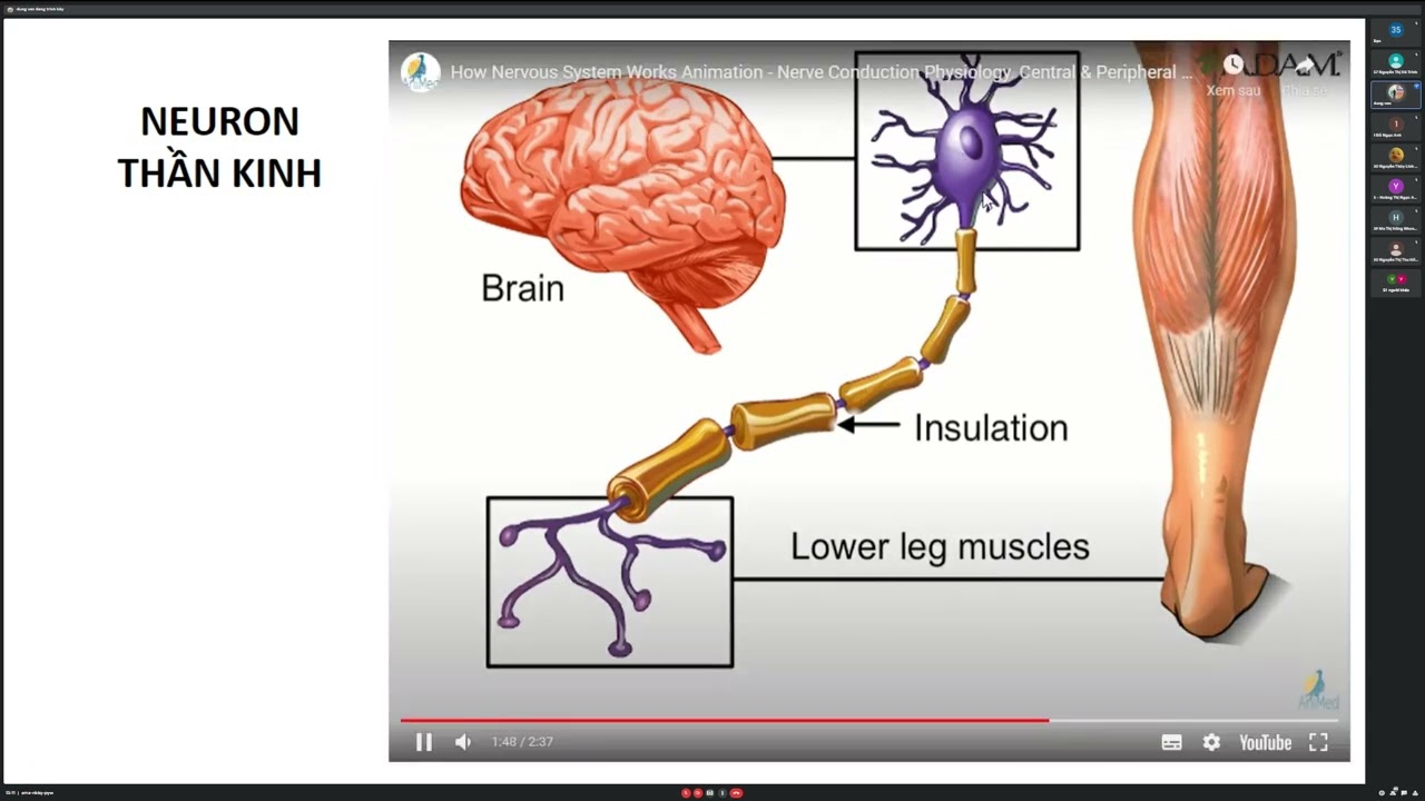



The anterolateral pathway (spinothalamic tract) conveys pain and temperature sensation through a three-neuron chain: primary neurons terminate in the posterior horn of the spinal cord, second-order neurons decussate at the spinal cord level and ascend to the thalamus, and third-order neurons project to the primary somatosensory cortex. This pathway contains two distinct sub-pathways: the fast neospinothalamic tract (using A-delta fibers) conveys sharp, pricking pain via glutamate neurotransmission, while the slow paleospinothalamic tract (using C fibers) conveys burning, throbbing pain via substance P. The body possesses a natural analgesia system involving the periaqueductal gray, nucleus raphe magnus, and enkephalin neurons that can suppress pain signals before they reach the brain.
Pain Pathways: Spinothalamic Tract, Analgesia & Endorphins
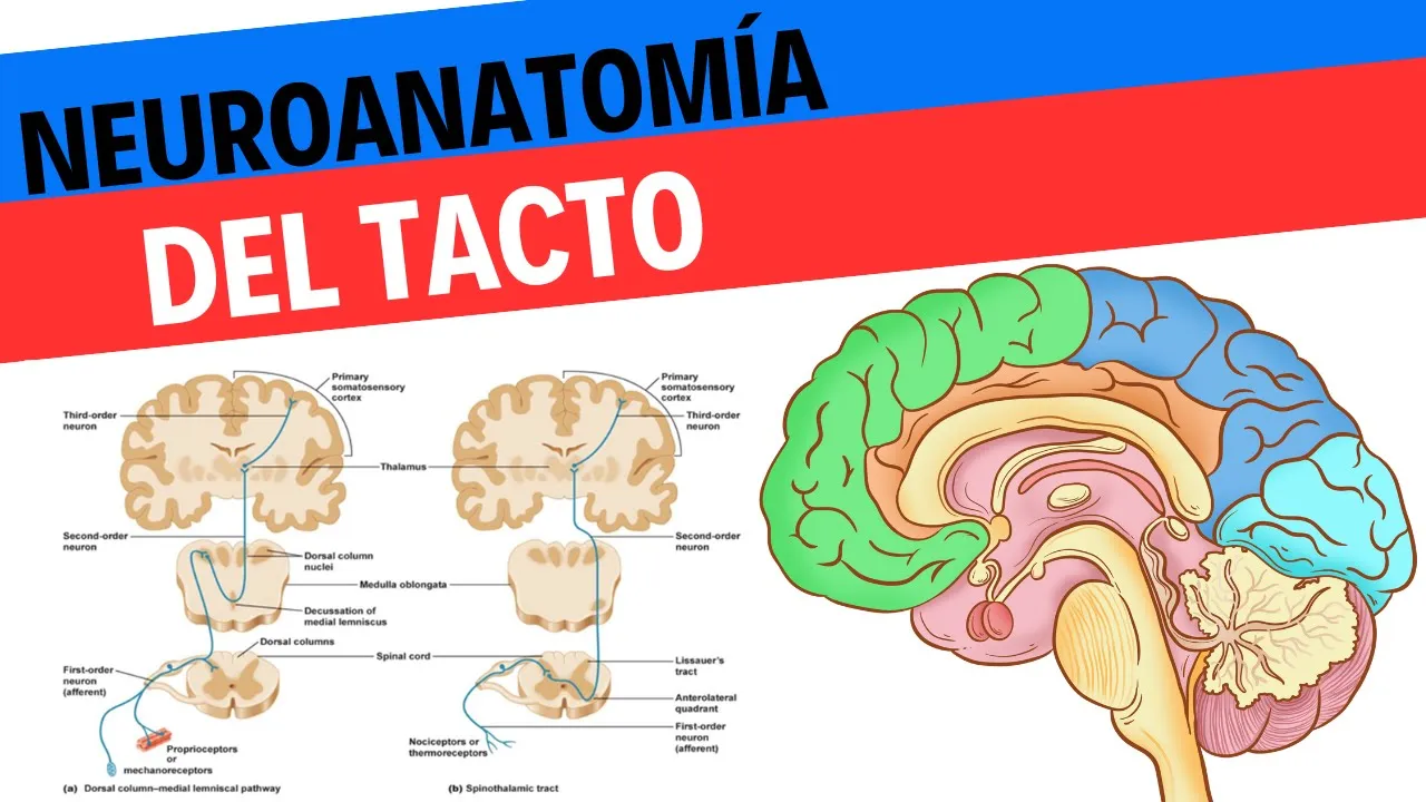
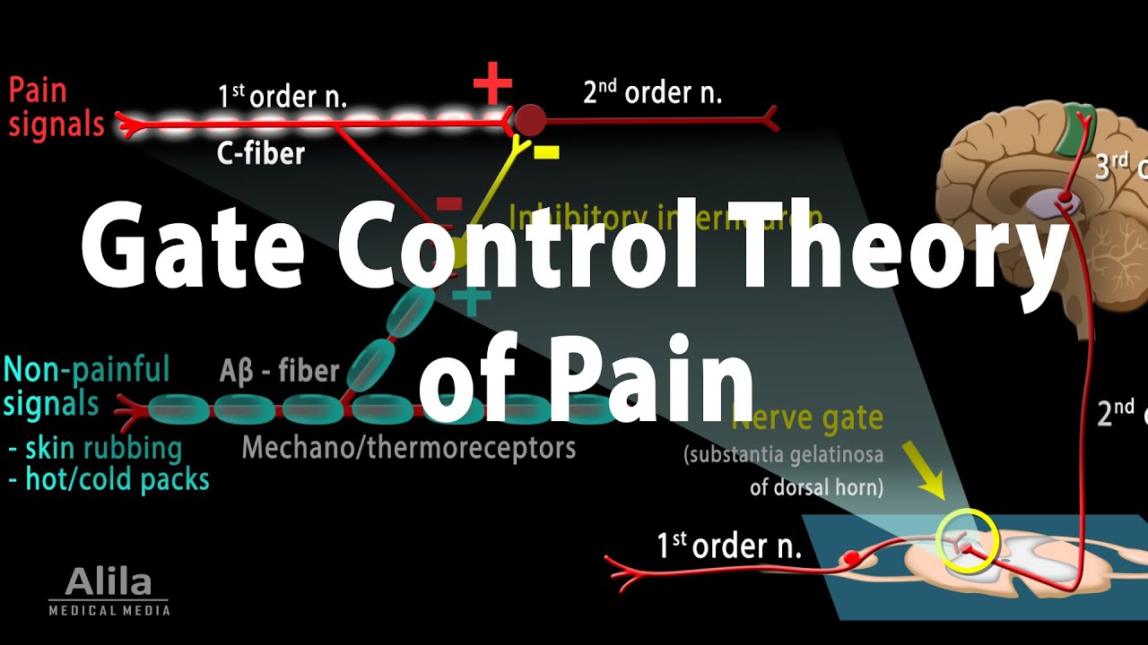
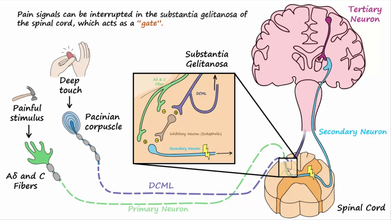
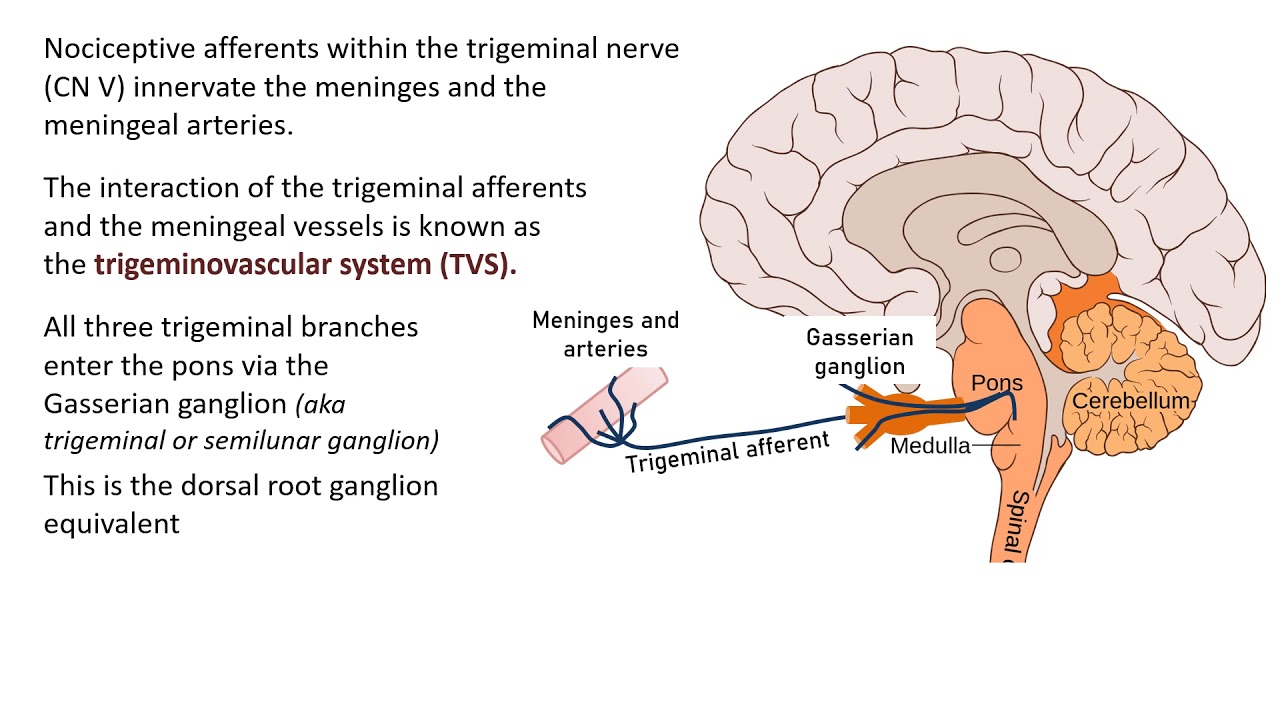
Added:hi class so it's time to continue on with our anterolateral pathway and the anterolateral pathway is also known as the spinothalamic pathway and this is going to consist of anterior and lateral white finicula don't let the word finicula confuse you it simply means a collection of axons inside the central nervous system now if you look carefully at this picture go all the way down to the bottom of the picture you're seeing that we have a three neuron chain we have the purple neuron shown here at the bottom of the picture synapsing on two different slightly colored blue neurons whose axons Ascend all the way up to the thalamus where they synapse on green neurons that are found in the um in the thalamus whose axons then are going to terminate ultimately on the primary somato sensory cortex the anterolateral pathway is primarily concerned with pain and temperature which is the primary focus of this lecture of course so by the words anterior and lateral we are simply mean meaning that there is a tract of white matter and remember white matter means axons that are melinated that are ascending up to the brain from the anterior portion of the spinal cord and the lateral portion of the spinal cord so if you look back down at the very bottom of this picture where it says pathway Direction you see an arrow and it's pointing to a little sliver of the spinal cord you'll see that there is a tract on the anterior portion of the spinal cord and then the slightly darker blue tract is slightly over to the side right so that's the lateral portion and forgive me but these terms are really really Technical and you probably don't remember them from Anatomy which is why I'm saying them again to you and hoping that your memory recall will be faster than when you learn these Pathways the first time when you were in anatomy so has the anterior lateral pathway different than the um the posterior finicula medial lisal pathway which boy is that a mouth word mouthful of words to say it's similar first of all and that it has a three neuron chain which so did the posterior piculus medial lisal pathway did but it's different if you look very carefully down at the bottom of the picture you'll notice that we're having crossing over decoation at the level of the spinal cord it's not up in the medulla like we saw in the posterior finicula medial lisal pathway which is for discriminative touch so that's a big difference here we're having Crossover at the level of wh the pain and temperature is being relayed from the anal lateral pathway is also important for crude touch and pressure but for our intensive purposes we're going to focus on pain and temperature now where the secondary neuron synapses on the tertiary neuron or third order neuron it's up here at the thalamus move your eyes upward and in our picture and you'll see that the blue axons are terminating on the green neurons now where this termination occurs the synapse this is where our first realization of pain is is going to occur but you don't know exactly where that pain is coming from you just know that you're hurting you can't really localize it and of course pain in order for it to be protected Ive or effective it needs to be localized so you know if you have pain in your foot you should move your foot if you have pain in your arm you should move your arm away from that painful stimulus so that's the protective effect of pain but you don't get that awareness of specificity when you have the second ordered neuron synapsing on the third ordered neuron in the thalamus it's not until the Third ordered neuron terminates in the primary somat sensory cortex that you get the idea of where the pain has originated from and I'll remind you from our first segment thanks to the review of the homunculus we know that the primary somat sensory cortex is designated to serve the needs of different body regions and that's already been mapped that's called somatrope and we also learned from our first segment that the larger the picture that was portrayed on the primary somatic cortex sorry primary stomato sensory cortex but that means more neurons are dedicated to serving that region of the body and interpreting the signals from that region of the body and I made a decently crude joke about the size of our genitalia on that somatotopy map and I hope you share my humor so what should you know about the anterolateral spinothalamic tract as it's called you should know it conveys pain and temperature it has three neurons you should know that the the primary neuron or as we say the um sorry I'm losing losing my voice here the first order neuron is going to come in from the periphery and terminate on the posterior horn this is where the second neuronal cell body is found and the second neuronal cell body the axon from it will immediately desiccate crossover to the opposite side again contralateral but this crossing over occurs much much earlier meaning more inferior on the on the spinal cord than the posterior piculus medial lemniscal path way that we saw in the previous segment the third ordered neuron or tertiary neuron is found in the thalamus and it's going to project onto the primary somat sensory cortex now in the next few pictures we're going to get pretty Technical and the reason why we're going to get pretty technical is because I have to explain to you that aarant fibers incoming sensory conveying fibers and the word fiber is a fancy way of saying axon so I'm going to have to tell you in the next few slides how we classify AER fibers they're either a or b or c fibers and then a fibers have further classification so I need to do that in the next slide and then the slide after that I have to tell you more about the posterior or dorsal gray horn that's the portion of the gray matter in the spinal cord that would be if you're thinking of a butterfly wing when you're looking at these pictures of the spinal cord the tops of the B the butterfly wings not the bottoms the vental gray horns so the dorsal gry horns are mapped or um according to anatomist they're arranged in layers and the word for an anatomist that means layer is lamina in plural is lamon so again to warn you it's going to get pretty technical with the vocabulary but I'm going to try and stop and say okay what does this mean what should you be focusing on okay I'm going to spend quite some time on this picture and it seems so straightforward but I want to make sure you're following along first of all look at this picture in terms of rows at the very top you should read that there is diameter and that means the diameter of the axon for each of these types of neurons so from the left side we're going from very large to very small on the right side and just to remind you that the larger the diameter the faster the rate of conduction for the action potential you can think of it of this in the following way if a freeway is very large such that you have eight Lanes in either direction you got to think about the flow of traffic being amazing but if you have a very highly traveled freeway where there's only two lanes on on either direction this is going to have high congestion and so the larger the diameter the f faster the rate of conduction now that's the second row you should be seeing here and if you look on the left side where the fiber is very large say 20 micrometers you're seeing a very fast conduction velocity whereas when it goes back down to 0.5 micrometers the very next row is showing you that it's very slow in conduction velocity okay so far so good now if you look at the third row it showing you General classification and it's basically showing you your a cell your a classification your B classification and your C classification now I've jumped down to sensory nerve classification now if you look down in your sensory nerve classification look you'll see a 1 A and A 1 B and then you'll notice that there is the number four which corresponds with the general classification above as C now I don't want this to get too too too crazy let's Whittle this down shall shall we we are going to be playing with either a fibers or C fibers let's let's just leave it as that now the a fibers you're noticing are reduced down to more subcategories Alphas betas gamas deltas and the C category just stays the same okay well let's address the C category those are just unmated very small fibers now if you took physiology at Saddleback you'll remember your physioex where we played with um a computer simulation where we played with um frog neurons and an earthworm do you remember that and and the axons that were the smallest and the unmated had the slowest conduction velocity all right so that's what this chart is here to show you so now let's add another layer to it some signals need to be transmitted to or from the central nervous system extremely rapidly otherwise the information would be useless an example of this is the sensory signals that apprise the brain tell the brain of the momentary positions of the legs at each fraction of a second during running it's a good idea to know where your legs are in space while you're running at The Other Extreme some types of sensory information such as pain aching pain do not need to be transmitted rapidly so slowly conducting fibers will suffice in this figure you should be seeing that nerve fibers come in all sizes between 20 micrometers in diameter very large to 0.5 very small the larger the diameter the greater the conducting velocity again how fast can you get home on an eight Lane freeway versus a two-lane freeway the range of conducting velocities is between 0.5 and 120 m/s now let's look at the general classification of nerves shown in this figure under General classification there is also below that a sensory nerve classification since we're talking about um ascending tracks we're talking about sensory nerves let's look at that you'll see excuse me in the general classification first of all there are fibers divided into types A and C okay and the type A fibers are further subdivided into Alpha Beta gamma and Delta okay great type A fibers are the typical large and mediumsized melinated fibers a spinal if you look all the way up at the top of this you're seeing melinated versus unmated type c fibers are the small unmated nerve fibers that conduct impulses at low velocities the C fibers constitute more than 1 half of the sensory fibers in most peripheral nerves the sizes velocities of velocities of conduction and functions of the different nerve fiber types are excuse me they are of importance to us we now want to look below and see this the next category that says sensor sensory functions please excuse me a cough is going around in my household and I feel like I'm choking now you're you should be looking at the blue boxes that are down in the sensory functions category you don't have to go down to motor function because we're not talking about outgoing commands we're only focusing on relaying incoming information now this is what I want you to realize there's your a alpha your a beta your a gamma and your a Delta and they're getting progressively smaller if you take that General classification and go down to the center sensory function I want you to realize that when we're talking about muscle stretch or tatile information like information from our meisner's um cor pusles or merkel's cor pusles or meinian cor pusles from our previous lecture those will all be conveyed through your a alpha and a beta fibers large fibers very fast you know when you're touching something very quickly now your deep pressure like your refines that's going to be your a gamma fibers they're going to be smaller and a little slower but now look at your a Delta notice that the a Delta go all the way down to where you first start seeing your blue boxes and you should see that a Del Delta is mostly that pricking pain this is your fast NEOS spinothalamic pain the more newer types of tracks to convey pain on the evolutionary scale now look at your C classification go all the way down and you'll realize that the C fibers that are smaller and slower notice they are primarily concerned with conveying information about pain aching throbbing pain temperature and tickle and again I want to remind you that there is a point where temperature can be perceived as pain so hey that's our Focus for right now our a Delta fibers that are larger and faster than the C fibers and our a Delta fibers are going to be conveying information about the fast occurring pricking pain and the Sea fibers that are unmyelinated are going to be conveying pain that is slower to come to our Consciousness and is going to be more longer lasting so to bring the story back to my initial story about when I burned myself let me ask you this which fibers were initially carrying impulses up toward my brain when I first had the Sensation that something was hurting but I didn't quite know where and it was very shortlived versus which fibers finally kicked in and conveyed information and I felt that throbbing burning pain so when I first touched the broiler those were my a Delta fibers relatively large m ated fibers I felt that sharp pricking pain but it was not very remarkable that's what Drew my attention I didn't quite know where it was what's going on and that's where I kind of watch my skin melt and then stick to the broiler I know it's gross but it's so true and then my sea fibers that were smaller still and unmyelinated once those kicked in that's when the signals reach my Thalamus and my my primary somata sensory cortex and that's when I registered that throbbing really aching pain and that's when I finally withdrew my hand so that's our Focus for the rest of this lecture okay my sweet class I'm going to spend a lot of time going through this slide and I hope my voice lasts for me but I'm recording this on a day when I had physiology and it was a big day of lecturing and I'm kind of losing my voice so I apologize in advance if I'm being irritating to you first I want you to look at the top right picture that's on your on your screen and I want you to realize that it's a section through the spinal cord you're seeing your traditional butterfly shape of the gray matter and I want you to look on the left side of that top picture and you will see in a dashed line a circle around two axons it says spinal nerve and you'll notice that one of them is labeled C and one is labeled a Delta up at the top left of that top picture now the C represents those small unmyelinated fibers I just described to you and the a Delta represents those somewhat larger and melinated fibers I just described to you now look at the the gray matter that's shown on that picture you'll notice it has Roman numerals on it and they're numbered one through Roman numeral nine so this is basically an anatomist putting theay matter into layers or lamina so there's lamina one through lamina 9 it just simply means layers again don't let the terminology scare you the plural for a lamina is lamin now if you look carefully in this picture for the C fiber or a Delta fiber you'll see that the primary neuron that's coming into the dorsal greyhorn will terminate on different layers or different lamin and I'm going to talk about where this primary neuron first initially terminates and those different lamin and again it's going to get very technical and I'm going to remind myself to stop and say here's what I want you to know or here's what I want you to pay attention to so let's start first by saying there are two Pathways for pain to be sent up or at least the sensation of pain to be sent up to the brain for interpretation we know there is the slow pathway or the paleos spinothalamic and the fast pathway or NEOS spinothalamic we're really talking about the interal lateral Pathways the a Delta fiber that I just introduced to you is part of the fast pathway the Neo spinothalamic Neo meaning more new new newer and evolutionary time the sea fiber smaller unmated is going to convey information that will be interpreted by your brain as the more burning throbbing aching pain this is the paleos spinothalamic pathway so let me say it again even though all pain receptors are free nerve endings these endings use two separate pathway ways for transmitting pain signals into the central nervous system the two Pathways mainly correspond to the two types of pain a fast sharp pain pathway neop spinothalamic and a slow chronic pain pathway paleos spinothalamic so we have fast and slow the fast sharp pain signals are elicited by either mechanic or thermal pain stimuli that basically means that if you were to Ram up against your desk that is enough to start the pain signals the action potentials to be conveyed through the alpha sorry the a Delta fibers but it also can be temperature remember temperature can be interpreted as pain once it gets Beyond 45° Centigrade so because of this double system of pain innervation a sudden pain stimulus often gives a double pain sensation what do I mean by that a fast sharp pain that is transmitted to the brain by the a Delta fiber pathway is followed a second or so later by a slow pain that is transmitted by the Sea fiber pathway sharp pain apprises the person rapidly of a damaging influence and therefore plays an important role in making the person react immediately to remove him or herself from the stimulus the slow pain tends to become greater over time this sensation eventually produces intolerable pain and makes the person keep trying to relieve the cause of the paint again both of these are going to have protective effects on entering the spinal cord the pain signals take two Pathways to the brain either one the neop spinothalamic tract or two the paleos spinothalamic tract if we focus on the neop spinothalamic tract for fast pain the fast type of a Delta pain fibers transmit mainly mechanical and thermal pain again you just run into your desk or you've touched the candle that's burning these primary neurons first ordered neurons terminate mainly if you look at this top right picture in lamina 1 this is in lamina one of the dorsal greyhorn and there they excite second order neurons of the neop neop spinothalamic tract these second order neurons give rise to Long fibers that cross immediately to the opposite side of the cord this is contralateral this is crossing over you'll notice that some of these fibers terminate um yeah sorry never mind I was getting hold of my ahead of myself you'll notice that these second ordered fibers are crossing over immediately and they are going to go up to the thalamus and then synaps on third ordered neurons which you cannot see in this picture but you'll see that the axons the tracks are circled as fast sharp pain fibers so again there's immediate crossing over and this is very different than our posterior phic medial lisal pathway that we learned previously now where do those second order fibers go if we look at the bottom picture we can kind of follow them up to the brain a few fibers of the neop spinothalamic tract terminate in the reticular areas of the brain stem the reticular area is a diffused area of the brainstem there's no one specific region but most of the neurons pass all the way to the phalus without interruption and they're going to terminate in the thalamus right and we see that up here now don't worry about the whole uh the ventral basil complex Etc but that's where they're going to go to and those third ordered fibers are then going to be transmitted to the somato sensory cortex now let's continue on with our story when the second order neurons terminated on the thalamus in the example of the neop spinothalamic pathway I just told you there can be a a conscious awareness of pain however the pain is not localized it's not until it's projected onto the primary somato sensory cortex that the pain can be exacted the next question is what are the neurotransmitters used in this NEOS spinothalamic pathway It is believed that glutamate is the neurotransmitter substance secreted in the spinal cord at the level of the a Delta pain fiber ending so where the a Delta pain fiber comes in and terminates on the first lamina it's releasing glutamate which is Prim it is a the largest widely used excitatory neurotransmitters in the central nervous system and it usually has a duration of action for about a few milliseconds but again this is excitatory so most certainly our Action potentials are going to be conveyed upwards towards the thalamus through the second the second ordered neurons because they will reach threshold spike in action potential and transmit it up to the third ordered neurons their cell bodies of course found in the thalamus now let's focus on the slow pain or the paleos spinothalamic pain now let's focus on the paleos spinothalamic pathway the paleos spinothalamic pathway like as said is a much older system and transmits pain from the slow un mated type c pain fibers in this pathway the peripheral fibers terminate if you notice in the top right picture in the spinal cord the dorsal greyhorn almost entirely in lamin 2 and three now lamin 2 and three are together called the substantia gelatinosa why would they be called such a thing so bizarre they're called that because when they were stained by early anatomist they stained the darkest of all the three layers and they had kind of a gelatinous look to them when they were stain so gelatinous substance means substantia gelatinosa so that's lamin 2 and three now most of the signals then pass through another set of short fiber neurons within the dorsal greyhorn itself on that side ipsilaterally onto lamina 5 and then from lamina 5 you'll notice then there's crossing over to the controlateral side where those technically second order neurons are now going up with the same tract as the fast ordered neurons or sorry the fast-tracked second ordered neurons so again crossing over occurs at level for the Paleo spinothalamic pathway now the initial cause of the action potential in the first ordered neuron is not so much a mechanical start as it is a chemical start for the action potential and this means that tissue damage has occurred now what chemical signals do tissues release when they become damaged Brady kyen potassium all remind you leaks from the cells acetal choline can even be used here histamine and serotonin now Brady kindan are really interesting um they've been studied for quite some time the reason why they're interesting is because they stimulate the Noor they they stimulate the free nerve ending Brady kinden have been around for a while maybe you've heard of a fine young gentleman from Uganda IDI Ami back in the early 19th century well he was in power when I was just a young child but he was well renowned for his torture of the people that he commandeered in Uganda and he actually had his some these people tortured by injecting Brady kinan into their skin which caused excruciating pain because again it was stimulating that paleos spinothalamic pathway way and he thought he was getting away with it because it left no overt marks a really fine chap I'm sure you would appreciate anyway with the c fibers and the slow pain pathway we're talking about chemical signals released by the damaged tissue leading to the action potentials in that first ordered neuron now let's continue on now from these C pain type c pain fiber terminals again the terminal bons are those primary or first order neurons entering the spinal cord research suggests that they release glutamate but also it suggests that primarily they're releasing substance called substance P transmitter known as just substance p and p doesn't stand for pain it's just substance P substance p is released much more slowly building up in concentration over a period of seconds or even minutes in fact it's been suggested that the double pain sensation one that one feels after a pin prick might result partly from the fact that the glutamate transmitter gives a faster pain sensation whereas the substance P transmitter gives a more lagging sensation regardless of the yet unknown details it seems clear that glutamate is the neurotransmitter most involved in transmitting fast pain into the central nervous system and substance p is concerned with a slow chronic paleos spinothalamic pathway now let's move on with the projection of the ca fibers in the second ordered fibers in the slow pain pathway the slow chronic paleo spinothalamic pathway terminates widely in the brain stem and only one tenth to 1/4 of the fibers pass all the way to the phalus where they would terminate on third order instead most of the second ordered neurons will terminate in one of the three areas again warning it's going to get technical either the reticular nuclei of the medulla ponds and Mez and sethlon and I'll remind you that nuclei means a collection of neuronal cell bodies inside the central nervous system so we're talking the reticular nuclei again the reticular um activating system is the system that helps arouse you from sleep helps you to remove background noise while you are studying it basically helps you keep alert and this is found in many regions of the brain stem but most notably here we're noting that we're going to Target the medulla ponds and mezen sephylon is the midbrain two the tactile area of the mezen sephylon deep to the superior and inferior calculi basically this is the region where your eye reflexes and ear reflexes are going to turn to any loud threatening object or sound so when you're in pain you want to turn to whatever is threatening you and three the per aqueductal gray region so the per aqueductal gray area um or per aqueductal gray um area is sometimes abbreviated as p a for per aqueductal gray and then area and this per aqueductal gray area is found surrounding the aqueduct of sylvus which is a old an Antiquated way of saying the cerebral aquaduct which connects of course the third ventricle to the fourth ventricle just to remind you of your Anatomy now again this slower pain pathway is older in evolutionary terms these lower regions of the brain are also older in evolutionary terms there are Reptilian Brain that you were learning in Paul McLean's Triune brain Theory and if we look at animals whose brains have been sectioned above the midbrain to block pain signals from reaching the cerebrum these animals although not consciously aware of the pain they still show undeniable evidence of suffering when any part of the body is traumatized so they may not be consciously aware of it but you you the researcher could very most certainly tell they are in pain from the brain stem pain areas multiple short fiber neurons relay the pain signals upward into the thalamus and then into the hypothalamus and the basil regions of the brain so what should you be taking home here this slower pain pathway is older in evolutionary terms it doesn't necessarily get to the cerebral CeX for conscious interpretation it is our more more and most primitive way of detecting pain and even when the cerebral cortex is disengaged in animals that have been studied when they are subjected to trauma they most certainly are displaying uh pain and it's sad that we're doing animal research I get that but it's important to know this and although they're feeling the pain they are not able to exactly localize the pain and interpret the pain quality that's what we need the cortex for and that's a later uh structure that has evolved in our brain so to can continue on with that localization of pain transmitted through this paleos spinothalamic pathway is imprecise for instance slow chronic pain can usually be localized only to a major part of the body like to one arm or leg but not to a specific point on the arm or leg this is in keeping with the multi synaptic diffuse connectivity of this pathway again we're going from the sea fibers to lamina 2 and three to lamina 5 then we're crossing over then we're going to the brain stem the ponds the medulla the midbrain then we're going to the area around the cerebral Aqueduct we're going to areas of deeper areas of the midbrain and then we're moving on to the hypothalamus and the thalamus and basil nuclei of the brain we're not even really getting to the cerebral cortex so again we're not going to or animals are not going to be able to discern exactly where that pain is coming from now what about these areas of the brain the reticular formation that I was talking about and their involvement in the appreciation of pain complete removal of these somato sensory areas of the cerebral cortex again does not destroy an animals ability to perceive pain therefore it is likely that pain impulses entering the brain particularly the brain stem the reticular formation the thalamus the hypothalamus and other lower brain centers cause conscious perception of pain this does not mean that the cerebral cortex has nothing to do with normal pain appreciation when electrical stimulation of the primary Soma sensory cortex areas used it does cause a human being to perceive mild pain from about 3% of the points that have been stimulated however it is believed that the cortex plays an especially important role in interpreting pain quality even though pain preception might be principally the function of the lower C centers again are more primal centers well now it's time to talk about caspin and that's how you pronounce it capin capis now this is a chemical that's found in hot peppers and it's an irritant and it activates heat receptors in your skin and in your mouth so when this irritant from hot peppers touches these Thermo receptors your brain brain because again the temperature goes the stimulus from this capin makes these thermal receptors detect quote unquote an increase in temperature Beyond 45 degrees your brain now interprets it as pain which is the reason why if you are a person who eats hot peppers and you don't like something hot it's because you have very sensitive Thermo receptors in your mouth now I for one love me some hot peppers and I grow all kinds of jalap well jalapeno peppers Habaneros no I don't grow ghost peppers they are very difficult to grow in Southern California and if you are growing them I'm very jealous of you but at the same time afraid of trying one anyway they put C capsus in in bird seed because birds don't have the same Thermo uh receptors as we do and so the idea is if they put capsus in in bird seed then other mammals like squirrels Chipmunks Etc will be less tempted to eat that bird seed now interestingly apparently capin if you take chili pepper and sprinkle it in your yard or your garden maybe because the neighbor's cat keeps coming over and pooping in your garden if you put that on there the capsus in irritates the PS on the cat's feet and they'd be less likely to go to the bathroom in your garden now that being said I personally have gone to the 99 cent store and purchased about I'd say 17 little plastic containers of chili powder sprinkled it in my garden kept the neighbor's cat away for maybe 12 hours and it didn't work now I don't I don't know if it's because it got absorbed in the soil the sprinklers came on I don't know but I'm willing to try it again or maybe the neighbor's cat just has very thick pads from being a street cat you know out on the road and not a house cat and you know has a lot of calluses on their little pads I don't know but cap capin is used in creams and patches and can be used for controlling the pain of arthritis and shingles and basically what you do is you know you've you've had that like Icy Hot cream you might have used it but you put it on your skin and the idea is that when the heat goes up it overwhelms your receptors so that you are no longer feeling the dull throbbing pain to put it another way it's almost like your brain has a different type of pain that is overwhelming it because stimulating so many more receptors because you've rubbed it all over your skin that it has more to contend with with this temperature change than the actual pain from the arthritis or shingles another way of saying that is it it's interfering with substance P the release of neurotransmitter from the primary or first ordered neuron it's going to deplete it it's going to cause the first order neuron to just dump it out and then there's nothing less to nothing more sorry to release on the second order neuron so now let's talk about our natural ways that our brain and spinal cord have the way to suppress pain or the conscious awareness of pain stopping the pain signals before they even reach our cerebral cortex this is our natural analgesia system pain suppression system now again I want to to warn you it's going to get pretty technical here the degree to which a person reacts to pain varies tremendously one person's itch is another person's slice Across the Skin okay so that's okay all of us have a threshold for pain I'm not trying to judge you shouldn't judge it just is this results partly from a capability of the brain itself to suppress input of pain signals to the nervous system by activating a pain control system called an analgesia system again technicalities the analgesia system is shown in this picture and it consists of three major components one the par aqueductal gray areas and this is found in the mezan sethlon and upper ponds okay per aqueductal gray area Perry around Aqueduct around the aqueduct shown up here at the top right of the picture you can see it in Gray around the aqueduct that's in between the third and fourth ventricle from Anatomy I'll remind you that the third ventricle is bounded by the ponds and the medulla the third ventricle is primarily found housed in the thalamus and the cereal Aqueduct is also known as the aqueduct of sylvus so we have a region of neurons that are found in this per aqueductal gray area and I'll remind you gray means unmyelinated you should be thinking cell bodies so you're seeing the red axon and red cell body that is in the per aqueductal gray area so that's the first area number two the rafy Magnus nucleus and the nucleus reticularis parag giganto cellularis wow I just said a whole bunch let me just break that down for you we're talking about cell bodies in the lower ponds upper medulla so if you look down here with a red axon from our neuronal cell body in the per aqueductal gray area is terminating on a second neuron it's in the lower region of the ponds and upper region of the medulla okay that's where it's just connecting to another neuron and you're seeing the axon of that orange neuron is going down to the spinal cord okay part three a pain inhibitory complex located in the dorsal grey horns of the spinal cord okay follow the orange neuron that is starting in the nucleus Rafi Magnus in this picture in the lower region of the ponds upper region of the medulla follow it all the way down the spinal cord to the dorsal greyhorn and you're seeing that it's terminating on another short short short red neuron that's labeled and keyin neuron and that's what I mean by the pain inhibitory complex so we need these three regions now through studies we we know that the analgesia signals can block pain before it is relayed to the brain and it's blocking pain through that enlin neuron Action potentials are going from the per aqueductal gry matter to the nucleus Rafi down to that enlin neuron who's going to release neurotransmitters on that primary neuron the the um first neuron conve information about a pain stimulus before it reaches the second order neuron and that little short and keflon neuron is going to interrupt the signal so it's like the Spy it's going to take the the message before the enemy can even detect what the message is we know this because electrical stimulation either in the per aqueductal gray area or in the rafy Magnus nucleus can suppress many strong pain signals entering by way of the dorsal spinal Roots also stimulation of areas at still higher levels of the brain that excite the per aqueductal gray area have also led to the suppression of pain these higher levels have been the hypothalamus just to name a few of I mean different areas of the hypothalamus I'll save you the technicalities several transmitter substances are involved in the analgesia system especially involved are those in keyins that I just told you about and serotonin many nerve fibers are going to be releasing ke and keyin from their endings and as a result this is going to block the transmission of substance P from the first ordered neuron to the second ordered neuron so again the enlon is believed to cause both the pre synaptic and post synaptic inhibition of incoming type c and type a Delta pain fibers where they synapse in the dorsal grey horns let me say it again that in keflon neuron when it's stimulated by the higher orders of the brain the per aqueductal gray area and the nucleus rapi or rapi sorry RFI Magnus is interrupting both type c and type a Delta fibers now in the next slide I'd like to talk a little bit more about the en keyins and these are very short five amino acid five amino acid peptides they um are abbreviated as t g GP l or t ggp met and I'll explain that in a little bit but there's some history to them so let's move on to that right now more than 40 years ago in the 60s and 70s I hate to say it but that's back when I was born it was discovered that injection of minute quantities of morphine either into the per ventricular nucleus around the third ventricle or into the per aqueductal gray area the brain stem caused an extreme degree of analgesia pain blocking in subsequent studies it has been found that morphine like agents mainly opiates like opium heroin morphine also act at many other points in the analgesia system including the dorsal gry horns of the spinal cord because most drugs that alter excitability of neurons do so by acting on synaptic receptors it was assumed that the morphing quote unquote receptors of the analgesia system must be receptors for some morphine like neurotransmitter that is naturally secreted in the brain so let me say this in plain English when scientists first started injecting these opiates into people either in their brain or in the spinal cord they had analgesia like effects and so the scientists correctly theorized that if these drugs from plants were working in the human then the human must have an endogenous chemical that was released that would bind to the same receptors that these plant chemicals were binding to so the research began searching for these morphine like drugs about a dozen such op like substances have now been found at different points of the nervous system all are breakdown products of three main protein molecules three main ones Pro opio melanocortin or Palm c pro enlin and proo dorphin prodor among the more important of these it like substance are beta endorphin met and kein L and keflin and dorphin so basically the Palm C proopiomelanocortin is going to be a precursor hormone maybe you remember this from your physiology it's a large protein and from it can be cut um adrenocorticotropin hormone melan it stimulating hormone but also cut from it is beta endorphin and from the beta endorphin or the pro enlin you can cut smaller pain relieving molecules like um the different en keins now the enlin two enlin are found in the brain stem and spinal cord in the portions of the analgesia system and the beta endorphin is present in both the hypothalamus and the pituitary gland the dorphin is found mainly in the same areas as the enlin but in much lower quantity so all of these are your natural painkillers is what I'm trying to say although the fine details the brain's opiate system are not well understood activation of the analgesia system by nervous signals entering the per aqueductal gray areas or an activation of the pain Pathways by morphine like drugs can almost totally suppress many pain signals entering through peripheral nerves so let me say this again endorphins were discovered almost by accident in the 1970s when scientists were carrying out research on drug addiction investigators had wondered for years why the human brain contained receptors for chemicals produced by the plant and they eventually discovered why the brain produces its own set of neurochemicals which are actually far more potent than morphine opium and heroin but they share the same neural receptors with those drugs the naturally produced brain chemicals called endorphins and an keyins and as I said a third class the dorphin are released in times of stress they can make a mangled accident victim as Serene as a Buddhist monk and they can also make an athlete feel great after an extremely vigorous workout the latter effect is sometimes referred to as the runner's high and the post exercise surge in endorphins helps to explain why many exercisers seem to become addicted to their sport their workouts become fixes which Mass the pain of everyday living and even injuries or illnesses can't stop the training process because the athlete is relentlessly searching for endorphin induced mood elevations and keins are five amino acids containing tyrosine glycine glycine phenol alanine and either Lucine or methionine that's what I said on the previous slide and I'm rating here on this slide they are the smallest of the molecules with painkilling or opiate activity and cins are found in the thalamus of the brain and in some parts the spinal cord that transmit brain pain impulses and the spinal cord and keyins inhibit painful Sensations by reacting with specific receptor sites on the sensory nerve endings again where the first ordered neuron is synapsing with the second second ordered neuron and that first ordered neuron is releasing substance P that en keflon neuron that I showed you that short little red one on the previous slide is releasing and keyins nerve endings of the central nervous system and the Adrenal medulla release these naturally occurring morphine like substances and cathlin bind to opiate receptors and release controlled or undo levels of pain L and kein is an endogenous Agonist for The receptors that are stimulated by opiate alkaloids it has multiple effects on the CNS L and keflon and and met and keflon are both like I said are natural pain killers met and keflon in particular is involved in a phenomenon associated with modulated pain perception regulation of memory and emotional conditions food and liquid consumption it also has an impact on digestive system motility gastric as well as pancreatic uh secretion Etc so the met and keflon is that Feelgood drug that is released when you eat food of sweet rewards you know your comfort food that's what is stimulating the release of the Met and keflon now again in addition to the runner's high and eating foods that you give that give you comfort we also see surges of these natural painkillers in both the mom and baby during birth because for both it's painful we see this occur in death as part of the reason why people when they have near death experiences they'll tell you that they feel no pain even though Mr Whitey great white shark bit off their leg but it's also important to note that in people who are taking exogenous morphine and they immediately withdraw from it they will have withdrawal um symptoms that are so intense in pain it's because the morphine is BL is occupying I should say these receptors and so the natural and keyins it's like a negative feedback effect are not being released so if the patient stops morphing cold turkey then their own body is not releasing their own pain killers and so the smallest stimulus can be perceived as very very painful on the scale of 9 to 10 so the drug naloxone is a way of basically working them down from that morphine so they don't have extreme withdrawal now the last part that I want to uh follow up with on this slide is THC Tetra hydro cannabinol which is basically uh marijuana now THC the substance found in marijuana causes a release of En keylons a natural release of En keylons so you get this analgesic effect but you also get that euphoric effect like taking opium or her so it's kind of a mixed event because they know that some of that a person can become addicted to that euphoric reaction and if you ask someone who's doing marijuana ask them are you addicted to it they'll say no I can quit any time and and it doesn't always work but we know on a scientific level that that drug te H C actually does stimulate the natural and cathline release and there's that kind of pain relief and euphoric uh Euphoria experience that they will uh report so there is a relationship be between enlin en keflin release and addiction so those who are saying that THC the substance found in marijuana is not addictive the science would suggest otherwise now to get back to your chapter and talking about problems with pain your chapter talks about pain threshold which is basically the minimum stimulus that can be perceived as painful there's also pain tolerance that means how much can you tolerate before you're going to pass out and usually this is on a scale of one to n when they first did the studies was actually on military personnel and since then in a clinical setting well we just round up to a scale of 1 to 10 we can further break down pain as no acceptive pain where the noors are aberant activated or neurop neur neuropathic pain or neuropathy and this can result from either direct injury to neurons or dysfunction of sensory neurons specifically the axons now we can take it a step further and say are these neurons over sensitive in which case we say neurobic hyperalgesia algesia means more sensitive to pain and this person with this neurobic hyperalgesia would be more sensitive to pain and the classic example of that is tick dillu now um tick D dulu is basically this neuroptic hyperalgesia and i' like to tell you a little bit about it it occurs in some people over one side of their face and the sensory area and it's usually because of cranial nerve five or nine so it's either due to trigeminal neuralgia or glossop feral neuralgia the pain feels like sudden electrical shocks and it may appear for only a few seconds at a time or may be almost continuous often it is set off by exceedingly sensitive trigger areas on the surface of the face Maybe in the mouth or inside the throat almost always by a mechano receptive stimulus meaning something touching rather than a pain stimulus like something they ate for example for instance when the patient swallows a Bolis of food as the food touches a tonsil it might set up a severe lanting pain in the mandibular portion of the fifth nerve trienal nerve the pain of tick dearu can usually be blocked by surgically cutting the peripheral nerve from the hypers sensitive area the sensory portion of the trial nerve is often sectioned immediately inside the cranium where the motor and sensory roots of this nerve would separate from each other so that means you're sparing the motor roots from the uh sensory part so the person won't have paralysis of their facial M muscles specifically the muscles of mastication which cranial nerve 5 would control now the opposite extreme would be neuropathic analgesia which can happen in diabetic neuropathy which we've already discussed in this case there is there is absence of pain from stimuli that most certainly should be painful and again to remind you pain is supposed to be protective and we store memories on it we're supposed to learn from it sort of like saying well I'll never do that again or when I saw that nice brown fuzzy creature I thought it would be something that I could play with but when it mauled me almost to death I realize now that that large bear is something that I should avoid neuroptic analgesia as I described in our just last week's lecture can happen in diabetic neuropathy because of the high blood glucose levels leading to vascular changes and neurons need blood vessels too for the delivery of nutrients and without these nutrients being delivered or oxygen being delivered the neurons become dysfunctional they do not relay the sensation of pain and I was telling you about new shoes with different pressure points the person is not aware of them and therefore the uh blisters turn into ulcers and they are poor for healing there isn't adequate blood flow so neuropathic uh neuropathy um that leads to inappropriate analgesia is not okay the that's the opposite of being hyp sensitive to paint now I'd like to conclude our talk on headache okay um headaches are a type of pain referred to the surface of the head from Deep head structures some headaches result from pains inside the cranium but others result from Pain arising outside the cranium such as from the nasal sinuses for example so I'd like to First Focus On Headache of intracranial origin now first of all I'd like to say the brain tissues themselves are almost totally insensitive to pain even cutting or electrically stimulating the sensory areas of the cerebral cortex only occasionally cause pain instead these Sensations cause prickly types of paresthesias on the area of the body represented by the portion of the sensory cortex stimulated and that goes back to the um experiments that I was telling you about Wilder Penfield with mapping the homunculus conversely tugging on the Venus sinuses around the brain damaging the tent torium which is a piece of the Duram Mater or stretching the dura at the base of the brain can cause intense pain that is recognized as headache Al also almost any type of traumatizing crushing or stretching stimulus of the blood vessels of the meninges can cause headache an especially sensitive structure is the middle menal artery and neurosurgeons are careful to anesthetize this artery specifically when performing brain operations under a local anesthesia types of intracranial headache can include headache of menitis one of the most severe headaches of all is that resulting from menitis which causes inflammation of all the menes the duramater the arachnoid the pamat including the sensitive areas of the dura and the sensitive areas of the Venus sinuses such intense intense damage can cause extreme headache and pain referred over the entire head headache can also occur by low cerebral spinal fluid in which the in which case the brain is not as buoyant as it should be it sinks to a more inferior position and this pulls the meninges taut and again can stimulate the receptors that are in the meninges migraine headache migraine headache is a special type of headache that may result from abnormal vascular phenomena although the exact mechanism is unknown migraine headaches often often begin with various um prodromal Sensations such as nausea loss of vision and part of one field uh visual Aura where they see spots and other types of sens hallucinations so there's like a preemptive warning alcoholic headache as many people have experienced a headache often follows excessive alcohol consumption con consumption oh my gosh consumption it is likely that alcohol because it is toxic to tissues of course directly irritates the meninges and it can directly irritate the meninges because it can cross the blood brain barrier and therefore causes the intracranial pain the dehydration of course also results from alcohol intake because alcohol inhibits the release of ADH and so that can lead to a loss of cerebral spinal fluid and pulling on the meninges so The Hangover from an alcoholic binge it's usually hydration that is the issue and hydrating can attenuate the uh the pain of an alcoholic headache but it does not abolish the headache and other symptoms of The Hangover so you can drink your water while you're drinking your wine and that might attenuate your hangover for the next day but it won't abolish it now extra cranial headaches these can be headaches that arise from muscle spasm emotional tension often causes many of the muscles of the head especially the muscles attached to the scalp and neck muscles um to become held in contraction and is postulated that this is one of the common causes of headache the pain of the head muscles supposedly is referred to the overlying areas of the head and gives one the same type of headache as intracranial lesions do again it's like a like a referred pain headache caused by irritation of nasal and accessory nasal structures have you ever been in the car with someone wearing perfume that just irritated your nasal cavity and you it led to a headache the mucous membranes of your nose and nasal sinuses are sensitive to pain but not intensely so nonetheless infection or other irrit IR irritative processes in widespread areas of your nasal structures can summate and cause headache that is referred to behind the eyes or in the case of frontal sinus infection to the frontal surfaces of your forehead and scalp so that person wearing the annoying uh perfume you get the headache and you squeeze your eyes together that's referred pain to behind your orbital area headache of course can be caused by eye disorders difficulty in focusing one's eyes clearly can cause excessive contraction of the eye ciliary muscles in an attempt to get your vision cleared and even though these muscles are extremely small it is believed that constant contraction of them can cause retroorbital headache meaning even behind the orbits so when you're studying it is suggested that you look up every 20 minutes look 20 feet away and blink 20 times over so that you're not holding your intrinsic eye muscles too extensively which can lead to eye strain vision problems and then headache so every 20 minutes a good optometrist will tell you the 2020 20 rule every 20 minutes look 20 feet away and blink 20 times Liv all right I hope you enjoyed your pain Pathways and discriminative touch Pathways and what can cause the perception of pain and how we block pain next week is our last lecture
Up Next
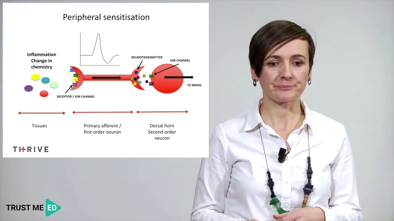
Peripheral Sensitization Explained | Physiology of Pain
@TrustmeImaPhysiotherapist1
20.4K views•2018-11-27

Graphic Medicine: Comics for Collaborative Healthcare Communication
@nationalpatientadvocate
189 views•2023-12-04

Neuroanatomy: Central and Peripheral Nervous System Divisions Explained
@AKLECTURES
136.2K views•2014-09-20

Stages of Labor and Vaginal Birth | Childbirth Animation
@nucleusmedicalmedia
52.1M views•2017-08-18
Related Study Plans & Knowledge Roadmaps
Structured learning paths in Medicine