Peripheral sensitization is a normal physiological phenomenon where inflammation causes increased sensitivity in injured tissues through two mechanisms: prolonged activation of existing ion channels and expression of new/different ion channels at nerve endings, leading to primary hyperalgesia (increased pain response at the injury site) and secondary hyperalgesia (sensitivity in nearby areas), which clinicians should recognize as part of normal healing but distinguish from chronic conditions like osteoarthritis or rheumatoid arthritis.
Peripheral Sensitization Explained | Physiology of Pain
Added:Deep Dive
Basic physiology of nociception, including the distinction between A-delta and C primary afferent fibers.

Fisiopatologia Dor
21K
Peripheral sensitization is the process where inflammatory mediators reduce the threshold for nociceptor activation, making them more responsive to stimuli. This involves activation of normally silent nociceptors and increased response to non-painful stimuli. Pain signals are transmitted via primary afferent fibers: A-delta fibers (thinly myelinated, rapid conduction, sharp localized pain) and C fibers (unmyelinated, slow conduction, dull poorly localized pain). These fibers synapse in the dorsal horn of the spinal cord, where information is processed before transmission to higher brain centers. The distinction between these fiber types explains the different qualities of pain perception.
Click to watch →
Pain vs. Nociception | Podcast
7.1K
Two main nociceptive fiber types transmit pain signals: A-delta fibers are myelinated and fast (6-30 m/s), conveying sharp, immediate pain for rapid withdrawal reflexes; C fibers are unmyelinated and slow (0.5-2 m/s), producing throbbing, ongoing pain. This speed difference creates distinct pain qualities and explains why initial injury causes sharp pain followed by aching pain as C fibers activate.
Click to watch →
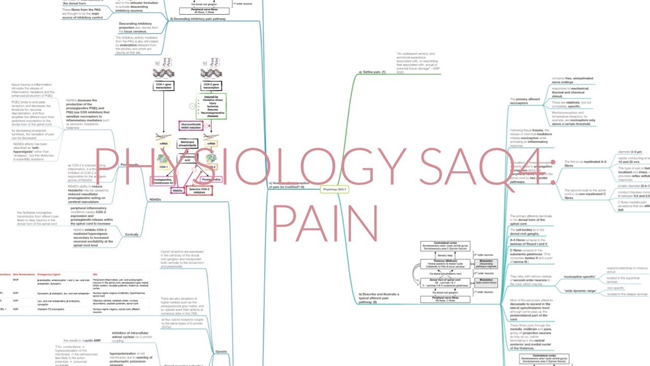
PHYSIOLOGY SAQ 4: PAIN
807
The complete afferent pain pathway involves multiple stages: (1) Primary nociceptors detect tissue damage through mechanical, thermal, or chemical stimuli; (2) Nociceptive signals travel via two parallel pathways—fast-conducting A-delta fibers (2-5 μm, 12-30 m/s) for sharp pain and slow-conducting C-fibers (0.4-1.2 μm, 0.5-2 m/s) for diffuse pain; (3) Signals synapse in the spinal cord dorsal horn, with A-delta fibers targeting laminae I/V and C-fibers targeting laminae II/III (substantia gelatinosa); (4) Second-order neurons relay signals via the lateral spinothalamic tract through the brainstem to the thalamus; (5) Thalamic nuclei project to somatosensory cortex (areas 1 and 2) and cingulate gyrus for emotional processing.
Click to watch →
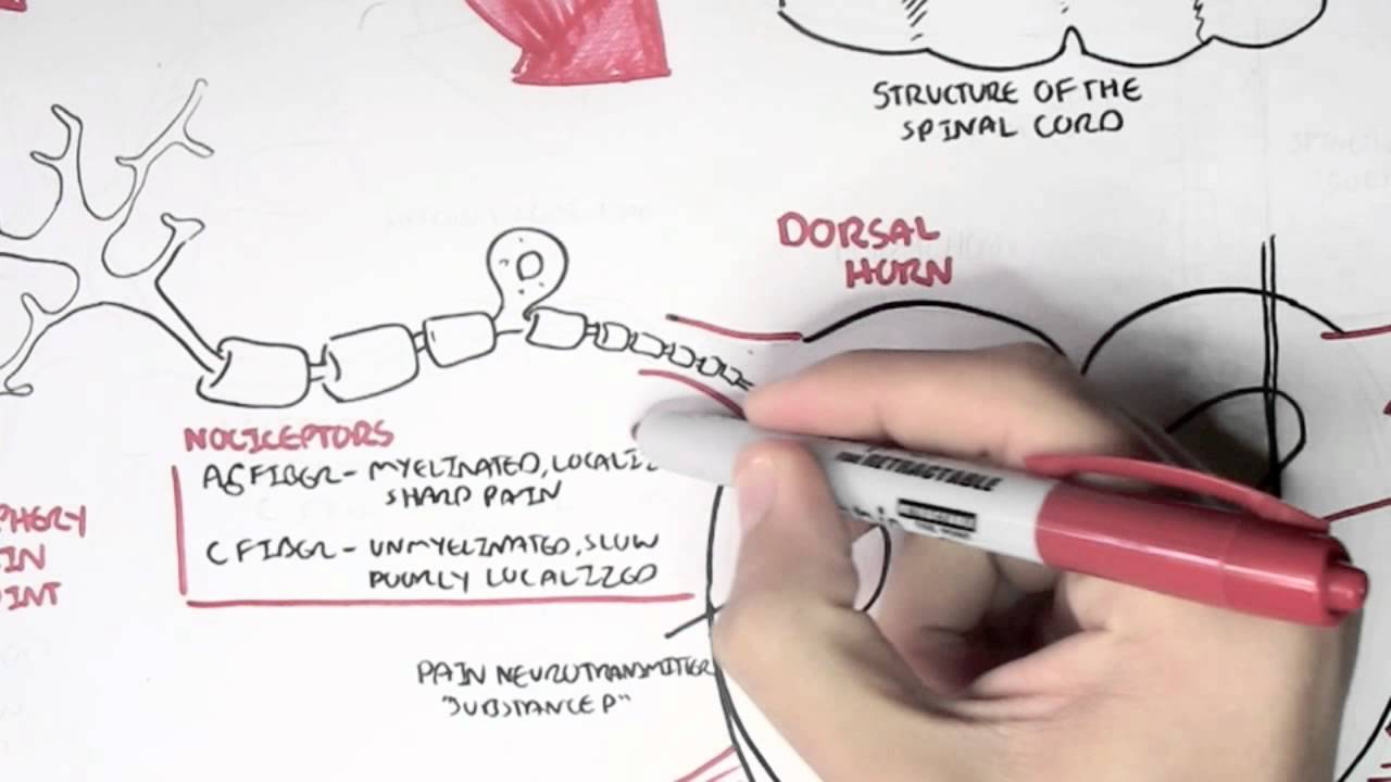
Nociceptors - An Introduction to Pain
1.4M
There are two main types of nociceptor fibers with different characteristics. Alpha delta fibers are small myelinated nerve cells that transmit fast, well-localized sharp pain sensations. In contrast, C fibers are smaller and unmyelinated, producing slow, poorly localized pain that feels burning or throbbing. These differences explain why some pain sensations are immediate and precise while others are delayed and diffuse.
Click to watch →

Fisiología y fisiopatología del dolor y la nocicepción - Dr. José Alejandro García
1.3K
Pain is defined as an unpleasant sensory and emotional experience associated with actual or potential tissue damage, transmitted through a three-neuron pathway (first-order neurons from receptors to dorsal horn, second-order neurons to thalamus, third-order neurons to somatosensory cortex) via Type A-Delta and Type C nerve fibers, with the process involving transduction, transmission, modulation, and perception, and modulated by endogenous opioid and non-opioid systems as well as the gate control theory.
Click to watch →
Deep Dive
The fundamentals of action potentials, ion channels, and synaptic transmission in the nervous system.
Human nervous system physiology
98.7K
Synaptic transmission involves three main steps: action potential arrival at axon terminal, neurotransmitter release into synaptic cleft, and receptor binding on postsynaptic membrane. When an action potential reaches the axon terminal, voltage-gated calcium channels open, allowing calcium influx. Increased intracellular calcium causes synaptic vesicles containing neurotransmitters to fuse with the presynaptic membrane, releasing their contents into the synaptic cleft by exocytosis. Neurotransmitters diffuse across the cleft and bind to receptors on the postsynaptic membrane. When neurotransmitters bind to postsynaptic receptors, ion channels open that remain active as long as neurotransmitter is bound. The type of channel opened determines whether the postsynaptic cell becomes excited (depolarized) or inhibited (hyperpolarized). Synaptic transmission terminates when neurotransmitters dissociate from receptors and are removed by reuptake, uptake by glial cells, or enzymatic breakdown.
Click to watch →
Ion channels and membrane potential
42.6K
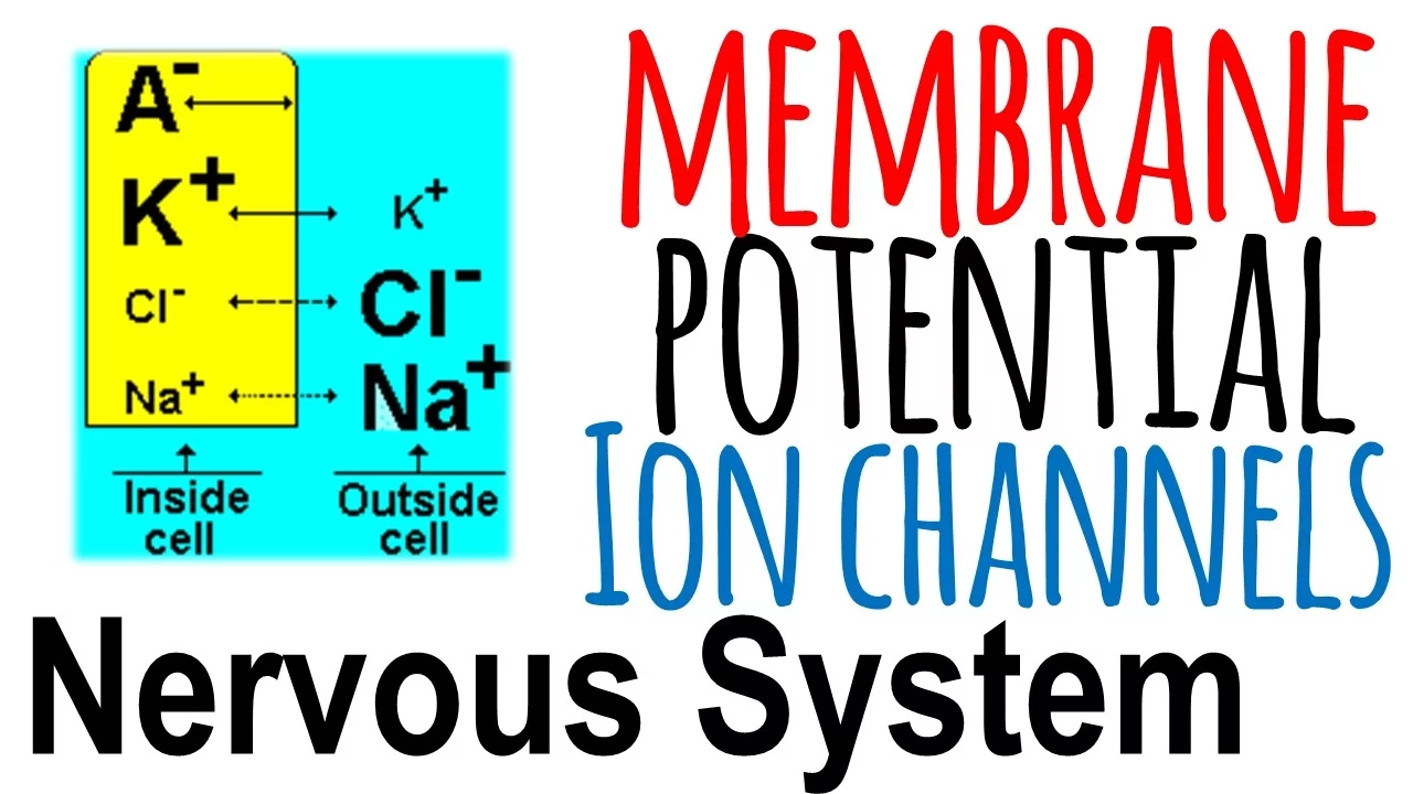
Action potentials are generated through coordinated voltage-gated channel activity: sodium channels open first causing depolarization, followed by potassium channels causing repolarization. Chemically-gated channels mediate synaptic transmission at dendrites and cell bodies, with excitatory neurotransmitters like acetylcholine promoting signaling and inhibitory neurotransmitters like GABA terminating it. Different channel types are regionally distributed—passive channels throughout, chemically-gated in dendrites/cell bodies, voltage-gated in axons. Myelinated axons concentrate channels at nodes of Ranvier for efficient signal propagation. This organization enables neurons to integrate inputs, generate outputs, and transmit signals efficiently.
Click to watch →
ZIDD SERIES PART-I | INTRODUCTION OF AUTONOMIC NERVOUS SYSTEM
158K
Synaptic transmission involves: (1) Impulse conduction to presynaptic terminal, (2) Neurotransmitter release via calcium-triggered exocytosis, (3) Neurotransmitter binding to postsynaptic receptors, (4) Generation of postsynaptic potentials, (5) Termination through metabolism, reuptake, or diffusion. Excitatory neurotransmitters increase sodium permeability, causing depolarization and generating Excitatory Postsynaptic Potentials (EPSP). Inhibitory neurotransmitters increase chloride permeability, causing hyperpolarization and generating Inhibitory Postsynaptic Potentials (IPSP). The resting membrane potential is approximately -70 mV, with sodium higher outside and potassium higher inside. Action potentials propagate by depolarizing adjacent membrane regions. When reaching the presynaptic terminal, calcium influx triggers synaptic vesicle fusion and neurotransmitter release. Neurotransmitter action is terminated through: (1) Local degradation by enzymes (acetylcholine by acetylcholinesterase), (2) Reuptake into presynaptic neuron (norepinephrine by norepinephrine transporter), (3) Diffusion away from synaptic cleft. Cotransmission involves release of multiple neurotransmitters from the same neuron, with cotransmitters modulating primary neurotransmitter effects and acting on presynaptic autoreceptors for feedback regulation.
Click to watch →

٦- محاضرة السيال العصبي⚡️ | هدية لطلاب تانية ثانوي ❤️
395K
Synaptic transmission involves: action potential arrival at presynaptic terminal, calcium channel opening, calcium influx, vesicle fusion, neurotransmitter release into synaptic cleft, diffusion across cleft, receptor binding on postsynaptic membrane, and postsynaptic depolarization. If threshold is reached, a new action potential is generated. Calcium ions play a crucial role by triggering vesicle fusion. Neurotransmitters bind to specific receptors, causing ion channels to open and potentially triggering a new action potential.
Click to watch →
Anatomy & Physiology Chapter 11 Part B: Nervous System and Nervous Tissue Lecture
64.2K
Neurons generate electrical signals through changes in membrane potential, where resting membrane potential (~-70 mV) is maintained by the sodium-potassium pump and differential ion permeability, and action potentials propagate through axons via voltage-gated sodium and potassium channels opening in sequence, with conduction velocity determined by axon diameter and degree of myelination.
Click to watch →
Deep Dive
The acute inflammatory response, specifically the role of chemical mediators (such as prostaglandins, bradykinin, and cytokines) released during tissue damage.

Pathology | Inflammation (1): The Acute Inflammatory Response بالتفصيل
648
The acute inflammatory response begins with tissue damage causing cell death (necrosis). Damaged cells release chemical mediators including histamine, bradykinin, and prostaglandins. These mediators act on endothelial cell receptors to produce inflammatory effects. The sequence includes: local tissue damage, chemical mediator release, vascular changes (vasoconstriction followed by vasodilation), increased capillary permeability, and cellular infiltration by immune cells. This coordinated response aims to eliminate pathogens and initiate tissue repair.
Click to watch →
Chemical mediators of inflammation
7.4K
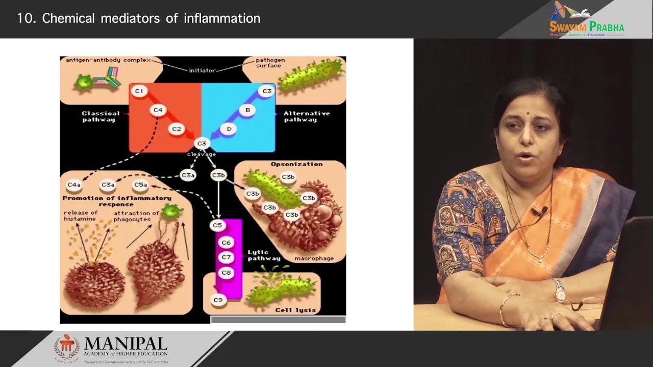
Chemical mediators of inflammation have specific effects: Vasodilation is caused by histamine, serotonin, prostaglandins, and nitric oxide; increased vascular permeability results from histamine, serotonin, prostaglandins, C3a, C5a (anaphylatoxins), and platelet activating factor; chemotaxis is mediated by C5a, leukotriene B4, and cytokines; pain is caused by prostaglandins and bradykinin; fever is mediated by cytokines. Acute inflammation has three outcomes: complete resolution when the offending agent is eliminated and tissue regenerates; fibrosis/scarring when significant damage prevents complete regeneration; chronic inflammation when injury persists, characterized by persistent tissue injury, fibrosis, and vascular proliferation with neutrophils/macrophages replaced by lymphocytes and plasma cells.
Click to watch →

8 general pathology, inflammation 1(definition, chemical mediators), Dr Ebtehal M
219
Key mediator functions in inflammation: (1) Vasodilation and increased permeability - histamine, prostaglandins, bradykinin, nitric oxide, leukotrienes, PAF, complement, cytokines, and bacterial products. (2) Chemotaxis - cytokines, chemokines, complement, and bacterial products attract white blood cells. (3) Pain and fever - prostaglandins (act on hypothalamus), bradykinin, and cytokines. (4) Tissue damage - lysosomal enzymes, reactive oxygen species, and nitric oxide. The inflammatory response requires balance, as excessive or prolonged inflammation can cause tissue damage.
Click to watch →

Inflammation | Vascular Events | Immunology | Dr Najeeb
686.3K
When tissue is injured, local cells release chemical mediators that control different steps of inflammation. These mediators include prostaglandins, histamine, leukotrienes, platelet activating factors, and cytokines (interleukin-1 and tumor necrosis factor). These substances are derived from injured cell membranes, granules, nuclear activation, and plasma protein breakdown products.
Click to watch →

Medical School Pathology, Chapter 2d
62.4K
Acute inflammation involves multiple chemical mediators working in concert. Platelet Activating Factor (PAF) is a phospholipid produced by neutrophils, basophils, platelets, and endothelial cells that stimulates both coagulation and inflammation. Cytokines (larger proteins from lymphocytes and macrophages) and chemokines (smaller proteins that attract PMNs) coordinate the immune response. TNF-α and IL-1 are key cytokines produced by macrophages that control other inflammatory mediators. Nitric oxide, synthesized from arginine by nitric oxide synthase, is one of the most potent vasodilators. Neutrophil granules mature from primary (acid hydrolases, lysozyme) to secondary granules (collagenase, alkaline phosphatase, lactoferrin, lysine). Free radicals like superoxide and hydroxyl radicals cause tissue damage. Neuropeptides such as Substance P and Neurokinin A, produced in the CNS, also participate in inflammatory processes.
Click to watch →
Deep Dive
The conceptual difference between acute (nociceptive) pain as a protective mechanism and chronic (maladaptive) pain.

Pain | Pathway and Analgesics (NSAIDs, Opioids, Antidepressants, and Anticonvulsants)
212.3K
Pain classification by duration: acute (<3 months, nociceptive, from tissue damage) versus chronic (>3 months, often neuropathic). Unlike other senses that desensitize, pain amplifies through central sensitization—a positive feedback loop where inflammation lowers pain neuron thresholds, creating persistent pain even without ongoing tissue damage. This explains why unmanaged acute pain can become chronic. Nociceptive pain serves protective purposes, while chronic neuropathic pain results from nerve dysfunction. This distinction guides treatment selection, as inflammatory pain responds to NSAIDs while neuropathic pain requires different approaches.
Click to watch →

CIÊNCIA DA DOR NA PRÁTICA: Série de 8 artigos da JOSPT (Grupo de estudos ReabDor)
102
Nociception functions as a protective mechanism for the organism, serving as a biological alarm system of high speed and precision. Acute pain represents the failure of nociception as a last resort protective mechanism. For example, when touching fire, the nociceptive response prompts withdrawal to prevent tissue damage. In chronic pain conditions, nociception may no longer be present for patients to experience pain, yet pain persists due to other mechanisms. This distinction is essential for understanding why pain can persist beyond tissue healing.
Click to watch →

Dolor crónico síntomas, tratamientos y soluciones efectivas | ALIVIAR y VIVIR sin DOLOR
142.7K
Acute pain is a natural protective mechanism that signals the body when something is wrong, such as a headache lasting one day. It serves an adaptive purpose by prompting us to identify and address the underlying cause, such as poor sleep, inadequate nutrition, or dehydration. Once the cause is resolved, acute pain typically disappears. In contrast, chronic pain persists beyond three months and has lost its useful, adaptive function. Chronic pain results from neural circuit imbalances where nerve cells become sensitized, causing even non-painful stimuli like a gentle touch to be perceived as painful.
Click to watch →

2026 07 17 Kapitel 7
433
Acute pain has a clear protective function and disappears once healing occurs. Chronic pain persists even after tissue has healed because the nervous system has learned to remain in a heightened protective state. The nervous system can learn, and like an alarm system that becomes over-responsive over time, it can continue to trigger pain responses even when the original threat has passed. This is not imagined pain but a real neurological response that can be changed through new experiences and retraining.
Click to watch →

Pain and the Brain
135.3K
Acute pain serves as a protective warning signal indicating tissue injury, prompting individuals to stop harmful activities and learn from the experience. Chronic pain, however, persists beyond normal healing time and represents a distinct disease of the nervous system rather than merely a symptom of another condition. A key clinical feature is allodynia—pain produced by normally non-painful stimuli like sunburned skin or arthritic joints during movement. The transition from acute to chronic pain involves maladaptive changes in the nervous system that amplify pain signals, making it fundamentally different from the original protective purpose of pain.
Click to watch →
Prerequisite Knowledge
- Concept 01Basic physiology of nociception, including the distinction between A-delta and C primary afferent fibers.
- Concept 02The fundamentals of action potentials, ion channels, and synaptic transmission in the nervous system.
- Concept 03The acute inflammatory response, specifically the role of chemical mediators (such as prostaglandins, bradykinin, and cytokines) released during tissue damage.
- Concept 04The conceptual difference between acute (nociceptive) pain as a protective mechanism and chronic (maladaptive) pain.
Subsequent Learning
- Step 01Central Sensitization, exploring how sustained peripheral input leads to neuroplastic changes in the dorsal horn of the spinal cord and the brain.
- Step 02Clinical pharmacology for pain management, focusing on how specific drugs (such as NSAIDs, local anesthetics, and membrane stabilizers) target peripheral transduction and transmission.
- Step 03The Biopsychosocial Model of chronic pain, analyzing how cognitive, emotional, and social factors influence nociceptive processing.
- Step 04Physiotherapy intervention strategies for sensitized nervous systems, including graded motor imagery, sensory desensitization, and pacing techniques.
Peripheral Sensitization
Chronic Inflammation
Peripheral Sensitization
0:00Playing Section
- 1
Explains inflammation-driven receptor activation and action potential generation.
- 2
Discusses ion channel changes that increase neural sensitivity.
- 3
Describes primary and secondary hyperalgesia as normal clinical phenomena.
Central Sensitization and the Neuromatrix Theory of Pain
While peripheral sensitization explains increased sensitivity at the site of tissue injury, modern pain neuroscience suggests that chronic pain is often driven primarily by central sensitization and the brain's 'neuromatrix.' The Neuromatrix Theory, developed by Ronald Melzack, proposes that pain is a multidimensional output produced by a widely distributed neural network in the brain, rather than a simple reaction to peripheral sensory inputs. In many chronic pain conditions, the central nervous system becomes hyperexcitable, amplifying signals and generating pain even in the absence of ongoing peripheral tissue damage or inflammation. Critics of an over-reliance on peripheral models argue that focusing too heavily on localized tissue and peripheral nerves can lead to ineffective treatments. Instead, this alternative perspective emphasizes that effective chronic pain management must target central nervous system processing, cognitive-emotional factors, and overall threat perception through a comprehensive biopsychosocial approach.
Deep Dive
Central Sensitization, exploring how sustained peripheral input leads to neuroplastic changes in the dorsal horn of the spinal cord and the brain.
CIOST 2019: Palestra: Terapia Manual e Dor - Dr. Rodrigo Vasconcelos
2.9K
The dorsal horn of the spinal cord undergoes neuroplastic changes that influence central sensitization. When nociceptive stimuli from peripheral receptors reach the dorsal horn, they excite postsynaptic neurons. In normal conditions, this produces appropriate responses, but in central sensitization, the behavior of these neurons is altered. The temporal summation of repetitive mechanical stimuli of the same intensity causes postsynaptic neurons to fire abnormally, altering information processing not only at the spinal level but also at the cortical level. This mechanism explains how repeated or persistent stimuli can lead to amplified pain responses.
Click to watch →

ION 328. ZOOM lecture, Nijs: Central Sensitization and Nociplastic Pain 13.03.23
1.5K
Central sensitization occurs when persistent nociceptive input leads to synaptic changes in the central nervous system. When sensory input (such as muscle tension from prolonged uncomfortable positions) continues for extended periods (like one week), synapses become more efficient through increased neurotransmitter production by presynaptic neurons and enhanced neurotransmitter capture by postsynaptic neurons. This results in amplified pain processing at both spinal cord and brain levels, where identical sensory inputs produce significantly greater pain output compared to normal conditions.
Click to watch →
Central Sensitization
11.9K
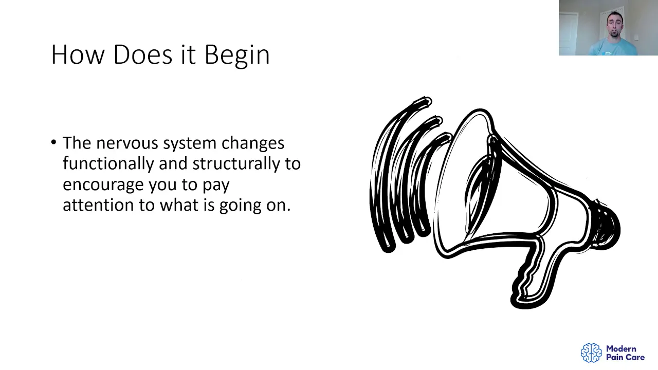
Central sensitization involves both structural and functional changes in the nervous system. Structurally, there is loss of inhibitory interneurons in the dorsal horn of the spinal cord and potentially further up the brainstem pathway. Functionally, these changes mean that full amounts of nociception reach the brain without being gated out, causing the brain to perceive increased threat and produce greater pain output. In heterosynaptic facilitation cases, light touch can cross over into nociceptive circuits due to lost inhibitory control, causing allodynia where non-noxious stimuli produce pain.
Click to watch →

Pain : Pathophysiology & Management By Dr Nishant Arya
6.2K
Central sensitization occurs at the level of the dorsal horn of the spinal cord. This is a key distinction from peripheral sensitization, which occurs at the level of peripheral nerve terminals. The dorsal horn is the site where neuroplastic changes lead to altered pain processing and conditions like allodynia.
Click to watch →

Peripheral and Central Pain Mechanisms: Finding Common Ground - Daniel Clauw and Michael Gold
533
Central sensitization involves persistent increases in spinal cord neuron responses to both high-threshold and low-threshold stimuli, producing hyperalgesia and allodynia. Unlike hippocampal memory, it requires continuous primary afferent input to maintain plastic changes. Windup represents temporal summation of repeated stimuli. Microglial activation contributes through cytokine release affecting GABA inhibition. Descending facilitation from RVM switches from inhibition to excitation. All central changes depend on peripheral afferent drive.
Click to watch →
Deep Dive
Clinical pharmacology for pain management, focusing on how specific drugs (such as NSAIDs, local anesthetics, and membrane stabilizers) target peripheral transduction and transmission.

Why does it hurts? Dr Ahmed Kafafy
362
Nociceptors exist everywhere except the brain. A-delta and C fibers carry pain signals. Peripheral sensitization occurs when inflammatory mediators (prostaglandins, serotonin) lower receptor thresholds. NSAIDs work by blocking cyclooxygenase enzymes, reducing prostaglandin production. Versatis (5% lidocaine patch) is a sodium channel blocker for post-herpetic neuralgia. Capsaicin cream (0.025%) depletes substance P for peripheral neuropathies. These drugs target the transduction and transmission stages of pain.
Click to watch →
Pharmacology of Pain Part 1 of 2
3.4K
This video explains the pharmacology of pain management through two key mechanisms: NSAIDs work by inhibiting cyclooxygenase enzymes (COX-1 and COX-2) in the arachidonic acid cascade, reducing inflammatory prostaglandins that cause pain and swelling, but this inhibition also affects protective prostaglandins leading to side effects like gastric irritation and renal issues; local anesthetics block sodium channels in nerve cells, preventing the transmission of pain signals from nociceptors to the spinal cord by binding to sodium channel proteins and interrupting the action potential wave, with examples including lidocaine (rapid onset, short duration) and bupivacaine (slower onset, longer duration); pharmacokinetics encompasses the five essential principles of drug handling: liberation (drug release), absorption (entry into bloodstream), distribution (spread throughout body), metabolism (liver breakdown), and excretion (kidney elimination).
Click to watch →

FARMACOTERAPIA DEL DOLOR - Eliberto Ruiz Ramirez
7.3K
Pain signals travel from damaged tissue through sensory neurons via voltage-gated sodium channels to the spinal cord and cerebral cortex. Local anesthetics block these channels for immediate surgical pain relief. As procedures continue, prostaglandins and leukotriens are released as inflammatory mediators that amplify pain signals. NSAIDs like naproxen, ibuprofen, diclofenac, and ketoprofen inhibit prostaglandin production (and leukotriens for some agents) to reduce amplified postoperative pain. Local anesthetics become less effective as tissue pH becomes more acidic during inflammation.
Click to watch →
vias del dolor y Farmacos analgésicos #Farmacia...farm2
4.2K
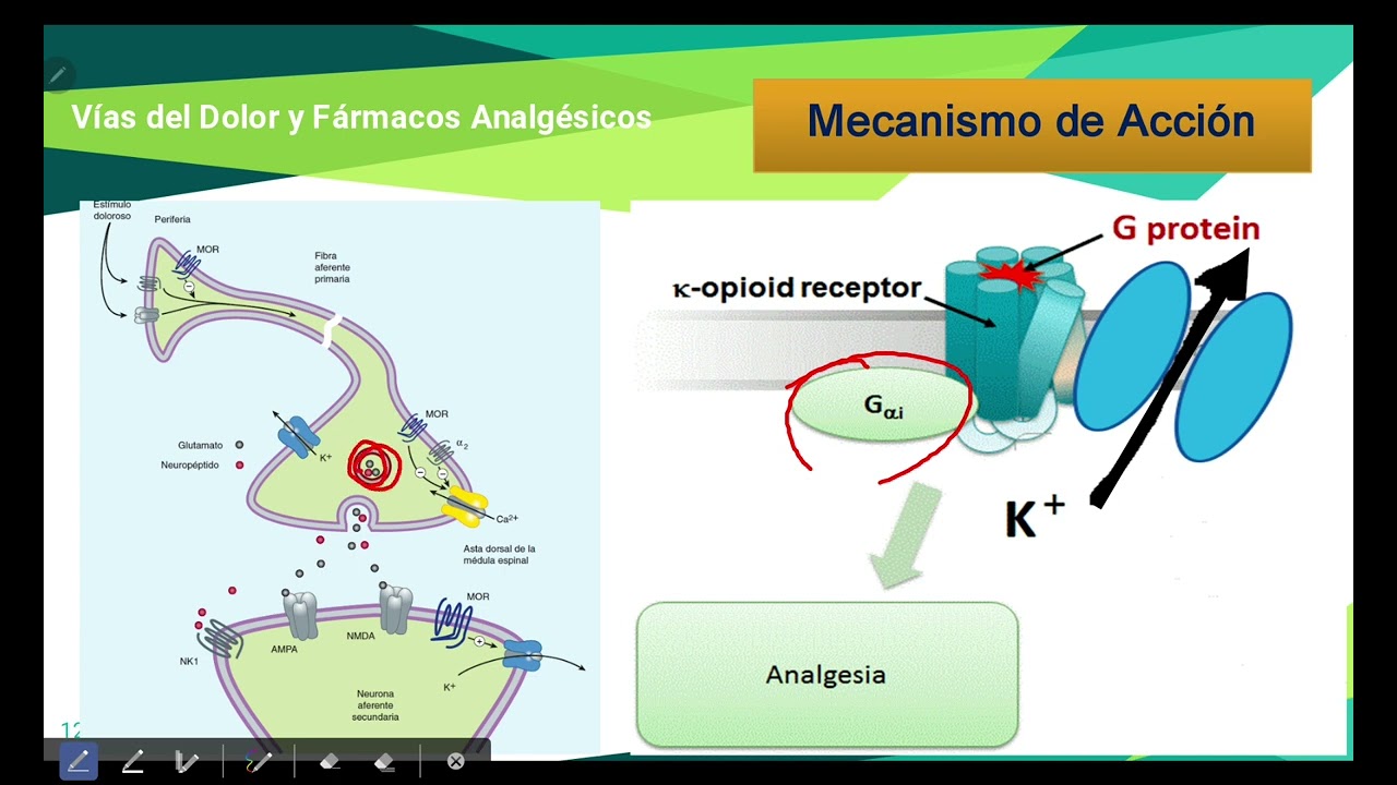
Pain is transmitted through a pathway where peripheral stimuli activate free nerve endings, releasing histamine, serotonin, and prostaglandins that travel via Type C fibers to the posterior root ganglion, where substance P and glutamate are released at the first synapse; signals then ascend 2-3 spinal segments, cross contralaterally, and reach the thalamus before processing in the somatosensory cortex. Analgesics are classified into two main groups: opioids (central-acting) that bind to mu, delta, and kappa receptors via Gi protein-coupled mechanisms to block calcium entry and prevent neurotransmitter release, and NSAIDs (peripherally-acting) that inhibit cyclooxygenase enzymes to reduce prostaglandin synthesis. Opioids include natural (morphine, codeine), semisynthetic (heroin, buprenorphine), and synthetic (fentanyl, methadone) types, with varying potencies and clinical applications from acute pain management to addiction treatment.
Click to watch →

How Painkillers (Paracetamol, NSAIDs, Opioids) Really Work | Pharmacology
167
Pain transmission begins when tissue injury triggers phospholipase A2 to release arachidonic acid, which is converted to prostaglandins by COX-1 and COX-2 enzymes. Prostaglandins sensitize nociceptors and drive inflammation. Signals travel through peripheral nerves to the spinal cord dorsal horn, where neurotransmitters (glutamate, substance P, CGRP) transmit pain. COX-2 activation in the spinal cord causes central sensitization, amplifying pain signals. In the brain, prostaglandins regulate temperature and pain perception. Three drug classes target this pathway at different points: paracetamol blocks central COX enzymes; NSAIDs inhibit both COX-1 and COX-2 peripherally and centrally; opioids bind to opioid receptors to block pain signal transmission.
Click to watch →
Deep Dive
The Biopsychosocial Model of chronic pain, analyzing how cognitive, emotional, and social factors influence nociceptive processing.

Chronische Schmerzen | Wie das System Schmerzen fördert
405.5K
The biopsychosocial model recognizes that chronic pain results from the interaction of three interconnected domains: biological factors (physical causes and nerve function), psychological factors (mental health, emotions, cognition), and social factors (relationships, work, societal attitudes). Effective chronic pain management requires addressing all three dimensions simultaneously, as ignoring any one component leads to incomplete treatment outcomes.
Click to watch →
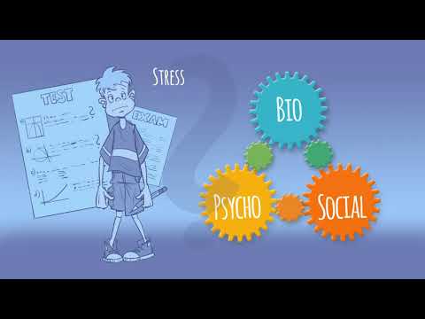
The biopsychosocial model - Explanation of chronic pain disorders
86K
The biopsychosocial model explains chronic primary pain as an independent disease arising from the interaction of biological, psychological, and social factors, where these three dimensions overlap and influence each other; effective treatment requires addressing all three dimensions through multimodal, non-pharmaceutical strategies that consider the whole person rather than focusing solely on physical causes.
Click to watch →

¿Por qué tu dolor de espalda NO DESAPARECE? Esto es lo que tienes que hacer
767
This segment introduces the biopsychosocial model (2007) as the modern approach to understanding chronic pain. Unlike the biomédical model, it considers physical, psychological, and social factors: habits, exercise, nutrition, rest, beliefs about pain, stress, anxiety, work environment, and support systems. The instructor explains that chronic back pain causes multiple changes in the body: muscle loss, joint stiffness, increased pain sensitivity, sleep disruption, fatigue, fear of movement, and emotional changes like anxiety and depression.
Click to watch →

30 minutos de massagem relaxante
158.9K
The biopsychosocial model of pain considers that pain is influenced by biological, psychological, and social factors. The video explains that emotional and psychological states can significantly affect pain perception and treatment outcomes. Psychological factors can contribute to the development of chronic pain conditions, and negative psychological states can negatively affect treatment outcomes. The nocebo effect demonstrates how negative expectations or suggestions can worsen pain symptoms. Effective pain management must address all three dimensions rather than focusing solely on physical symptoms.
Click to watch →
How to Get Bulletproof Tendons ft. Professor Keith Baar | Doctor Yaad Podcast
151.5K
The biopsychosocial model explains that pain perception involves biological (tissue damage), psychological (brain interpretation), and social (environmental context) components. Anxiety and negative social environments increase pain perception, while positive coaching and external focus techniques reduce it. This model supports early movement after injury, as immobilization and fear-based messaging can perpetuate chronic pain states. The eight-week isometric rehabilitation course is essential—many patients prematurely discontinue treatment after feeling better, risking recurrence.
Click to watch →
Deep Dive
Physiotherapy intervention strategies for sensitized nervous systems, including graded motor imagery, sensory desensitization, and pacing techniques.

What to do... you've had your neck assessed/treated for headache or migraine, and things are worse..
157
Graded motor imagery is a strategy that can help retrain a sensitized nervous system. This approach involves imagining movements or using mirrors to trick the brain into performing more movements. It helps reduce nervous system sensitivity before manual therapy can be safely applied.
Click to watch →

How Neurologic Changes Drive Chronic Pain — And What Actually Helps
424
Research shows that normalizing neurologic adaptations requires strategically integrating multiple evidence-based techniques in the right order at the right time. Key techniques include: Pain Neuroscience Education (PNE), which is evidence-based and can lower pain levels on its own; Graded Motor Imagery (GMI) including laterality training, explicit motor imagery, and mirror therapy, which improves sensory processing and weakens automatic pain responses; Neurodynamic nerve drills, which are specific movements targeting nerves to reduce peripheral sensitivity; Sensory discrimination strategies using non-painful tactile inputs to improve brain-body communication; and Pacing, which finds the sweet spot between overdoing and underdoing to build activity tolerance while reducing pathway sensitivity. Introducing all techniques at once is a common mistake that can cause the pain system to react negatively. Instead, techniques should be introduced in phases: Phase 1 - Understanding neurologic adaptations; Phase 2 - Setting the foundation for recovery; Phase 3 - Expanding activity tolerance; Phase 4 - Normalizing hypersensitive pain pathways; Phase 5 - Rebuilding physical strength safely once sensitivity is reduced.
Click to watch →
Pain Science Basics for Clinical Practice Webinar: What Pain Is (and What It Isn’t)
121
Advanced interventions for central sensitization include graded motor imagery progressing through lateralization, visualization, and mirror therapy. Traditional desensitization may heighten alarm responses, so graded approaches are preferred. Work injuries create higher threat levels for the nervous system because they threaten livelihood and earning capacity. Without addressing the heightened nervous system state first, functional training fails. Osteoarthritis is common but shouldn't be framed as devastating; education must be gentle. Complex Regional Pain Syndrome represents the highest end of the central sensitization spectrum. Sensory testing comparing opposite body sides helps identify dysfunction. Clear communication that this is a sensory system problem empowers patients to understand their condition.
Click to watch →

The #1 Reason You’re Not Recovering From Chronic Fatigue & Sensitives
114
Pacing differs fundamentally from graded exercise therapy (GET), which uses a fixed schedule of increasing aerobic activity. While some trials suggest GET was moderately effective, most patients report it causes significant long-term harm when pushed by therapists who don't understand PEM. The key difference is that GET uses rigid schedules while pacing uses the patient's body signals as the guide. Pacing is the crucial first step that stops constant crashes and stabilizes the nervous system. Once stabilized, patients can gradually expand their life. This approach works by retraining the limbic system, which can get stuck in high alert and cause fatigue, brain fog, and sensitivities. By combining pacing with calming exercises, patients teach their nervous system that normal activities are no longer threats, calming the fight-or-flight response and helping with PEM more than anything else.
Click to watch →

Congress 2014 - Behaviour Change in Physiotherapy, Marsha Macrae
369
Graded exposure involves identifying patient fears in a hierarchy and systematically exposing patients to feared activities to decrease fear responses. Pacing teaches patients to regulate their activity levels rather than pushing through pain. Both techniques address the physical behaviors that perpetuate chronic pain. Many patients lack awareness of how to pace themselves appropriately, requiring physiotherapists to help identify barriers and establish realistic activity goals.
Click to watch →
Peripheral Sensitization
Chronic Inflammation
Peripheral Sensitization
0:00- 1
Explains inflammation-driven receptor activation and action potential generation.
- 2
Discusses ion channel changes that increase neural sensitivity.
- 3
Describes primary and secondary hyperalgesia as normal clinical phenomena.
Central Sensitization and the Neuromatrix Theory of Pain
While peripheral sensitization explains increased sensitivity at the site of tissue injury, modern pain neuroscience suggests that chronic pain is often driven primarily by central sensitization and the brain's 'neuromatrix.' The Neuromatrix Theory, developed by Ronald Melzack, proposes that pain is a multidimensional output produced by a widely distributed neural network in the brain, rather than a simple reaction to peripheral sensory inputs. In many chronic pain conditions, the central nervous system becomes hyperexcitable, amplifying signals and generating pain even in the absence of ongoing peripheral tissue damage or inflammation. Critics of an over-reliance on peripheral models argue that focusing too heavily on localized tissue and peripheral nerves can lead to ineffective treatments. Instead, this alternative perspective emphasizes that effective chronic pain management must target central nervous system processing, cognitive-emotional factors, and overall threat perception through a comprehensive biopsychosocial approach.
Basic physiology of nociception, including the distinction between A-delta and C primary afferent fibers.
The fundamentals of action potentials, ion channels, and synaptic transmission in the nervous system.
The acute inflammatory response, specifically the role of chemical mediators (such as prostaglandins, bradykinin, and cytokines) released during tissue damage.
The conceptual difference between acute (nociceptive) pain as a protective mechanism and chronic (maladaptive) pain.
Central Sensitization, exploring how sustained peripheral input leads to neuroplastic changes in the dorsal horn of the spinal cord and the brain.
Clinical pharmacology for pain management, focusing on how specific drugs (such as NSAIDs, local anesthetics, and membrane stabilizers) target peripheral transduction and transmission.
The Biopsychosocial Model of chronic pain, analyzing how cognitive, emotional, and social factors influence nociceptive processing.
Physiotherapy intervention strategies for sensitized nervous systems, including graded motor imagery, sensory desensitization, and pacing techniques.
20.4K views146likes2:10@TrustmeImaPhysiotherapist1Original Release: 2018-11-27
so if we look at something that's really common peripheral sensitization and take for example inflammation somebody is an ankle sprain the ankle is inflamed there changes to the tissue chemistry is inflammation within the tissues and so these receptors or ion channels will be activated an action potential will be generated and transmitted through the nervous system a couple of things happen that allow the system to be more sensitive or these action potentials to be generated more easily one of them is that these ion channels open and get plugged open for longer so they're just much more easier they're much easier to to be activated the second thing that can happen is that we get more and different ion channels expressed at the end of the primary afferent neuron so this is again we'll sensitize the the nervous system and allow action potentials to be generated more easily again these are really normal phenomenon if you think about your your person with an ankle sprain you'll see things like primary hyperalgesia hyperalgesia is just pain response to something you would normally think would produce pain so if I pressed on your ankle over the lateral ligament and I press hard enough it would generate a pain response primary hyperalgesia just means that in the area of the injury and the tissue is more sensitive and produces a painful response more easily we might also see secondary hyperalgesia or sensitivity in nearby areas and in this case it's usually secondary to the spread of inflammation it's a really normal phenomena that we see every day in clinical practice and so if demands that we're really aware of the stages of inflammation across acute subacute injuries and that we know what's predictable for tissue healing so that we know when it's not following that pathway and that we're cognizant of what happens with chronic or recurrent inflammatory conditions so think of osteoarthritis or rheumatoid arthritis
Up Next

58:26
Spinothalamic Tract: Ascending Tracts Explained (Part 1)
@DoctorNajeeb
953.6K views•2011-02-13

48:47
Graphic Medicine: Comics for Collaborative Healthcare Communication
@nationalpatientadvocate
189 views•2023-12-04

10:28
Neuroanatomy: Central and Peripheral Nervous System Divisions Explained
@AKLECTURES
136.2K views•2014-09-20

3:05
Stages of Labor and Vaginal Birth | Childbirth Animation
@nucleusmedicalmedia
52.1M views•2017-08-18
Related Study Plans & Knowledge Roadmaps
Structured learning paths in Medicine
medicine
Abnormal LFTs: Diagnostic Workup & Pathology
01Liver Anatomy and Basic Physiology
02Introduction to Liver Function Tests (LFTs)
03Hepatocellular vs. Cholestatic Patterns
+ 3 more learning modules
12h6 modules
Explore Roadmap
medicine
Analyzing Blood Smears: RBC, WBC & Platelets
01Introduction to Blood Components & Smear Preparation
02Normal Morphology of Blood Cells
03Erythrocyte Pathologies & Morphological Variants
+ 2 more learning modules
12h5 modules
Explore Roadmap
medicine
Mechanical Ventilation: Waveforms & Alarms
01Anatomy and Physiology of Breathing
02Introduction to Mechanical Ventilation & Core Modes
03Ventilator Settings and Waveform Interpretation
+ 2 more learning modules
12h5 modules
Explore Roadmap
medicine
Neuro Exam: Localization & Stroke Syndromes
01Foundations of Neuroanatomy and Vascular Supply
02The Neurological Examination: Step-by-Step
03Principles of Neurological Localization
+ 3 more learning modules
18h6 modules
Explore Roadmap