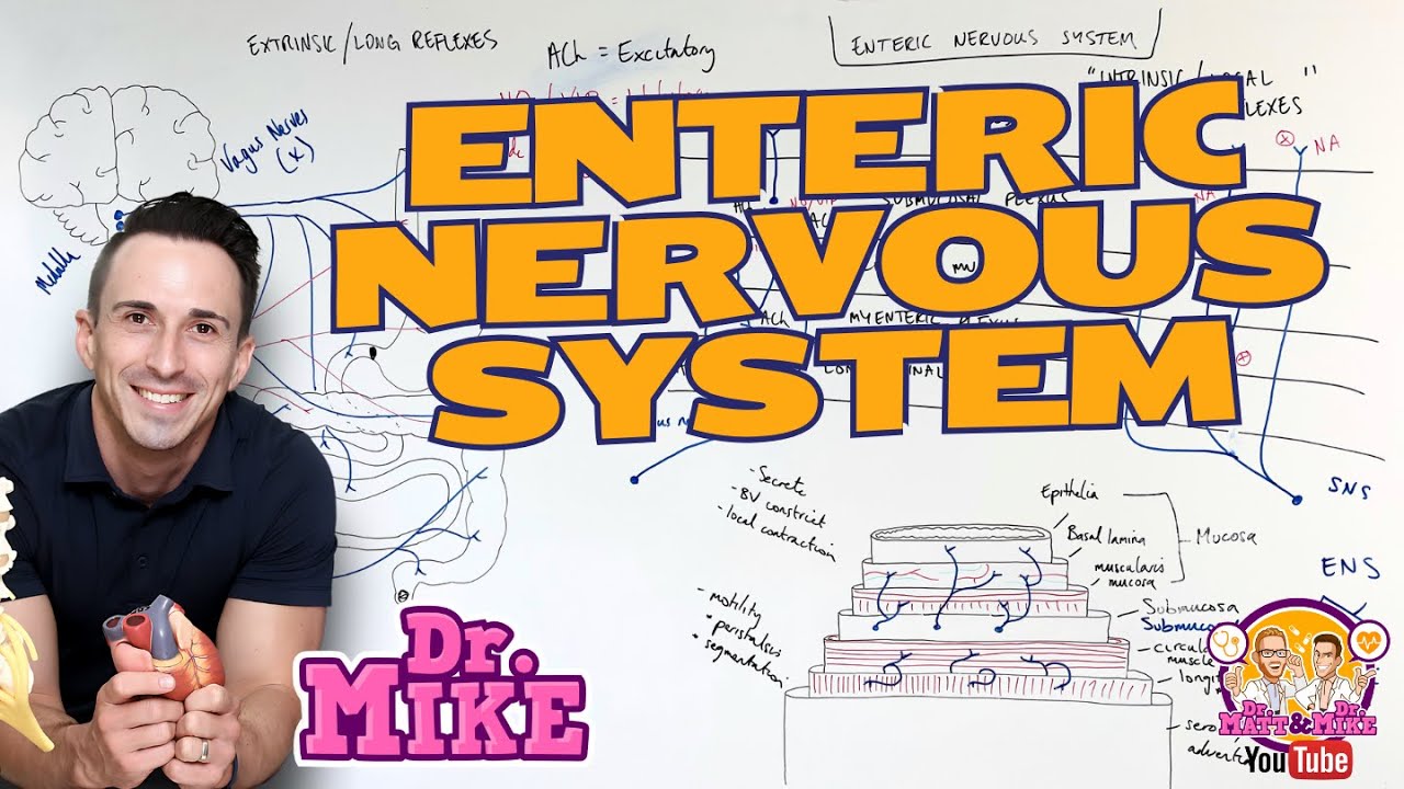
The enteric nervous system, often called the 'little brain of the intestine,' consists of two major plexuses: the myenteric plexus (located in the muscularis propria layer) and Meissner's plexus (located in the submucosa layer), which work together to regulate gastrointestinal motility and secretion; this system receives parasympathetic input primarily from S2-S4 spinal segments and sympathetic input from the inferior mesenteric ganglion, with the parasympathetic system promoting secretion and motility while the sympathetic system inhibits these functions.
Enteric Nervous System: Myenteric & Meissner's Plexus
Added:Deep Dive
The histological organization of the gastrointestinal tract, specifically the four concentric layers: mucosa, submucosa, muscularis externa, and serosa.
Sistema Digestório 1/5: Introdução, Funções, Órgãos e Histologia | Anatomia e etc
1.2M
The gastrointestinal tract is histologically organized into four concentric layers from the inside out: (1) Mucosa - the innermost layer composed of simple epithelial tissue in direct contact with food, plus underlying loose connective tissue (areolar tissue); (2) Submucosa - a layer of loose connective tissue that joins the mucosa to the muscular layer; (3) Muscularis externa - a thick layer of smooth muscle responsible for peristaltic movements; (4) Serosa (visceral peritoneum) - the outermost layer composed of epithelial and connective tissue that secretes serous fluid allowing organs to slide against each other.
Click to watch →

Травна система: травна трубка (глотка, стравохід, шлунок та кишечник). Гістологія
61.9K
The gastrointestinal tract has a consistent four-layer structure across all segments: (1) Mucosa - epithelium, lamina propria, and muscularis mucosae; (2) Submucosa - connective tissue with blood vessels, nerves, and lymphoid tissue; (3) Muscularis externa - smooth muscle arranged in circular and longitudinal layers; (4) Adventitia/Serosa - connective tissue with mesothelium when covered by peritoneum. The epithelium varies by segment: anterior and posterior divisions have stratified squamous non-keratinized epithelium (ectodermal type), while the middle division has simple columnar epithelium (endodermal type). The esophagus is an exception as it belongs to the anterior division but develops from endoderm.
Click to watch →
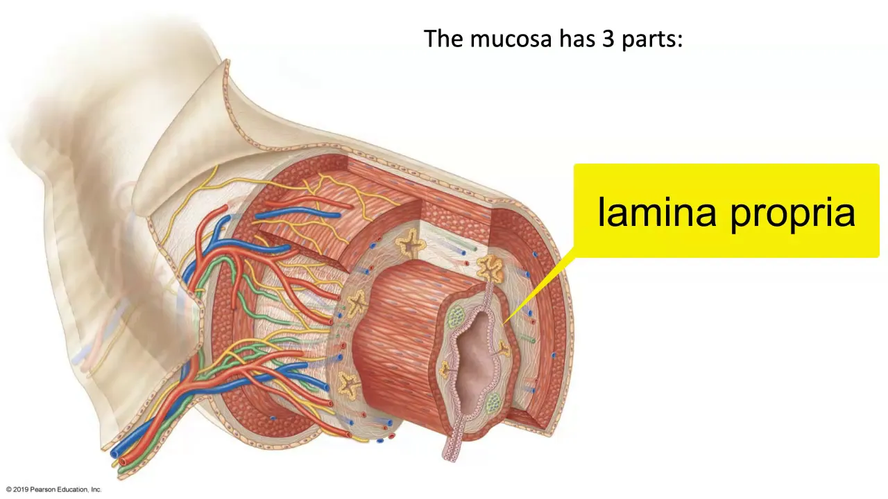
Layers of the GI Tract
95.1K
The gastrointestinal tract is a muscular tube extending from the mouth to the anus, consisting of four concentric layers: the mucosa (innermost layer with epithelium, lamina propria, and muscularis mucosae), the submucosa (connective tissue containing blood vessels, nerves, and glands), the muscularis externa (smooth muscle with inner circular and outer longitudinal coats), and the serosa/adventitia (outer protective layer); the epithelial type varies between stratified squamous at the ends and simple columnar in the middle sections.
Click to watch →
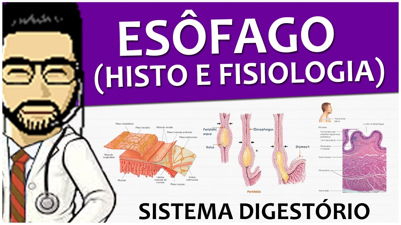
Sistema Digestório 09 - Esôfago P2 + Histologia do canal alimentar
44.5K
The gastrointestinal tract from esophagus to anal canal consists of four main layers: mucosa, submucosa, muscular layer, and serosa/adventitia. The mucosa has three sublayers: epithelium, lamina propria (connective tissue with vessels and cells), and muscularis mucosae (inner circular and outer longitudinal smooth muscle). The submucosa contains dense connective tissue, blood vessels, lymphatics, and Meissner's plexus (submucosal plexus) for secretion control. The muscular layer has circular and longitudinal smooth muscle with Auerbach's plexus (myenteric plexus) for motility control. The outermost layer is serosa (peritoneal lining) except in the esophagus, which has adventitia.
Click to watch →
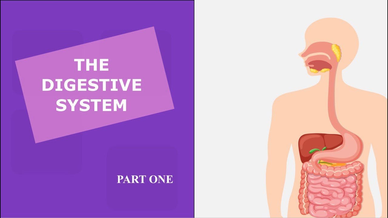
THE DIGESTIVE SYSTEM (PART I) - HISTOLOGY | الجهاز الهضمي (الجزء الاول) - انسجة
769
The GI tract wall consists of four layers: (1) Mucosa - contacts food, contains epithelium (stratified squamous in mouth/esophagus/colon, simple columnar in stomach/intestine), lamina propria, and muscularis mucosae; (2) Submucosa - connective tissue with blood vessels, lymphatic vessels, and nerve plexuses controlling muscle movement and gland secretion; (3) Muscularis externa - two layers of smooth muscle (inner circular, outer longitudinal) with nerve plexuses; (4) Serosa/adventitia - outermost layer, serosa when surrounded by mesothelium (abdominal organs), adventitia when not (thoracic organs).
Click to watch →
Deep Dive
Fundamentals of the Autonomic Nervous System (ANS), including the functional and anatomical differences between the sympathetic and parasympathetic divisions.
![[#1] SISTEMA NERVOSO AUTÔNOMO PARASSIMPÁTICO E SIMPÁTICO: Diferenças anatômicas | MK Fisiologia](https://i.ytimg.com/vi/7OIIYv2oXJw/maxresdefault.jpg)
[#1] SISTEMA NERVOSO AUTÔNOMO PARASSIMPÁTICO E SIMPÁTICO: Diferenças anatômicas | MK Fisiologia
108.5K
The autonomic nervous system (ANS), the efferent division of the visceral nervous system, controls internal organs like the heart, lungs, gastrointestinal tract, and glands to maintain homeostasis. The ANS is divided into three main systems: parasympathetic (craniosacral), sympathetic (thoracolumbar), and enteric. The key anatomical differences are: (1) Preganglionic neuron origin - parasympathetic neurons arise from brainstem nuclei and sacral spinal cord segments S2-S4, while sympathetic neurons originate only from thoracic and lumbar spinal cord segments T1-L2; (2) Ganglion location - parasympathetic ganglia are located close to or within target organs, whereas sympathetic ganglia are arranged in paravertebral chains along the spine; (3) Axon length - parasympathetic preganglionic axons are long and postganglionic axons are short, while sympathetic preganglionic axons are short and postganglionic axons are long; (4) Adrenal gland exception - the adrenal medulla is directly innervated by preganglionic sympathetic neurons without synaptic relay.
Click to watch →
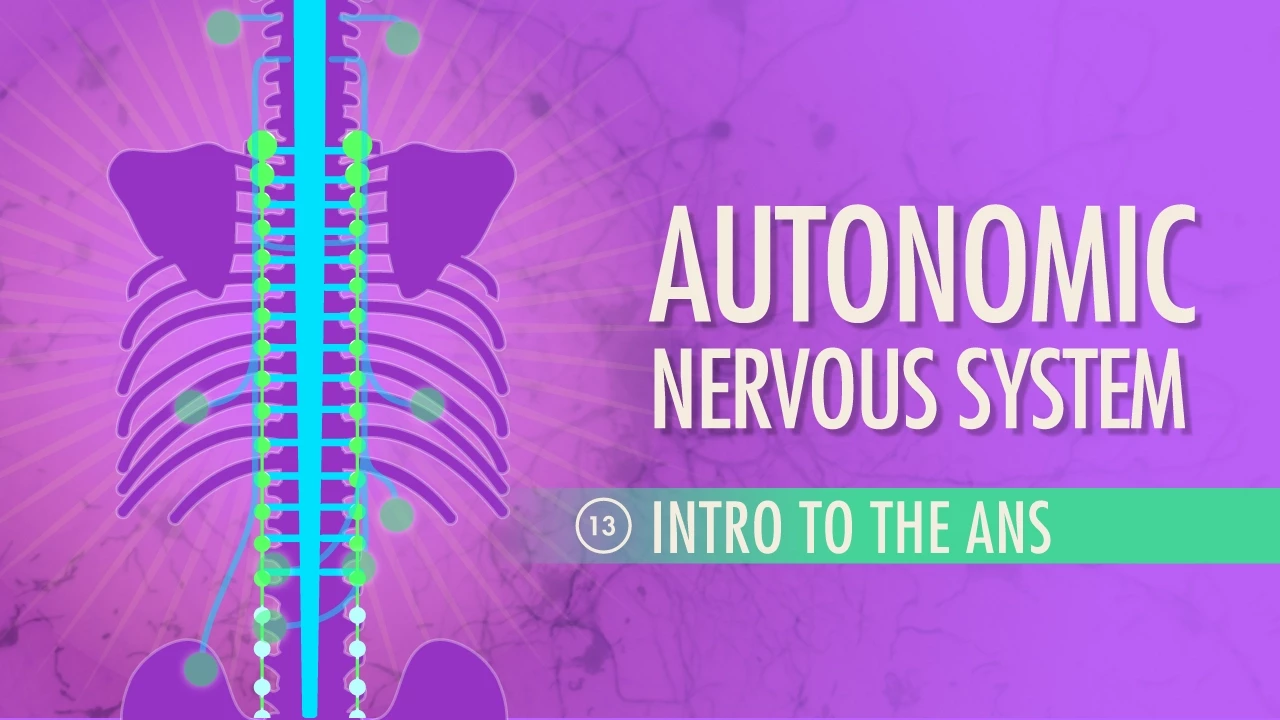
Autonomic Nervous System: Crash Course Anatomy & Physiology #13
3.4M
The autonomic nervous system consists of two complementary divisions—the sympathetic nervous system (which triggers the 'fight or flight' response by preparing the body for activity) and the parasympathetic nervous system (which promotes 'resting and digesting' by maintaining bodily functions and conserving energy)—that work together through three key anatomical differences: the sympathetic system originates from thoracolumbar regions and has ganglia near the spinal cord with short preganglionic fibers and long postganglionic fibers, while the parasympathetic system originates from craniosacral regions and has ganglia near or within effector organs with long preganglionic fibers and short postganglionic fibers.
Click to watch →
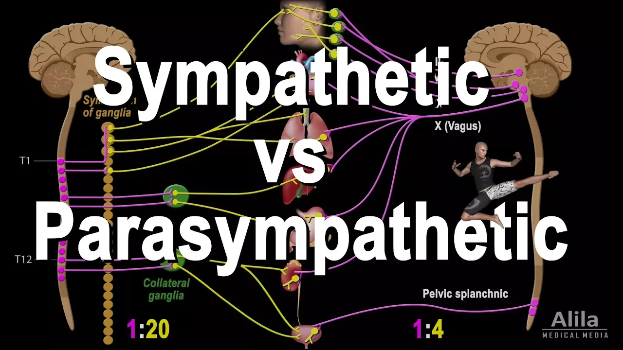
Autonomic Nervous System: Sympathetic vs Parasympathetic, Animation
1.5M
The autonomic nervous system (ANS) regulates internal organ activities through two opposing divisions: the sympathetic division (fight or flight response) that increases cardiac output, accelerates breathing, releases stored energy, and dilates pupils during emergencies, while the parasympathetic division (rest and digest state) slows heart rate, decreases respiratory rate, stimulates digestion, and stores energy during ordinary conditions; despite their opposing effects, both systems are simultaneously active in most organs, maintaining a balance called autonomic tone, with the sympathetic system having a high degree of neuronal divergence (one preganglionic neuron can synapse with up to 20 postganglionic neurons) producing widespread effects, whereas the parasympathetic system produces more specific, localized responses due to shorter postganglionic fibers and lower divergence.
Click to watch →

Autonomic Nervous System (ANS) Overview: Sympathetic vs. Parasympathetic | Dr. Priyanka Sachdev
98.2K
The Autonomic Nervous System (ANS) is the motor division of the peripheral nervous system that controls involuntary organs through two opposing divisions: the sympathetic (thoracolumbar distribution, ganglia near CNS, post-ganglionic neurotransmitter norepinephrine) and parasympathetic (craniosacral distribution, ganglia near organs, post-ganglionic neurotransmitter acetylcholine) systems. Both divisions use two neurons (preganglionic and post-ganglionic) with acetylcholine at the preganglionic level, but differ in their post-ganglionic neurotransmitters and ganglion positions. In most organs, these systems have opposite actions (e.g., pupil constriction vs dilation, increased vs decreased heart rate), except in genital organs where they have complementary actions (erection vs ejaculation).
Click to watch →
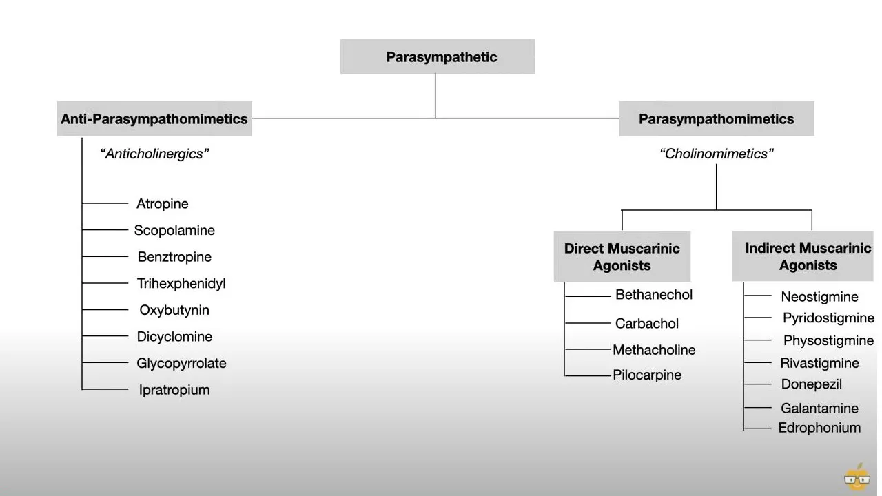
Autonomic Nervous System (Pharmacology, Receptors, and Physiology)
783.5K
The autonomic nervous system comprises two opposing divisions: the sympathetic nervous system (fight or flight) and the parasympathetic nervous system (rest and digest). The sympathetic system activates during extreme stress, preparing the body for fight or flight by releasing hormones and activating large muscle groups. The parasympathetic system activates during rest and digestion, allowing the body to process food and recover energy. These systems work in opposition - when one is activated, the other is inhibited. The parasympathetic system has craniosacral outflow, originating from cranial nerves and extending to sacral nerves. Four cranial nerves have specific parasympathetic functions: CN III (Edinger-Westphal nucleus) controls miosis and accommodation; CN VII (superior salivary nucleus) controls lacrimal gland salivation; CN IX (inferior salivary nucleus) controls parotid gland salivation; CN X (dorsal vagal nucleus) controls GI and lung secretions. This foundational understanding is essential for grasping autonomic pharmacology.
Click to watch →
Deep Dive
Basic neurobiology, including neural signaling, synaptic transmission, and primary neurotransmitters like acetylcholine.

The Synapse - Neuro - Biology and Physiology - Nervous System
43.4K
This section covers the foundational concepts of how neurons communicate. A synapse connects one neuron to another, consisting of the soma, axon, and axon terminalis. Neurotransmitters are synthesized in the soma, stored in vesicles, and released via exocytosis requiring ATP. The nerve impulse flows unidirectionally from soma to axon terminals (orthodromic). Three synapse types exist: neuro-neural (nerve-nerve), neuromuscular (nerve-muscle), and neuroglandular (nerve-gland). The cholinergic system uses acetylcholine, while the adrenergic system uses norepinephrine. Acetylcholine is synthesized from acetyl-CoA and choline by choline acetyltransferase, while norepinephrine follows the pathway: phenylalanine → tyrosine → dopa → dopamine → norepinephrine.
Click to watch →
(C2.2) - Neural Signalling - IB Biology (SL/HL)
18.8K
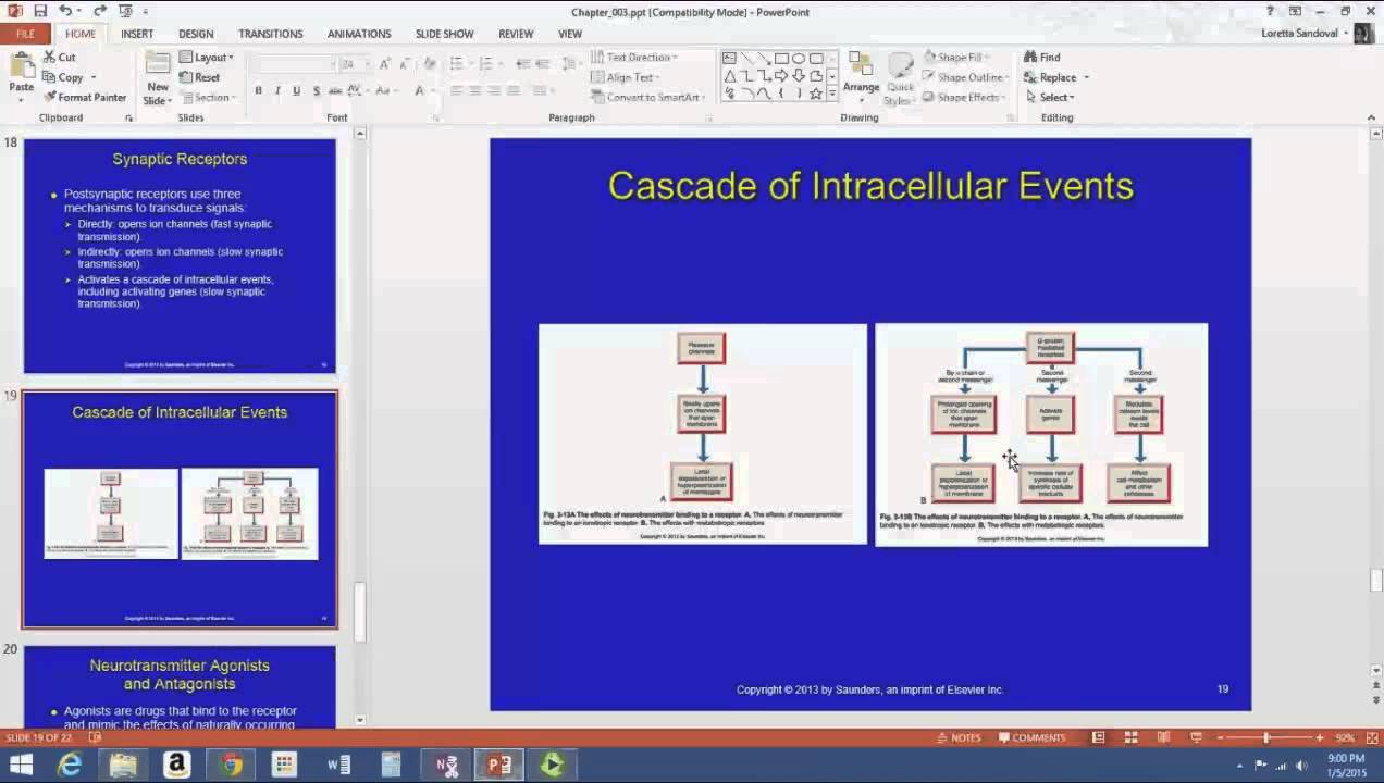
Synaptic transmission involves six key steps: (1) The action potential arrives at the presynaptic terminal, (2) Voltage-gated calcium channels open and calcium ions diffuse in, (3) Calcium causes vesicles containing neurotransmitters to fuse with the presynaptic membrane, releasing neurotransmitters via exocytosis, (4) Neurotransmitters diffuse across the synaptic cleft to the postsynaptic membrane, (5) Neurotransmitters bind to chemically-gated ion channels, opening them and allowing sodium to flow in, depolarizing the postsynaptic neuron, (6) Enzymes break down neurotransmitters to terminate the signal and allow recycling. Acetylcholine is a key neurotransmitter involved in this process, binding to receptors and opening sodium channels to transmit signals between neurons.
Click to watch →
Chapter 3 Part 4 Neurotransmitters Neuromodulators and Receptors
1K
Acetylcholine is an excitatory neurotransmitter used at the neuromuscular junction and throughout the autonomic nervous system. Amino acid neurotransmitters include glutamate (excitatory), glycine, and GABA (both inhibitory). These basic neurotransmitters form the foundation of neural signaling in the nervous system.
Click to watch →

Examen Simulador EXANI II: 40 Reactivos + Explicación Paso a Paso | Área de la Salud
3.3K
Neurotransmitters are chemical messengers that transmit signals between neurons. Acetylcholine (acetilcolina) is the primary neurotransmitter responsible for synaptic transmission. Myelin (mielina) is a lipid-rich substance that insulates neurons and speeds up signal transmission but is not a neurotransmitter. Epinephrine (adrenalina) is also a neurotransmitter but is not the primary one for synaptic transmission.
Click to watch →
![PSICOLOGÍA - Bases biológicas de la conducta [CICLO FREE]](https://i.ytimg.com/vi/l5Bxh7DHiRo/maxresdefault.jpg)
PSICOLOGÍA - Bases biológicas de la conducta [CICLO FREE]
39.6K
Neural communication occurs at synapses, the gaps between neurons. Presynaptic terminals release neurotransmitters into the synaptic cleft, which bind to postsynaptic receptors. Two transmission types exist: electrical (direct ion flow) and chemical (neurotransmitter release). Key neurotransmitters include acetylcholine (memory), adrenaline (fight-or-flight), serotonin/dopamine/oxytocin (pleasure/relaxation), glutamate, and norepinephrine. These chemicals serve excitatory or inhibitory functions, enabling complex neural signaling that underlies all mental processes and behaviors.
Click to watch →
Deep Dive
General embryology, specifically the origin, differentiation, and migration of neural crest cells during early fetal development.
Embryology:(Wk9)(Win21): Finishing Neurulation, Neural Crest Cells & Mesoderm Differentiation - I.
327
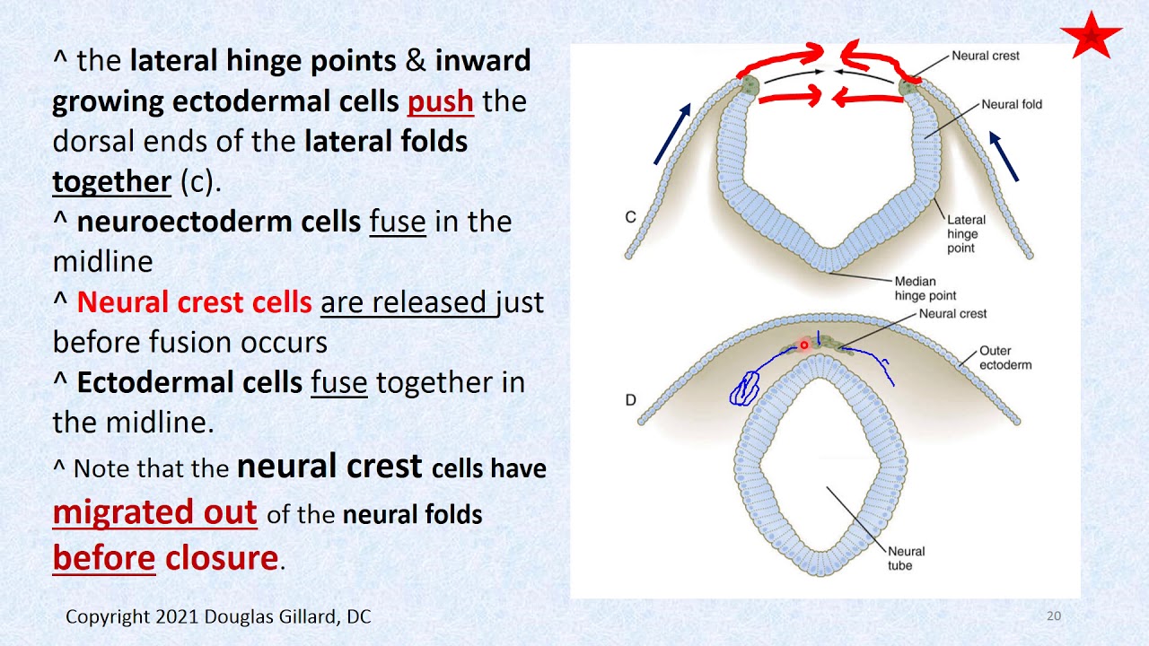
Neural crest cells arise from neural ectoderm at the boundary between BMP-secreting ectoderm and non-BMP-secreting neuroectoderm, expressing intermediate BMP levels that activate key transcription factors (msx, dlx5, pax, sox10). They undergo epithelial-mesenchymal transformation through snail, twist, and foxd3 activation, detaching via cadherin downregulation. Before neural tube closure, they migrate along the basal lamina, guided by robo, slit, semaphorin, and ephrin signals. They become the fourth germ layer, giving rise to peripheral nerves, adrenal medulla, melanocytes, leptomeninges, craniofacial structures, and carotid bodies.
Click to watch →

neural crest; Origin and Formation, Migration and Differentiation, Derivatives of Neural Crest Cells
143
The neural crest is a transient embryonic structure that arises at the border between the neural plate and non-neural ectoderm during neurulation; after the neural tube closes, these cells undergo epithelial-to-mesenchymal transition and migrate extensively throughout the embryo to differentiate into diverse tissues including peripheral nervous system components (sensory neurons, Schwann cells, adrenal medulla), melanocytes, craniofacial cartilage and bone, enteric nervous system, and cardiac septa; defects in neural crest development can lead to disorders such as DiGeorge syndrome, Waardenburg syndrome, Hirschsprung's disease, and craniofacial dysplasia.
Click to watch →

Origin And Fate Of Neural Crest Cells Part 1 By Dr Ranjana Agrawal-1
516
Neural crest cells are a unique population of migratory cells arising at the boundary between the neural tube and overlying epidermis during embryonic development. They are neither part of the neural tube nor the epidermis but represent a distinct lineage. Migration begins after neural tube closure and is regulated by transcription factors like Snail proteins and signaling molecules including FGFs and BMPs. The process involves dissociation of cell-cell adhesion molecules, enabling cells to become motile. As cells migrate away from the neural tube, they lose primitive properties and begin expressing new markers. These cells then differentiate into diverse tissue types including cartilage, bone, neurons, and connective tissue, depending on their destination. In the head region, they contribute to facial structures, teeth, and sensory ganglia; in the trunk, they form parts of the peripheral nervous system and various connective tissues.
Click to watch →

PARALELO DE EMBRIOLOGÍA-CLASE 4.
1K
Neural crest cells form transiently in the midline dorsal position and then rapidly migrate laterally. These cells have high migratory capacity and invade many regions of the embryo. In each region, they receive different signals that determine their differentiation into various cell types. This explains how neural crest cells can give rise to such diverse structures as neurons, glia, melanocytes, and connective tissues. The neural crest cells undergo an epithelial-mesenchymal transition, leaving the ectoderm and entering the mesenchymal compartment. Neural crest cells migrate to form spinal ganglia, paravertebral ganglia, prevertebral ganglia, and the adrenal medulla. They also contribute to the enteric nervous system, form melanocytes in the skin and iris, and contribute to craniofacial development through cranial neural crest cells.
Click to watch →

Embryology of CVS part 1
1.4K
Neural crest cells are a transient population of cells that migrate from the neural tube during embryonic development. These cells give rise to diverse structures including the peripheral nervous system, melanocytes, craniofacial cartilage and bone, and parts of the heart and face. Neural crest cells migrate along specific pathways and differentiate into various cell types depending on their location. The neural crest is often described as the 'fourth germ layer' due to its extensive contributions to multiple body systems. The somites develop as paired blocks of mesoderm along the neural tube, differentiating into sclerotome (vertebrae and ribs), myotome (skeletal muscle), and dermatome (dermis of the skin).
Click to watch →
Prerequisite Knowledge
- Concept 01The histological organization of the gastrointestinal tract, specifically the four concentric layers: mucosa, submucosa, muscularis externa, and serosa.
- Concept 02Fundamentals of the Autonomic Nervous System (ANS), including the functional and anatomical differences between the sympathetic and parasympathetic divisions.
- Concept 03Basic neurobiology, including neural signaling, synaptic transmission, and primary neurotransmitters like acetylcholine.
- Concept 04General embryology, specifically the origin, differentiation, and migration of neural crest cells during early fetal development.
Subsequent Learning
- Step 01The physiological coordination of gastrointestinal motility patterns, such as peristalsis and segmentation, by the myenteric plexus.
- Step 02How the submucosal (Meissner's) plexus regulates glandular secretion, nutrient absorption, and localized mucosal blood flow.
- Step 03The clinical diagnosis and surgical management of Hirschsprung's disease, including rectal suction biopsy analysis and pull-through procedures.
- Step 04The 'Gut-Brain Axis', exploring bidirectional communication between the enteric nervous system, the central nervous system, and the gut microbiome.
- Step 05Other functional and motility-related disorders of the gut, such as Achalasia, Gastroparesis, and Irritable Bowel Syndrome (IBS).
Enteric NS
Independence
Parasymp Supply
Symp vs Parasymp
Conclusion
Enteric NS
0:08Playing Section
- 1
Discusses the enteric nervous system as the brain of the gut.
- 2
Notes the number of neurons present in the spinal cord.
- 3
Describes the enteric system as an independent neural network.
The Non-Neuronal and Microbiome-Centric Paradigm of Gut Regulation
While traditional gastroenterology frames the Enteric Nervous System (ENS)—specifically the myenteric and Meissner's plexuses—as an autonomous 'second brain' regulating motility and secretion, a rising counter-perspective challenges this neuro-centric model. Critics argue that focusing primarily on neural pathways oversimplifies gut physiology. Instead, emerging research suggests that the gut microbiota, mucosal immune system, and enteroendocrine cells do not merely feed information to the ENS, but often bypass it entirely to directly regulate intestinal motility, barrier function, and local blood flow. Furthermore, enteric glial cells—long dismissed as mere support structures—are now recognized as active regulators of synaptic transmission and immune response. In conditions like Hirschsprung's disease, focusing solely on aganglionosis (the absence of nerves) overlooks critical microenvironmental and immunological anomalies that contribute to the pathology. This shift suggests that the ENS is not an autonomous controller, but rather one node within a highly decentralized, non-neuronal system dominated by the microbiome and the immune system.
Deep Dive
The physiological coordination of gastrointestinal motility patterns, such as peristalsis and segmentation, by the myenteric plexus.
Fisiología 2: Absorción y digestión + Motilidad Gastrointestinal
390
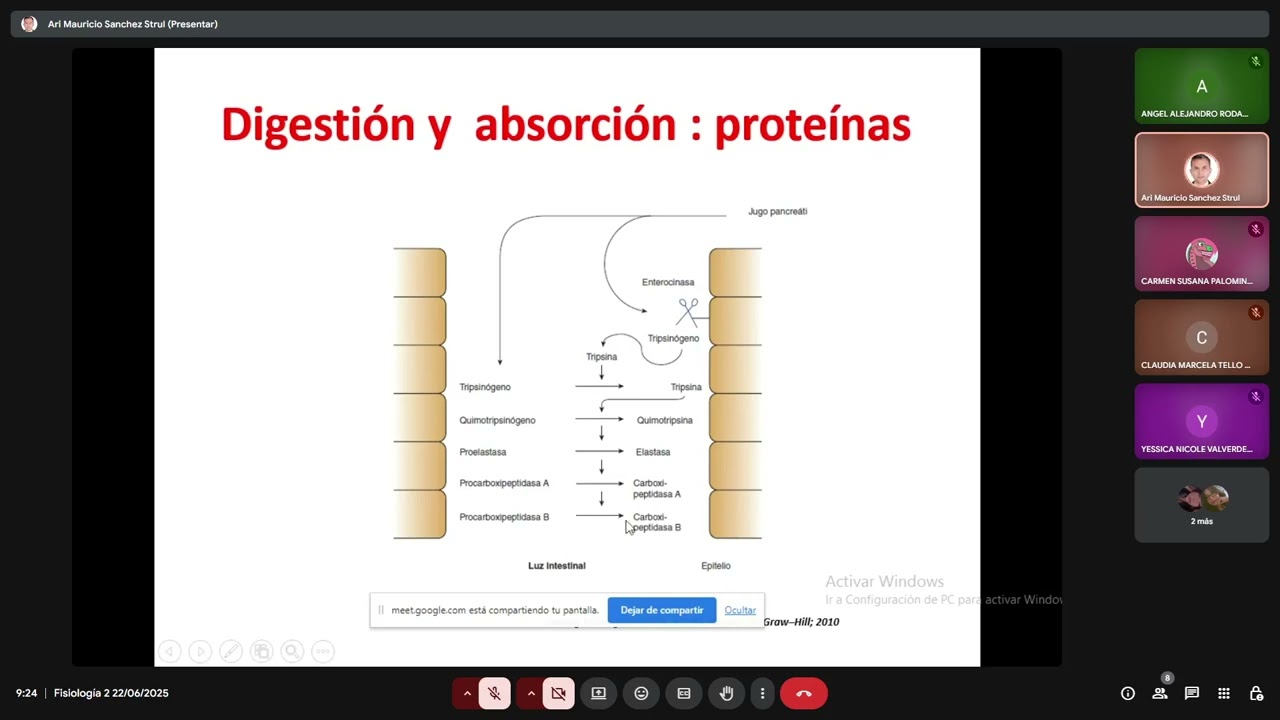
Gastrointestinal motility serves three functions: mixing, propulsion, and exposure to secretions. Peristaltic movements are reflex responses to distension, involving constriction behind food and relaxation ahead, resulting in caudal propulsion. This is mediated by the enteric nervous system: sensory neurons release serotonin, stimulating motor neurons that release acetylcholine/substance P (contraction) or nitric oxide/VIP (relaxation). Segmental movements involve contraction of intestinal segments with simultaneous constriction in the middle, creating retrograde flow that mixes contents without propulsion. Both movements are coordinated by the basic electrical rhythm (BER), generated by interstitial cells of Cajal (pacemaker cells). The BER frequency varies: stomach (4/min), duodenum (12/min), jejunum (8/min), ileum (9/min), colon (6/min).
Click to watch →
Enteric Nervous System | Neural Regulation of the Digestive System
47.8K
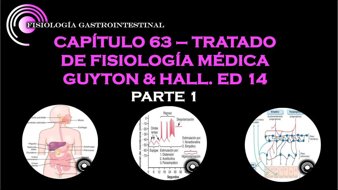
The myenteric plexus is located between the circular and longitudinal muscle layers. This neural network controls the contraction of both muscle layers to produce peristalsis and segmentation. The myenteric plexus is primarily responsible for motility functions in the digestive tract, working in coordination with the submucosal plexus.
Click to watch →
GUYTON & HALL CAP 63. PRINCIPIOS GENERALES DE LA FUNCION GASTROINTESTINAL . PARTE 1.
16K
The myenteric plexus controls intestinal movement through both excitatory and inhibitory mechanisms. It contracts muscle ahead of food to propel it forward and inhibits sphincters (pyloric, ileocecal) to allow passage. This creates the peristaltic wave pattern. The submucosal plexus controls intestinal secretion and absorption, and produces contraction of the submucosal muscle layer to increase absorption surface area. Both plexuses work together to coordinate GI function.
Click to watch →
Large Intestine & Histology | Digestion and Absorption | Biology | Class 11
2.6K
The myenteric plexus (Auerbach's plexus) controls the motility of the entire alimentary canal by regulating the contraction and relaxation of the muscularis externa. This nerve network coordinates peristaltic movements, segmentation contractions, and other motor patterns throughout the digestive tract, enabling coordinated movement of contents from mouth to anus.
Click to watch →

FISIOLOGÍA DIGESTIVA: cap 1 y 2
117
GI motility is controlled by the enteric nervous system, which coordinates peristalsis and segmentation. Peristalsis involves contraction of circular muscle behind the bolus and relaxation ahead of it, propelling contents forward. Segmentation involves alternating contraction and relaxation that mixes contents with secretions. Both movements require the integrity of the myenteric plexus for coordination.
Click to watch →
Deep Dive
How the submucosal (Meissner's) plexus regulates glandular secretion, nutrient absorption, and localized mucosal blood flow.

Clase de Refuerzo Histologia Sist. Digestivo pt1
171
Meissner's plexus (submucosal plexus) regulates all functions of the mucosa. It controls glandular secretion (stimulating or inhibiting mucous glands), regulates blood flow to the mucosa, and influences the muscularis mucosae to produce movements related to nutrient assimilation. The plexus receives input from both sympathetic and parasympathetic nervous systems.
Click to watch →

Principios generales de la función gastrointestinal fisiologia YouTube
3.8K
The submucosal plexus integrates sensory signals and regulates: (1) Secretion of digestive enzymes and fluids; (2) Blood flow to the digestive tract; (3) Local absorption of nutrients; (4) Mucosal blood flow. This plexus acts as a local control center that coordinates the secretory and absorptive functions of the digestive tract based on the presence of food and other stimuli.
Click to watch →
Fisiología GASTROINTESTINAL Pt. II - Sistema Nervioso ENTÉRICO (Plexo de Meissner y Auerbach)
19.1K
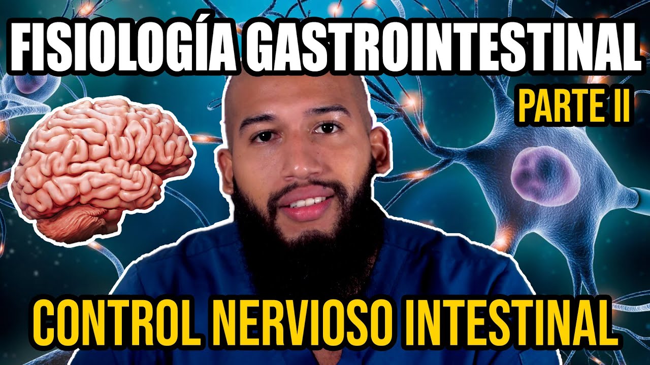
The submucosal plexus of Meissner is located in the submucosa of the gastrointestinal tract. Its primary function is to control intestinal secretion. The neurons in this plexus connect to glands present in the gastrointestinal epithelium, thereby controlling intestinal secretion, absorption, and local blood flow. Since the submucosa contains the most important blood vessels that nourish all layers of the intestine, this plexus plays a crucial role in regulating local blood supply.
Click to watch →

Enteric nervous system
17K
The submucosal plexus (Meissner's plexus) contains sensory neurons (including stretch and chemoreceptors), motor neurons that regulate glandular secretion, and neurons controlling blood vessel smooth muscle. It extends from the esophagus to the rectum and manages local gut functions including secretion of mucus and digestive enzymes, as well as blood flow regulation to the gastrointestinal tract.
Click to watch →

Control of GI Functions | Mustafe Ali | GIT 113
877
The submucosal plexus (Meissner's plexus) is located in the submucosal layer and is primarily responsible for the secretion of glandular components including hormones, enzymes, and other secretions. It also regulates intestinal absorption and controls local blood flow via the muscularis mucosa.
Click to watch →
Deep Dive
The clinical diagnosis and surgical management of Hirschsprung's disease, including rectal suction biopsy analysis and pull-through procedures.

Hirschsprung Disease
110K
Diagnosis begins with barium enema showing proximal colon dilation meeting a narrowed distal rectum, but definitive confirmation requires rectal biopsy demonstrating absence of ganglion cells. Treatment involves surgical pull-through procedure where the aganglionic colon segment is removed and the healthy bowel is pulled through to the rectum. Clinical vignettes demonstrate classic presentations including failure to pass meconium, persistent abdominal distension, vomiting, and temporary relief from digital rectal exam without sustained stool passage.
Click to watch →

20 de marzo de 2026
160
Diagnosis requires clinical suspicion combined with histopathological confirmation via rectal biopsy demonstrating complete absence of ganglion cells. Barium enema may show characteristic transition zones. Manometry demonstrates absence of rectal inhibitory reflex. Treatment involves resection of the aganglionic segment with primary anastomosis placed as low as possible (typically 7 cm above pectinate line). Modern techniques allow primary pull-through procedures without initial colostomy, performed laparoscopically or openly. Postoperative care focuses on preventing infection and ensuring proper anastomotic healing. The goal is to restore normal bowel function while minimizing complications.
Click to watch →

Hirschprung Disease || TNC Bullet Series # 195|| Most Important Topic|| Daily 7.00 AM|| NURSING EXAM
9K
Diagnosis involves: (1) Clinical suspicion based on ribbon-like stool and chronic constipation, (2) Abdominal X-ray showing dilated bowel loops and air-fluid levels, (3) Barium enema to identify the transition zone between affected and normal bowel, and (4) Rectal biopsy - the gold standard test showing absence of ganglion cells and hypertrophied nerve trunks. Rectal manometry demonstrates absence of the recto-anal inhibitory reflex. Intestinal obstruction shows presence of ganglion cells, distinguishing it from Hirschsprung disease. The primary treatment is surgical. The pull-through procedure involves removing the aganglionic segment and anastomosing the normal bowel to the anus. Types include: (1) Swenson procedure - direct removal and connection, (2) Duhamel procedure - creates a pouch from normal bowel, (3) Soave procedure - mucosal stripping and anastomosis. In severe cases or neonates with enterocolitis, initial management involves colostomy (right or left). After stabilization, definitive surgery is performed.
Click to watch →

IAPS NextGen #28 Hirschsprung's disease
390
This segment covers the histopathological diagnosis and surgical management of Hirschsprung disease. Histopathological findings include absence of ganglion cells, hypertrophic nerve fibers (cutoff 40 micrometers), and increased acetylcholinesterase activity. Calretinin staining identifies ganglion cells (absent in Hirschsprung disease). Preoperative workup includes reviewing all records and biopsies, confirming the biopsy was taken from the correct site (stoma opening), and ensuring the biopsy shows hypoganglionosis. If a stoma biopsy shows hypoganglionosis but the child is passing stools normally, this may indicate the biopsy was taken from a transition zone. During definitive surgery, the last 3 cm of the pull-through should be discarded to ensure only ganglionated bowel is brought down. The three main pull-through procedures are Duhamel, Swenson, and Soave. Duhamel is commonly performed, easy to learn, and considered to have the best postoperative outcome. Swenson is the most anatomical procedure but is technically demanding. The choice of procedure should be based on surgeon comfort, hospital facilities, and patient factors.
Click to watch →

INI-CET July 2021 Exam Discussion by Surgery Expert, Dr. Pritesh Singh
12.2K
Hirschsprung's disease is characterized by absence of ganglion cells in rectal biopsy. The affected segment is contracted while proximal segment is dilated. Management: confirm diagnosis with rectal biopsy, temporary colostomy, then definitive procedures (Swenson, Duhamel, or Soave procedures).
Click to watch →
Deep Dive
The 'Gut-Brain Axis', exploring bidirectional communication between the enteric nervous system, the central nervous system, and the gut microbiome.
Gut-Brain link
111.1K
The gut-brain axis describes the bidirectional communication network between the brain and the enteric nervous system in the gut, involving approximately 100 million neurons and the vagus nerve as the primary connection; this axis is regulated by the gut microbiome containing trillions of microorganisms that influence mood, behavior, immune function, and susceptibility to diseases such as inflammatory bowel disease, type 2 diabetes, and anxiety/depression, with evidence showing that fecal microbiota transplants can transfer these effects between individuals.
Click to watch →
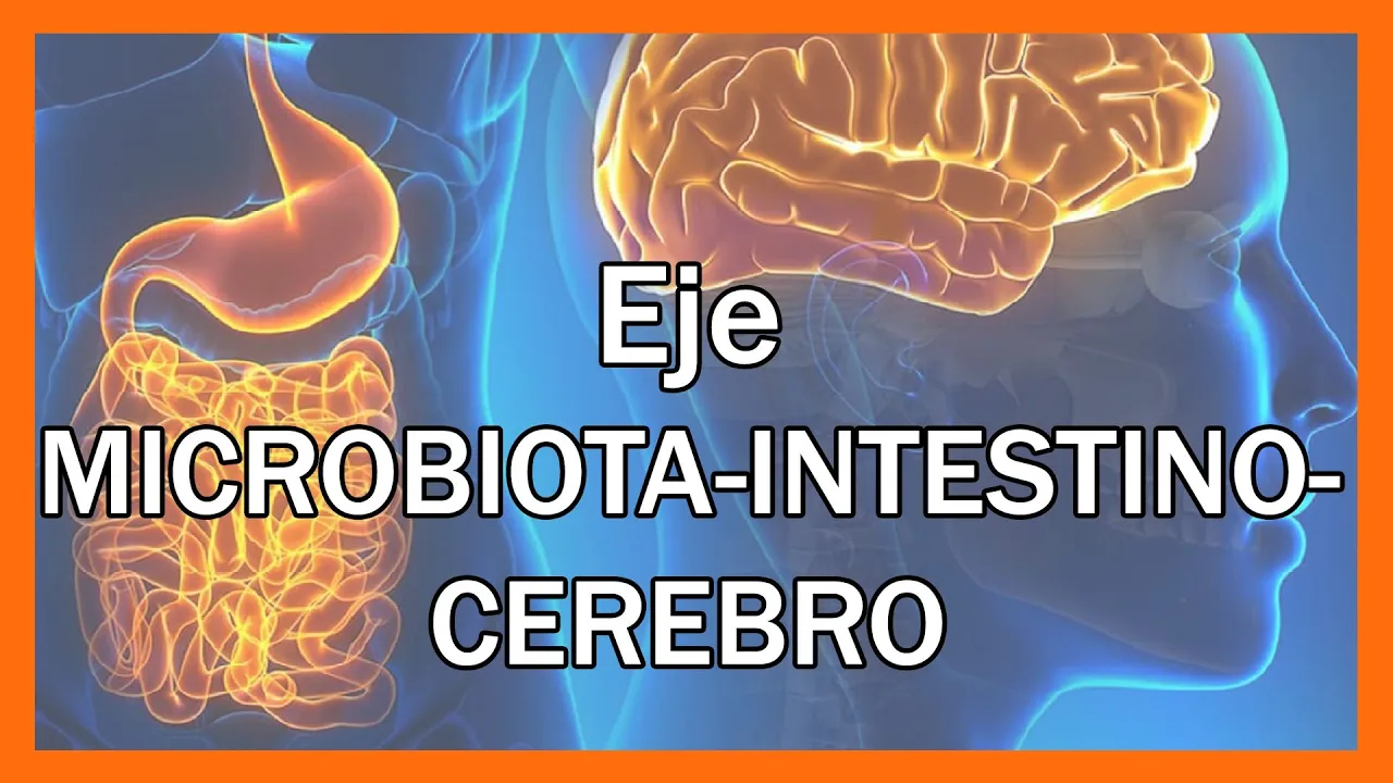
Aprende Cómo Funciona el EJE MICROBIOTA-INTESTINO-CEREBRO, una compleja comunicación.
109.1K
The gut-brain axis represents a complex bidirectional communication system between the cranial brain and enteric nervous system (the 'second brain' with 100 million neurons). The enteric nervous system controls basic gastrointestinal functions and produces 80% of serotonin and 50% of dopamine. Communication occurs through three pathways: vagal (direct neural), circulatory (metabolites like short-chain fatty acids), and immune (cytokines). The microbiota serves as a communication hub connecting nervous, endocrine, and immune systems. Each system has receptors for mediators of other systems, and cells can produce mediators of other systems. The microbiota produces neurotransmitters and hormones, with specific bacterial species producing specific neurotransmitters like serotonin, GABA, and dopamine. The microbiota affects the HPA axis, regulating cortisol release and stress response. It presents molecular patterns to the immune system, activating immune cells that produce cytokines affecting brain neurons and glial cells. The microbiota can facilitate cytokine release that stimulates neurogenesis. Clinical studies with prebiotics and probiotics have been conducted in neurological diseases including encephalopathy, multiple sclerosis, constipation in cerebral palsy, and epilepsy. This demonstrates that neurodegenerative pathologies and neurological alterations relate to intestinal microbiota state, whether as cause or consequence.
Click to watch →
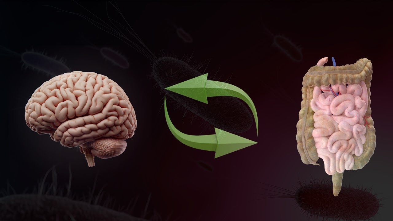
Eixo intestino-cérebro
10.8K
The gut-brain axis represents the bidirectional communication between the intestinal microbiota and the central nervous system. The enteric nervous system contains approximately 500 million neurons and operates with significant independence, controlling intestinal functions. The dense innervation of the intestines is partly dependent on the intestinal microbiota itself. Intestinal bacteria produce an infinite variety of molecules including neurotransmitters, vitamins, secondary bile acids, amino acids, and short-chain fatty acids. These molecules reach the brain through the bloodstream or nervous system, particularly via the vagus nerve. Short-chain fatty acids, produced from fiber fermentation, are involved in neuroplasticity, gene expression, and immune modulation. The typical Western diet is poor in fiber, weakening the intestinal wall and facilitating pathogen entry. Several neurotransmitters are produced by the intestinal microbiota, including GABA and serotonin. The intestine is the main producer of serotonin in the body, acting primarily locally.
Click to watch →
![[Sách Nói] Ăn Gì Bổ Não - Chương 1 | David Perlmutter](https://i.ytimg.com/vi/3cKb0DW9cqk/sddefault.jpg)
[Sách Nói] Ăn Gì Bổ Não - Chương 1 | David Perlmutter
245.7K
The gut and brain communicate bidirectionally. The vagus nerve, the longest of the 12 cranial nerves, is the main communication channel between hundreds of millions of neurons in the enteric nervous system and the central nervous system. The enteric nervous system of the digestive tract is so rich in neurons that many scientists call it the 'second brain.' It controls muscles, immune cells, and hormones, and produces substances like serotonin. Approximately 80-90% of the body's serotonin is produced by neurons in the gut, more than the brain in the head. Neurologists and psychiatrists are recognizing that the gut brain may not be secondary at all—it may function independently from the main brain and control many functions without commands or assistance from the brain.
Click to watch →
Дубынин. Секреты мозга, диеты и дофамин.
1.2M
The gut contains a nervous system (enteric nervous system) with nearly as many neurons as the spinal cord, capable of learning and even influencing dreams. The gut produces melatonin, which synchronizes different intestinal sections rather than primarily regulating sleep. This gut-brain axis involves bidirectional communication between the digestive system and the brain, with the gut producing hormones and neurotransmitters that affect mood, cognition, and overall health.
Click to watch →
Deep Dive
Other functional and motility-related disorders of the gut, such as Achalasia, Gastroparesis, and Irritable Bowel Syndrome (IBS).
5 Minutes in GI: What common GI disorders are associated with the disruption of GI motility?
1.9K
Gastrointestinal motility disorders disrupt the coordinated neuromuscular functioning of the GI tract, leading to various conditions: slow transit constipation results from poor stool movement through the colon, pelvic floor dysfunction involves dis-coordinated muscles preventing stool expulsion, diarrhea-associated motility disorders involve hyperactive colon muscles, GERD occurs when the esophageal sphincter is too relaxed allowing stomach acid reflux, gastroparesis delays stomach emptying due to muscle discoordination, and achalasia involves hyperactive sphincter muscles making food passage difficult.
Click to watch →

Treatment of Motility Disorders and Functional Gastrointestinal Disorders
42.9K
Functional gastrointestinal disorders include dyspepsia (indigestion), gastroesophageal reflux disease (GERD), gastroparesis (delayed stomach emptying), irritable bowel syndrome (IBS), chronic constipation, pelvic floor disorders, and achalasia (esophageal motility disorder). These conditions involve dysfunction of the GI tract without identifiable structural abnormalities.
Click to watch →
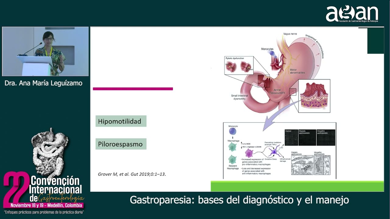
Gastroparesia, bases del diagnóstico y el manejo - Dra. Ana María Leguízamo
749
Gastroparesis is associated with other motility disorders: (1) Constipation - present in 40% of patients; (2) Intestinal transit disorders - present in up to 64% of patients; (3) Achalasia - rare association. These associations suggest that patients with gastroparesis may have broader gastrointestinal motility dysfunction.
Click to watch →

Функциональная гастроэнтерология - как справиться с симптомами и ничего не пропустить
479
Esophageal motility disorders include achalasia cardia (failure of LES to relax and absence of peristalsis), diffuse esophageal spasm (simultaneous non-peristaltic contractions), and scleroderma esophageal involvement (loss of smooth muscle peristalsis). Only the lower esophageal sphincter can develop achalasia because it is under autonomic nervous system control (parasympathetic stimulation via vagus nerve), while the upper esophageal sphincter is under somatic control. Pseudoachalasia is caused by extrinsic compression (most commonly by esophageal cancer). Gastric motility disorders include gastroparesis (delayed gastric emptying) and functional dyspepsia. Functional dyspepsia is divided into epigastric pain syndrome and postprandial distress syndrome. Treatment follows a stepwise approach: test for H. pylori and treat if positive; if negative, empiric PPI for 8 weeks; if no response, consider tricyclic antidepressants, prokinetics, or psychological interventions.
Click to watch →

Syndromes of GI Motor Function Distubancies (MA groups)
148
GI motor function disturbances encompass three main categories: esophageal motility disorders (including achalasia, diffuse esophageal spasm, and CREST syndrome), gastric motility disorders (delayed gastric emptying/gastroparesis, rapid gastric emptying/dumping syndrome, and functional dyspepsia), and intestinal motility disorders (including constipation, irritable bowel syndrome, and malabsorption syndromes). These conditions result from primary motility defects or secondary causes such as diabetes, scleroderma, hypothyroidism, infections, medications, and psychological factors. Diagnosis relies on clinical evaluation, imaging (barium studies), endoscopy, manometry, and specialized tests like the smart pill wireless motility capsule. Treatment approaches vary by condition but commonly include lifestyle modifications, dietary adjustments, pharmacotherapy, and in some cases, endoscopic or surgical interventions.
Click to watch →
Enteric NS
Independence
Parasymp Supply
Symp vs Parasymp
Conclusion
Enteric NS
0:08- 1
Discusses the enteric nervous system as the brain of the gut.
- 2
Notes the number of neurons present in the spinal cord.
- 3
Describes the enteric system as an independent neural network.
The Non-Neuronal and Microbiome-Centric Paradigm of Gut Regulation
While traditional gastroenterology frames the Enteric Nervous System (ENS)—specifically the myenteric and Meissner's plexuses—as an autonomous 'second brain' regulating motility and secretion, a rising counter-perspective challenges this neuro-centric model. Critics argue that focusing primarily on neural pathways oversimplifies gut physiology. Instead, emerging research suggests that the gut microbiota, mucosal immune system, and enteroendocrine cells do not merely feed information to the ENS, but often bypass it entirely to directly regulate intestinal motility, barrier function, and local blood flow. Furthermore, enteric glial cells—long dismissed as mere support structures—are now recognized as active regulators of synaptic transmission and immune response. In conditions like Hirschsprung's disease, focusing solely on aganglionosis (the absence of nerves) overlooks critical microenvironmental and immunological anomalies that contribute to the pathology. This shift suggests that the ENS is not an autonomous controller, but rather one node within a highly decentralized, non-neuronal system dominated by the microbiome and the immune system.
The histological organization of the gastrointestinal tract, specifically the four concentric layers: mucosa, submucosa, muscularis externa, and serosa.
Fundamentals of the Autonomic Nervous System (ANS), including the functional and anatomical differences between the sympathetic and parasympathetic divisions.
Basic neurobiology, including neural signaling, synaptic transmission, and primary neurotransmitters like acetylcholine.
General embryology, specifically the origin, differentiation, and migration of neural crest cells during early fetal development.
The physiological coordination of gastrointestinal motility patterns, such as peristalsis and segmentation, by the myenteric plexus.
How the submucosal (Meissner's) plexus regulates glandular secretion, nutrient absorption, and localized mucosal blood flow.
The clinical diagnosis and surgical management of Hirschsprung's disease, including rectal suction biopsy analysis and pull-through procedures.
The 'Gut-Brain Axis', exploring bidirectional communication between the enteric nervous system, the central nervous system, and the gut microbiome.
Other functional and motility-related disorders of the gut, such as Achalasia, Gastroparesis, and Irritable Bowel Syndrome (IBS).
125.8K views3.8Klikes16:41@EduClubOfficialOriginal Release: 2020-10-15
the topic we are going to discuss in this video is enteric nervous system so coming to the neural total number of neurons present in spinal cord is enteric nervous system little brain of intestine be sub [Music] so what is basically the centric nervous system [Music] system is an independent system but it is inferior mesenter gallium layer now coming to the parasympathetic supply this is the parasympathetic supply foreign um most s2 s3 we know that sympathetic nervous system releases norepinephrine or yeah sympathetic nervous system decreases now coming to parasympathetic supply parasympathetic supply co mainly divided [Music] so this was all about enteric nervous system if you like this video please hit the like button do share with your friends and please subscribe our channel and please press the bell icon to get notified get notifications for more such videos you can also follow us on facebook and instagram for more beautiful contents on such topics link given in the description below thank you
Up Next

63:58
Vagus Nerve Function: Assessment & Optimization with Dr. Navaz Habib
@designsforhealth3090
568 views•2024-06-05

50:12
Bacterial Communication: Quorum Sensing and Biofilm Formation Explained
@JHUAAP
12.6K views•2011-06-28

5:35
Enteric Nervous System Explained: The Gut's Brain | Neurobiology Lecture
@alumniu6029
438 views•2018-09-12

7:08
Bacteriophages: Earth's Deadliest Killers and Future Antibiotics
@kurzgesagt
34.6M views•2018-05-13
Related Study Plans & Knowledge Roadmaps
Structured learning paths in Biology
biology
RNAi: Biogenesis, RISC & Gene Silencing
01Molecular Biology Foundations: DNA to Protein
02Introduction to RNA Interference (RNAi)
03Molecular Biogenesis of miRNA and siRNA
+ 2 more learning modules
10h5 modules
Explore Roadmap
biology
Mycorrhizae: Signaling, Biophysics & Ecology
01Foundations of Mycorrhizal Symbiosis
02Molecular Signaling and Root Colonization
03Biophysics and Bioenergetics of Nutrient Exchange
+ 1 more learning modules
11h4 modules
Explore Roadmap
biology
Circadian Clocks: TTFL, SCN & Periphery
01Foundations of Molecular Biology & Gene Expression
02Introduction to Chronobiology and Actograms
03The Core TTFL: Molecular Architecture of the Clock
+ 3 more learning modules
14h6 modules
Explore Roadmap
biology
Population Genetics: Math, Drift & Selection
01Genetic & Probability Foundations
02The Hardy-Weinberg Principle and Baseline Models
03Mathematical Modeling of Natural Selection
+ 3 more learning modules
18h6 modules
Explore Roadmap