This video demonstrates a modular approach to creating tissue-specific hydrogel bioinks by combining commercially available components—including tissue-specific extracellular matrix digests, hyaluronic acid hydrogel base materials, and a 0.1% photo initiator solution—in a systematic process that enables the fabrication of functional 3D tissue constructs for applications such as drug screening and disease modeling.
Bioprinting Tissue Constructs with Hydrogel Bioinks: A Protocol
Added:Deep Dive
Fundamentals of cell biology and tissue engineering, including cell-extracellular matrix (ECM) interactions and 3D cell culture principles.
To grow the cell outside the body
19.4K
Cell culture technology requires understanding underlying philosophy, chemistry, physics, and biology rather than blindly following techniques. Cells require proper gaseous exchange with sufficient oxygen for survival, as anaerobic conditions cause cell death. Carbon dioxide must be removed to prevent acidification. Cells are categorized as adherent (attach to surfaces) or non-adherent (circulate in suspension). Adherent cells secrete cementing materials forming the extracellular matrix (ECM), composed of proteins, carbohydrates, and metal ions. The ECM serves as a critical structural feature ensuring cells remain at specific locations and acts as a cellular 'barcode' determining where cells can grow.
Click to watch →

BME2104 Lecture 7 ECM Part 1 Spring 2023
106
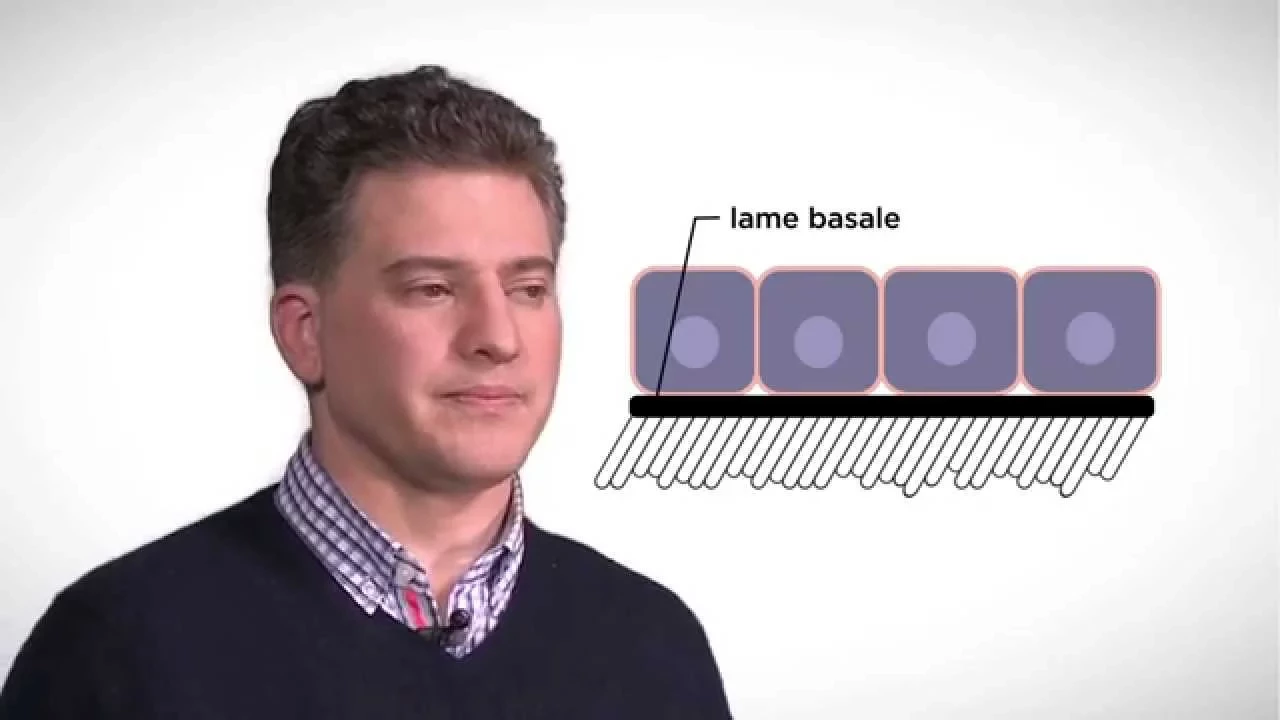
Tissue engineering and regenerative medicine leverage the extracellular matrix to create functional tissues and organs. Tissue engineering evolved from biomaterials science, pioneered by Bob Langer's lab at MIT in 1993, demonstrating that scaffolds could support cell growth to create tissue structures. These fields combine scaffolds with cells and biologically active materials, with FDA-approved examples including artificial skin and cartilage. Regenerative medicine focuses on leveraging natural self-healing properties (liver, bone) while addressing tissues lacking healing capacity (nerve). The extracellular matrix (ECM) is a combination of proteins and polysaccharides secreted by cells, classified as basement membrane (thin sheet under epithelium/endothelium) or connective tissue (fewer cells in larger matrix). The ECM provides structural scaffolding, tensile strength (via collagen), cushioning, molecular filtering, and biological cues for cell polarity, adhesion, morphogenesis, and differentiation. ECM proteins are essential for life, as knockout of many leads to lethal outcomes.
Click to watch →
MOOC côté cours : Le rôle de la matrice cellulaire
101.7K
The extracellular matrix (ECM) is a complex network of macromolecules secreted by cells that fills the space between cells in tissues, serving multiple critical functions including mechanical support, filtration of small molecules, cell signaling, and regulation of cellular processes like differentiation and migration; the ECM contains approximately 70% water along with structural proteins such as collagen (which provides tensile strength through its hierarchical organization from alpha chains to tropocollagen rods, fibrils, and fibers) and elastin (which gives tissues elastic properties allowing them to stretch and recoil), and glycoproteins that act as adhesives between matrix components and between the matrix and cells, with the ECM composition being tissue-specific and subject to continuous remodeling throughout life.
Click to watch →
Novel 3D Culture Technologies Utilizing Patient-Derived Tissue
252
Conventional 2D cell culture produces flat monolayers that fail to recapitulate 3D human tissue architecture. Historical development began in 1800 with Roux maintaining chicken embryo cells, progressed through Evelyn's first cell line in 1922, Warburg's 3D tissue slice work in 1923, and culminated in Minna Bissell's 1980s demonstration that extracellular matrix composition and three-dimensionality are essential for proper tissue biology. Multiple methods create 3D aggregates: hanging drops, magnetic levitation, synthetic scaffolds, and alginate scaffolds. However, extracellular matrix composition is absolutely critical—correct matrices (laminin and collagen) enable proper organoid formation with correct polarity and function, while incorrect matrices completely disrupt tissue organization. For hematopoietic cells, conventional matrices fail while bone-reconstructed matrices enable 28-day viability with native tissue organization.
Click to watch →

3D Cell Culture | 3D Culture Techniques
12.1K
This comprehensive section establishes the theoretical foundation of 3D cell culture technology. It begins by defining 3D cell culture as an artificial environment enabling cells to grow in all three dimensions, contrasting sharply with traditional 2D monolayer cultures. The section then delves into the extracellular matrix (ECM), explaining its composition including basement membrane and interstitial matrix containing collagen, laminin, fibronectin, glycosaminoglycans, and growth factors. Key concepts covered include ECM elasticity variations across tissues, tumor-associated stiffness due to collagen cross-linking, and molecular concentration gradients affecting cell behavior. The section transitions to comparing 3D versus 2D systems, demonstrating how 3D cultures enable natural cell-cell and cell-matrix interactions, develop physiological gradients, and better represent in vivo tumor architecture. Finally, it introduces scaffold-based approaches where cells are embedded in matrices that can be biological (collagen, gelatin, alginate) or synthetic, engineered to mimic ECM properties for tissue engineering applications.
Click to watch →
Deep Dive
Basic polymer chemistry and hydrogel crosslinking mechanisms, such as thermal, chemical, and photo-crosslinking.

How Are Hydrogels Crosslinked? - Chemistry For Everyone
321
Hydrogels are formed through crosslinking processes that connect polymer chains into three-dimensional networks; physical crosslinking uses weak non-covalent interactions like hydrogen bonding and ionic bonds that can be disrupted by environmental changes, while chemical crosslinking creates permanent covalent bonds using crosslinking agents that react with functional groups on polymer chains, requiring monomers, initiators, and crosslinking agents for proper network formation, with crosslink density directly affecting hydrogel properties such as stiffness and flexibility.
Click to watch →
Hydrogels
24.3K
Chemical cross-linking creates permanent covalent bonds between polymer chains using cross-linkers like glutaraldehyde, epichlorohydrin, adipic acid dihydrazide, and citric acid. These react with functional groups (hydroxyl, carboxyl, amine) on polymer backbones. Grafting polymerizes monomers onto preformed polymer backbones, activated by chemicals or radiation. Radiation grafting uses gamma rays, electron beams, or X-rays to create radicals that initiate polymerization. Radiation methods retain polymer biocompatibility, avoid chemical additives, and achieve simultaneous modification and sterilization, though polysaccharides degrade completely under radiation.
Click to watch →

Hydrogels- Introduction, Synthesis, Characterisation and Applications
4.4K
Hydrogels are three-dimensional networks of hydrophilic polymers that can swell in water and hold large amounts of solvent without dissolving. The term combines 'hydro' (water) and 'gel' (jelly-like consistency). The key property enabling water retention is crosslinking—chemical or physical bonds that hold polymeric chains together. Physical crosslinking uses methods like UV radiation to create weaker, reversible bonds, offering high biocompatibility and low toxicity. Chemical crosslinking uses covalent bonds that are very strong and difficult to break, resulting in insoluble hydrogels. Hydrogels are classified into natural types (hyaluronic acid, chitosan, alginate, fibrin) and synthetic types (PVA, polyethylene glycol, acrylics). Mechanical properties can be tuned by varying crosslinker concentration—increasing crosslinkers creates more rigid materials, while decreasing them produces more flexible hydrogels.
Click to watch →

Hydrogels as Processed Polymers - A Biofabrication Lecture
1.4K
Hydrogels are polymer networks capable of absorbing significant water (97-98%), making them ideal for integrating cells, drugs, and biologicals. They maintain structure through three crosslinking mechanisms: physical entanglements (reversible), chemical crosslinks (permanent anchor points), and hydrogen bonds (dynamic). Swelling is a thermodynamic equilibrium driven by water-polymer interactions, where chain stretching reduces entropy and creates restoring forces. Molecular weight dramatically affects hydrogel behavior—the same polymer produces vastly different properties at different molecular weights. Swelling impacts surface roughness (potentially causing inflammation), mechanical stiffness (dramatically reduced from dry to swollen states), and optical clarity (critical for contact lenses). Applications include drug delivery (encapsulating drugs in dried hydrogels for controlled release) and food industry (protecting flavors).
Click to watch →
Crosslinking of Hydrogels for Bioprinting
2.1K
Hydrogel crosslinking in bioprinting encompasses three main mechanisms: physical crosslinking (non-covalent interactions like ionic bonding, hydrogen bonding, and thermal responses), chemical crosslinking (covalent bond formation between polymer chains), and photocrosslinking (light-induced polymerization); each method offers distinct advantages and disadvantages in terms of reversibility, mechanical strength, biocompatibility, and processing requirements, making them suitable for different bioprinting applications depending on specific needs such as rapid gelation, structural stability, or cell viability preservation.
Click to watch →
Deep Dive
Introduction to fluid mechanics and rheology, specifically shear-thinning behavior and printability of viscoelastic materials.
![Rheology - introduction to the course [presented by Dr Bart Hallmark, University of Cambridge]](https://i.ytimg.com/vi/ft8z24SjSdo/hqdefault.jpg)
Rheology - introduction to the course [presented by Dr Bart Hallmark, University of Cambridge]
3.5K
Rheology is the branch of science that studies the deformation and flow of matter, particularly non-Newtonian fluids and viscoelastic materials, which exhibit complex behaviors such as shear-thinning (paint), viscoelasticity (polypropylene), viscoplasticity (clay), and thixotropy (breakfast cereal), making them essential for understanding industrial processes like extrusion, injection molding, and inkjet printing.
Click to watch →

To understand how to make Bacterial Ink for 3D printing, we first need to understand ketchup
218
Shear thinning (also called pseudoplastic behavior) describes materials that become less viscous when force is applied, allowing them to flow easily under shear stress but remain solid when at rest. Ketchup exhibits this property—it requires shaking to flow out of a bottle but holds its shape once deposited. This behavior is ideal for 3D printing because the material needs to flow through the printer nozzle during extrusion but must maintain its shape after being deposited to preserve the printed structure.
Click to watch →
Bizarre liquid jets explained - the Kaye effect
885.9K
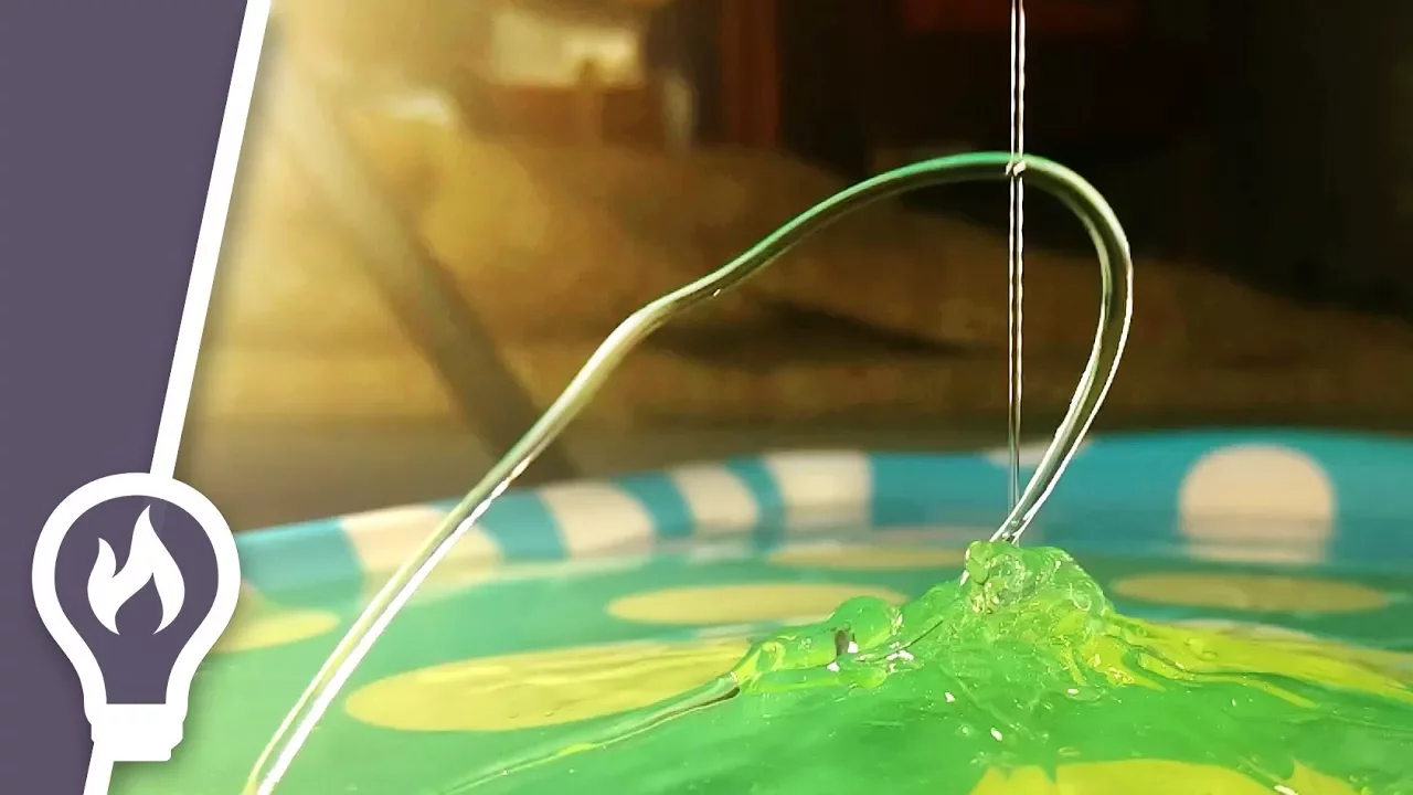
Shear-thinning is a property of certain liquids where their viscosity decreases when shear force is applied. In liquids, shear force occurs when different layers move at different velocities, creating a velocity gradient through the liquid. When shear force is applied to shampoo, its viscosity goes down, making it flow more easily.
Click to watch →

Geopolymer 3D Printing: When Simulation Meets Robotics and Reality
549
Rheology—the study of material deformation and flow under stress—is critical for 3D printing success. Key requirements include shear-thinning behavior for easy extrusion, recovery rate ≥85% for strand shape retention, large Linear Viscoelastic Region (LVER) for strain resistance, viscoelastic solid/gel-like behavior, and high particle association. Four numerical simulation methods exist: Finite Element Method (FEM) for part performance; Discrete Element Method (DEM) for particle flow; Smoothed Particle Hydrodynamics (SPH) for fluid flow; and Computational Fluid Dynamics (CFD) for flow behavior prediction. CFD was selected for its direct parameter optimization capability without requiring new tool development.
Click to watch →

SSC JE Crash Course 2024 - Safalta Batch | Fluid Mechanics | Fluid Properties | Civil Engineering
54.7K
Viscosity is the internal resistance of a fluid to flow, caused by molecular cohesion in liquids and molecular collisions in gases. Newton's Law of Viscosity states that shear stress is directly proportional to the velocity gradient (τ = μ × du/dy). The SI unit of dynamic viscosity is Pascal-second (Pa·s), while kinematic viscosity (ν = μ/ρ) has units of m²/s. For liquids, viscosity decreases with temperature, whereas for gases, it increases. Fluids are classified as Newtonian (following Newton's law, e.g., water, air) or Non-Newtonian (following power law τ = K(du/dy)^n), with further subcategories including dilatant (shear-thickening), pseudoplastic (shear-thinning), Bingham plastic (yield stress required), thixotropic (viscosity decreases with time), and rheopectic (viscosity increases with time) fluids.
Click to watch →
Deep Dive
Core concepts of additive manufacturing, including CAD modeling, slicing software, and basic extrusion-based 3D printing.

Additive manufacturing (3D Printing)- Lecture 1- Basic principles of additive manufacturing
20.2K
Additive manufacturing represents a paradigm shift from subtractive manufacturing by adding material layer by layer rather than removing it. The formal definition describes converting 3D designs into real products through programmed material deposition. Known as 3D printing in the market, this process distinguishes itself from traditional methods. The workflow involves CAD design, slicing into planar layers, and printer execution. Key benefits include personalization, reduced tooling, complex geometry capability, and eco-friendliness. Applications span industrial prototyping, architectural modeling, customer personalization, and medical uses. Slicing converts solid models into executable layer instructions, while coordinate systems (Cartesian, Delta, Polar) determine spatial accuracy in material deposition.
Click to watch →

Conferencia: Fundamentos de la Manufactura Aditiva, LDI. Juan Pablo González Berjón
285
This comprehensive section covers the foundational concepts of additive manufacturing and 3D printing. The presenter distinguishes additive manufacturing (the broader family of processes that unite materials to fabricate parts from a 3D model, typically layer by layer) from 3D printing (a specific type involving material deposition using a nozzle). Chuck Hall is recognized as the father of 3D printing for inventing the first stereolithography (SLA) machine in 1986. The technology evolved from industrial equipment to desktop devices through MakerBot (founded 2009) and Formlabs (Kickstarter success in 2014). Essential components include a 3D printer, a 3D model (created using software like SolidWorks, Rhino, Maya, Fusion, or Blender), and slicing software (like Cura). Three methods exist for obtaining 3D models: designing from scratch, downloading from repositories (Thingiverse, MyMiniFactory, Cults3D), or scanning with 3D scanners.
Click to watch →

From Concept to Creation Exploring 3D Printing's Present and Future Pathways
342
Manufacturing is divided into subtractive (removing material through machining) and additive (adding material layer by layer). Additive manufacturing, technically called 3D printing, joins materials from 3D model data using layers. The workflow involves creating a CAD model, generating an STL file, slicing to create G-code, and post-processing. Key advantages include no cutting tools, ability to produce porous/dense parts, complex geometries, and customization. Only CAD modeling skills are required as most processes are automated in software.
Click to watch →

Additive Manufacturing Basic Concepts and Processes _ Dr. Rupshree Ozah, Asst. Prof., Mech., MITS
486
The additive manufacturing process involves several steps: 1) Design using CAD software (like AutoCAD) or 3D scanning. 2) Slicing the design into layers using slicing software (like Ultimaker Cura). 3) Setting parameters including layer thickness, print speed, infill percentage, and support structures. 4) Generating code from slicing software. 5) Feeding code to the printer. 6) Printing the object. 7) Removing the part from the build platform. 8) Removing support structures if used.
Click to watch →
Conceptos básicos de manufactura aditiva
7.5K
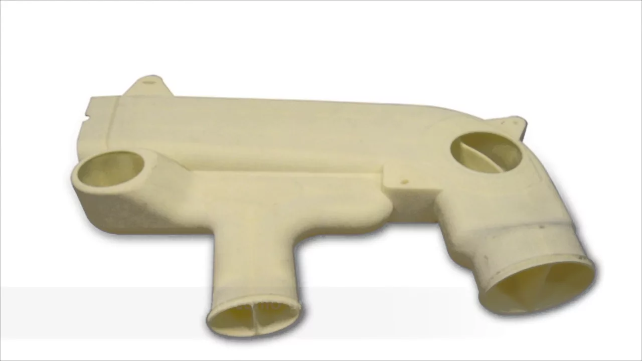
Additive manufacturing (3D printing) is a production method that builds parts layer by layer by fusing material, enabling the creation of complex geometries like internal channels and lattice structures that traditional manufacturing cannot achieve, while also allowing customization without dedicated tooling and reducing assembly requirements; it serves as a complementary process to machining and molding rather than a replacement, with applications spanning aerospace, medical devices, and mold components.
Click to watch →
Prerequisite Knowledge
- Concept 01Fundamentals of cell biology and tissue engineering, including cell-extracellular matrix (ECM) interactions and 3D cell culture principles.
- Concept 02Basic polymer chemistry and hydrogel crosslinking mechanisms, such as thermal, chemical, and photo-crosslinking.
- Concept 03Introduction to fluid mechanics and rheology, specifically shear-thinning behavior and printability of viscoelastic materials.
- Concept 04Core concepts of additive manufacturing, including CAD modeling, slicing software, and basic extrusion-based 3D printing.
Subsequent Learning
- Step 01Applications of bioprinted constructs in high-throughput drug screening, toxicology assays, and personalized disease modeling.
- Step 02Advanced strategies for vascularizing thick 3D tissue constructs to support cellular survival and nutrient diffusion.
- Step 03Translational regenerative medicine, focusing on preclinical in vivo transplantation and FDA regulatory pathways for bioprinted tissues.
- Step 04Multi-material and coaxial bioprinting techniques to fabricate complex, heterogeneous tissue interfaces (e.g., bone-to-tendon).
Bioink Design
Bioink Design
0:00Playing Section
- 1
Demonstrates a modular approach for designing extrudable hydrogel bioinks.
- 2
Uses commercial components to fabricate 3D tissue constructs like liver, muscle, and colon.
- 3
Starts by preparing ECM digest and dissolving a photo initiator for hydrogel formation.
Scaffold-Free Assembly and Organ-on-a-Chip Systems
While hydrogel-based 3D bioprinting is popular, critics point out significant limitations: exogenous hydrogel matrices can restrict cell-to-cell communication, alter natural cellular signaling, and often suffer from low resolution and poor vascularization. A major alternative paradigm is scaffold-free tissue engineering, which utilizes self-assembling multicellular spheroids or organoids. This approach allows cells to secrete their own native extracellular matrix, leading to more physiologically accurate cell densities and interactions. Additionally, microfluidic 'organ-on-a-chip' platforms offer a more robust alternative for in vitro screening. These systems simulate dynamic physiological shear stress and fluid flow far better than static, printed hydrogel constructs, leading to more reliable drug toxicity and efficacy data without the printability-versus-viability compromises inherent to hydrogel bioinks.
Deep Dive
Applications of bioprinted constructs in high-throughput drug screening, toxicology assays, and personalized disease modeling.

3D Bioprinting and Organoids
977
3D bioprinting enables the creation of personalized organoids from patient-derived induced pluripotent stem cells, allowing researchers to generate functional 3D tissue models that mimic specific organs such as neural tissues, testicular tubules, and cardiac tissues; these bioprinted constructs can incorporate drug-releasing microspheres for sustained factor delivery, maintain viability for extended periods (up to 45+ days), and exhibit functional characteristics like electrical activity, making them valuable tools for high-throughput drug screening and disease modeling.
Click to watch →
REPLAY | 3D Systems: Drug Screening with Biofabricated Tissue Models
203
3D biofabricated tissue models, created through bioprinting technology, offer a transformative approach to drug discovery by providing human-relevant, physiologically accurate tissue constructs that can replace animal testing and improve drug screening efficiency; these models enable researchers to screen hundreds to thousands of compounds in medium-to-high throughput formats while maintaining complex tissue architecture, physiological features like vasculature, and disease-specific characteristics, ultimately helping to address the challenge that less than 10% of compounds entering clinical trials become approved drugs due to poor translation from animal models to human patients.
Click to watch →

Bioprinting for Skin Model Systems. University of Wisconsin-Madison Department of Dermatology.
161
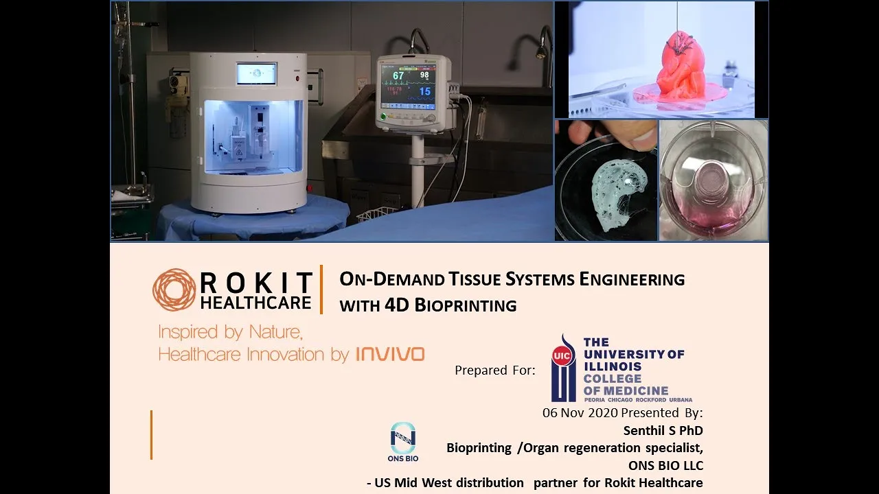
The bioprinting process involves five steps: (1) Preparation - designing experiments with appropriate biomaterials, cell types, and 3D models; (2) Printing - loading materials, slicing models, and depositing bio-inks; (3) Post-printing - cross-linking (temperature, UV), allowing cell differentiation; (4) Evaluation - assessing structure and functionality; (5) Application - in vitro disease modeling, compound testing, or in vivo transplantation. HSE applications include: toxicological evaluation following OECD guidelines (TG431 for corrosion, TG439 for irritation), absorption/permeability tests measuring transdermal moisture loss, sensitization tests for immune reactions, drug screening, phototoxicity testing, and aging skin models created by manipulating dermal layers with different cell types representing different age groups.
Click to watch →
Bioprinting for Basic and Translational Research Applications. UCI College of Medicine Rockford.
103
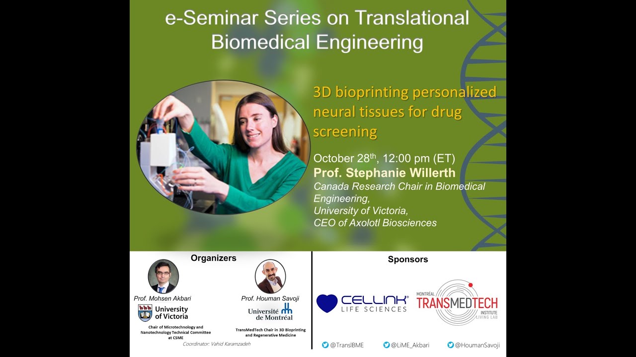
Bioprinting applications span three categories: (1) In vitro efficacy testing for toxicological screening, offering alternatives to animal testing; (2) Disease modeling using 3D constructs that better replicate human physiology than 2D cultures; (3) Transplantation and regeneration for addressing organ shortages. Specific examples include: self-healing hydrogels for cartilage repair, nanoporous nerve conduits with aligned collagen for axon regeneration, 3D cancer models showing superior drug efficacy assessment, and customized bone implants manufactured in GMP conditions. These applications demonstrate systematic translation from bench research to clinical potential.
Click to watch →
e-Seminar Series on Translational Biomedical Engineering with Prof. Stephanie Willerth (2020-10-28)
568
3D bioprinted tissues enable sophisticated disease modeling and drug screening applications. For Alzheimer's disease, patient-derived induced pluripotent stem cells differentiate into cortical neurons that recapitulate disease-specific phenotypes including tau tangles and amyloid beta accumulation over time, enabling personalized drug screening. For glioblastoma, the most aggressive brain tumor with 32-36 week survival, 3D bioprinted models better replicate invasive tumor behavior than 2D cultures—cells become more tumorigenic, form additional spheroids, and express elevated CD133 and DCX markers. Novel n-cadherin antagonists, which disrupt tumor cell adhesion, demonstrate selective cytotoxicity against cancer cells while sparing healthy astrocytes in co-culture models. These applications validate 3D bioprinted tissues as superior platforms for evaluating therapeutic efficacy in clinically relevant tumor microenvironments, addressing the critical need for better predictive models in oncology research.
Click to watch →
Deep Dive
Advanced strategies for vascularizing thick 3D tissue constructs to support cellular survival and nutrient diffusion.
From 3D Printing to Cyborg Tissues: Redefining Tissue Engineering: Prof. Tal Dvir
124
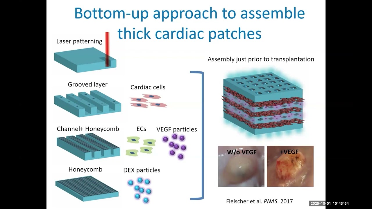
Thick tissues (1 cm like human heart) face oxygen diffusion limitations causing core cell death. The bottom-up approach solves this by creating thin vascularized layers separately: electrospun fibers with laser-patterned grooves for aligned cardiomyocytes, endothelial cell channels, and drug-releasing particle structures. These layers are cultured independently then assembled into thick vascularized tissues just before transplantation. This ensures proper oxygenation and vascularization throughout the engineered tissue.
Click to watch →

Stanford Pediatric Grand Rounds: The Long and Winding Road: Steps Towards 3D Printing a Heart
563
Without vascularization, only the outer 500-800 microns of thick tissue survives. Printed vascular networks enable oxygen perfusion throughout dense cellular tissues. Cardiac organoids (80% cardiomyocytes, 20% stromal cells) beat asynchronously when isolated but synchronize when assembled and perfused. By writing coronary artery-like vessels into cardiac tissue and flushing sacrificial material, researchers create perfusable channels. After 16 days of perfusion, centimeter-tall cardiac tissue beats visibly. This demonstrates creating functional, perfused cardiac constructs capable of drug testing and eventual implantation.
Click to watch →
Phd Defence of Maria Gabriella Fois
244
Electrical impedance spectroscopy enables non-invasive, real-time monitoring of 3D cell cultures by measuring electrical impedance responses to applied currents, distinguishing between 2D and 3D cultures and detecting biomaterial presence. For vascularization modeling, two approaches exist: non-porous microwells enable faster vascularization through direct cell contact, while porous microwells provide controlled interaction where pore density determines vascularization extent. Both approaches show abundant vascularization with endothelial cells forming 3D sprout-like structures. This vascularization capability is essential for modeling highly vascularized tissues like bone, enabling simultaneous study of multiple biological processes in engineered tissue constructs.
Click to watch →

Will Tissue Engineering Change Human Nature?
244
Tissue engineering develops biological substitutes to restore, maintain, or improve function, requiring cells, scaffolds, and bioactive molecules. Oxygen diffusion limits thick tissue viability—cells die beyond 100 microns from capillaries. Two approaches address vascularization: bottom-up prevascularization using endothelial cells on alginate beads forming vessel networks requiring feeder cell stabilization, which successfully connects to host vasculature upon implantation; and top-down 3D bioprinting using photopolymerization or extrusion to create defined vascular architectures. Mechanical stimulation enhances tissue development—chondrocytes in dynamically stimulated alginate disks develop greater stiffness, and perfused bone constructs show increased mineralization. These technical foundations enable progressing from simple constructs toward complex functional tissues.
Click to watch →
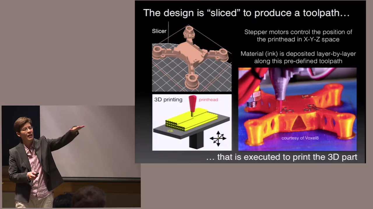
3D Printing: Making the Future: Fall 2015 Donoho Colloquium - Jennifer A. Lewis
977
Vascularized tissue engineering combines fugitive ink technology with cell-laden bioinks and extracellular matrix materials. The process involves printing sacrificial channels that are later removed, leaving empty lumens lined with endothelial cells (human umbilical vein endothelial cells). Multiple cell types including fibroblasts can be co-printed and subsequently populated into separate vascular compartments. Initial experiments showed some cell degradation during printing, but optimized matrices now achieve 95% cell viability. The resulting constructs demonstrate confluent endothelial lining with tight junctions, proper barrier function preventing uncontrolled diffusion, and support for angiogenesis when growth factors are introduced. These advances enable creation of centimeter-thick tissues with pervasive vascular networks suitable for long-term perfusion studies.
Click to watch →
Deep Dive
Translational regenerative medicine, focusing on preclinical in vivo transplantation and FDA regulatory pathways for bioprinted tissues.
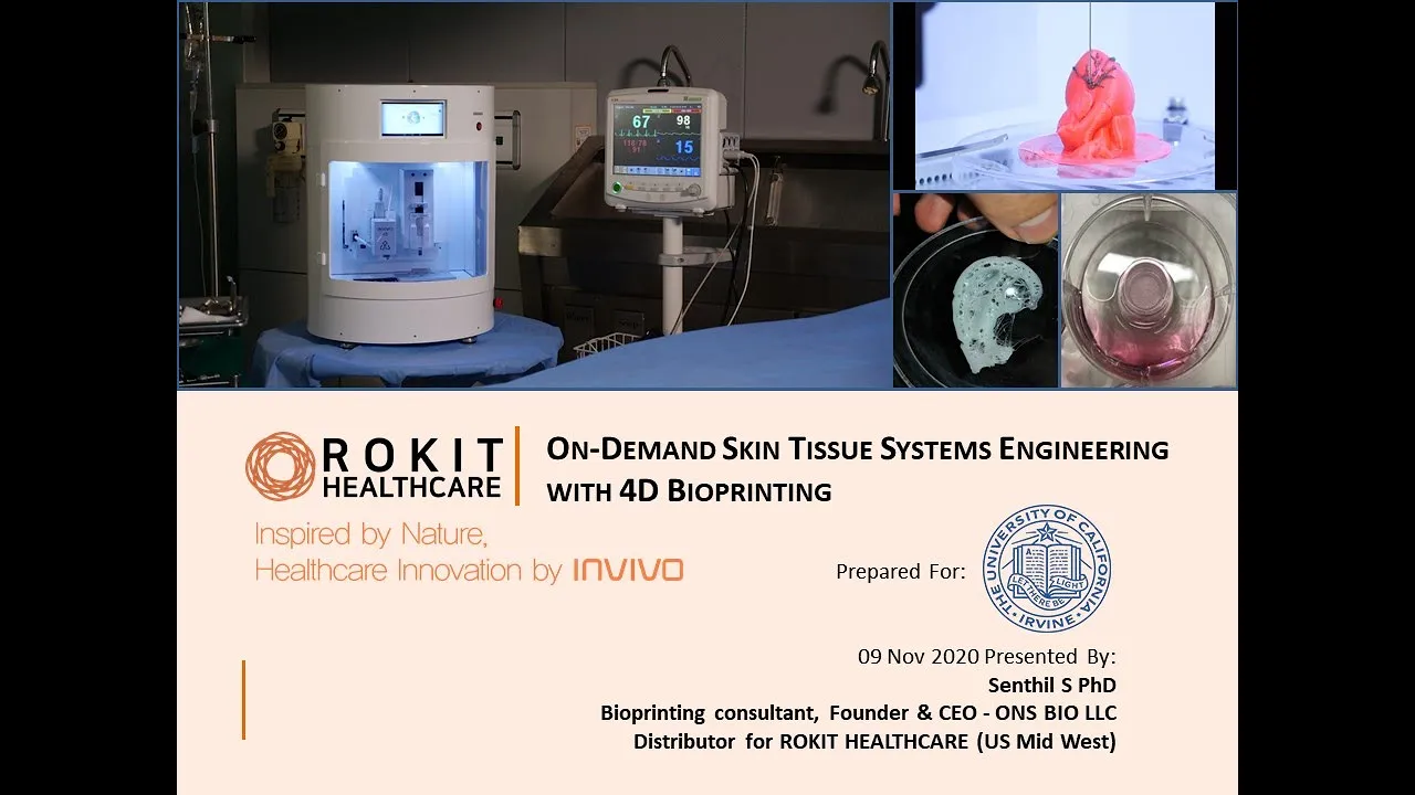
Bioprinting for Skin Regeneration. University of California, Irvine
179
Two approaches exist for FDA regulatory pathways for bioprinted therapies: (1) For standardized bone implants, the process occurs in GMP facilities with FDA approval for printing standardized versions; (2) For autologous therapies like diabetic foot ulcer skin regeneration, the in-hospital manufacturing process approval pathway is used, which involves minimal manipulation of patient-derived stem cells (not cultured but applied directly to wound beds to prime regeneration). This approach allows faster clinical application without lengthy biologics or cell therapy approval processes. Clinical studies are ongoing in India, Korea, California (USC), Turkey, and Egypt.
Click to watch →
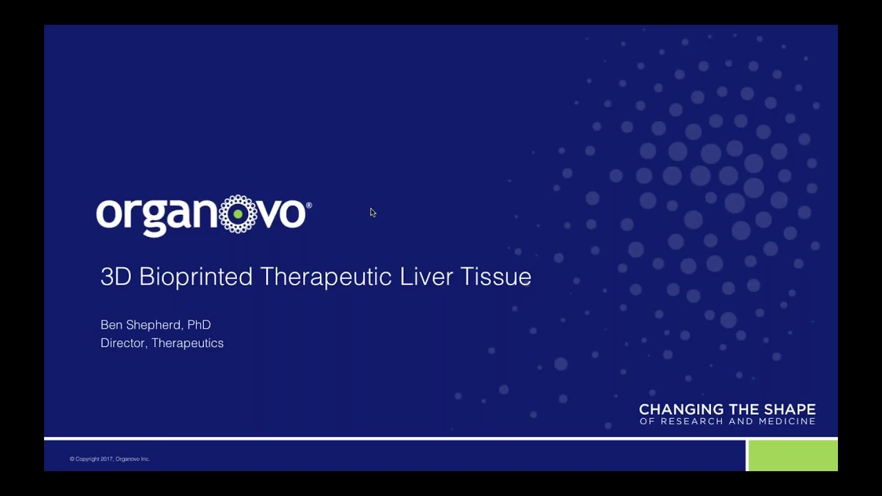
Webinar: 3D Bioprinted Therapeutic Liver Tissue
4.9K
Bioprinted therapeutic tissues represent a novel regulatory category neither purely drug nor medical device, fitting within FDA's Office of Tissues and Advanced Therapies (OTAT) purview. The pathway involves filing an Investigational New Drug (IND) application, anticipated around calendar year 2020, followed by clinical trial initiation. Scaling strategies involve two approaches: multiple patches or single patient-specific patches. Modular design enables fabrication to size based on individual anatomy, particularly valuable for rare orphan diseases where patient-specific construction is anticipated.
Click to watch →
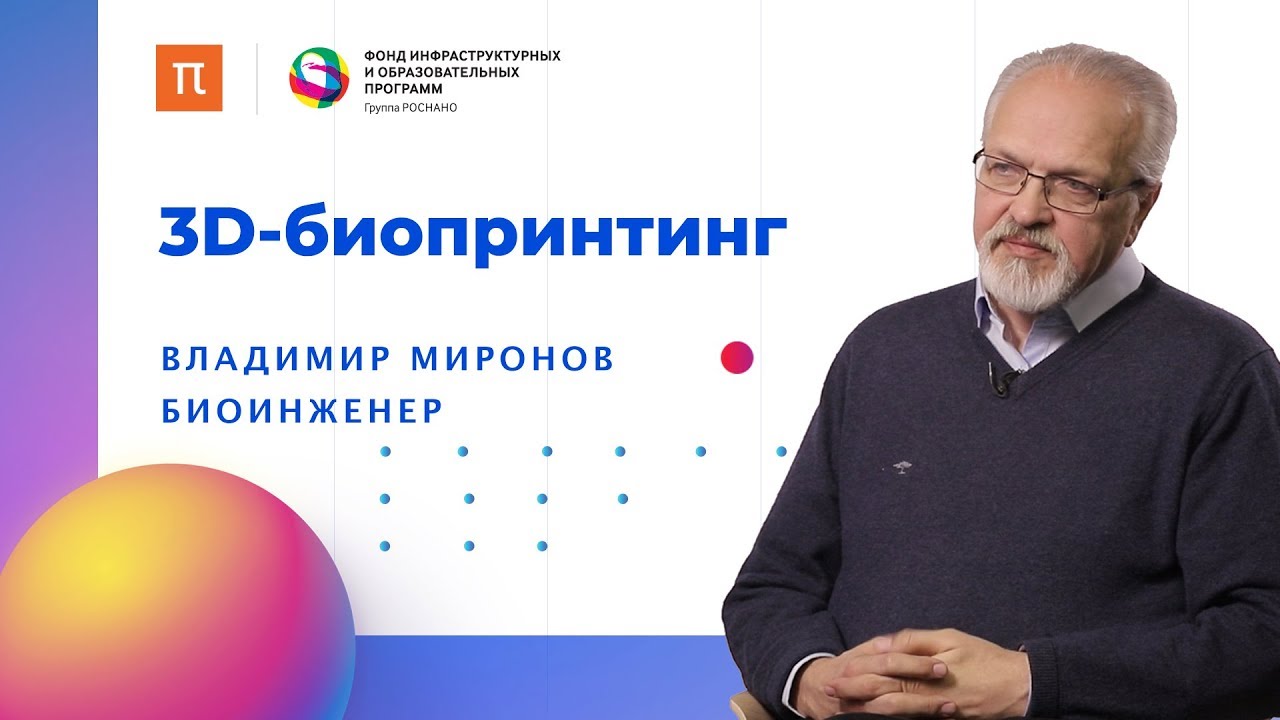
3D-биопринтинг // Биоинженер Владимир Миронов на ПостНауке
9.6K
Translating bio-printing technology to clinical practice faces multiple barriers: the need for multidisciplinary teams combining expertise in computer science, chemistry, cell biology, and mechanical engineering; the absence of clinically certified bio-printers for medical use; and complex regulatory frameworks that create high barriers to entry. The regulatory environment, designed for traditional medical products, creates challenges for bio-printed tissues that may not fit existing categories. Additionally, the technology requires significant investment (5-10 years) that both academic research and industry may be reluctant to provide. Emerging applications include beta-cell replacement for diabetes treatment, where researchers have created insulin-producing cells responsive to glucose concentration that can be transplanted under the kidney capsule. Hair follicle regeneration represents another promising application, with stem cells grown in specialized bioreactors and implanted using robotic systems.
Click to watch →
Organovo
949
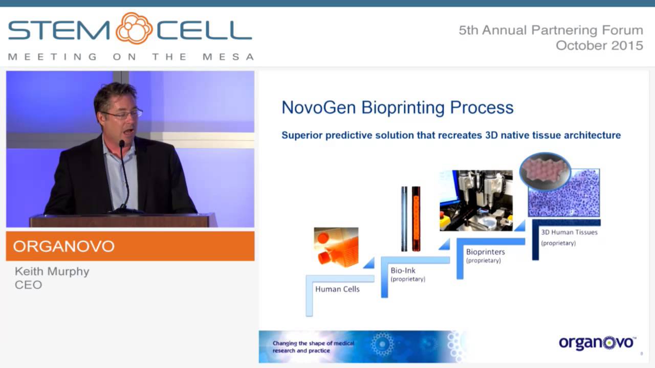
Organovo's technology serves two primary application categories. In vitro applications focus on creating human tissue models for pharmaceutical research, including preclinical safety testing and disease modeling. The company operates a service model where pharmaceutical companies send drug candidates to be tested on bioprinted tissues, with results ranging from basic survival indicators to detailed histological pathology analysis. In vivo applications involve developing therapeutic tissues for transplantation, with the goal of creating tissue sections that could deliver 10-15% of organ function to delay or avoid transplantation needs. Potential applications include liver tissue, blood vessels, nerve grafts, and heart muscle patches. The company allocates approximately 10% of R&D to early preclinical animal research in this area.
Click to watch →
3D Bioprinting with Bioinks for Printing Neural and Cardiac Tissues | Axolotl Biosciences
193
Bioprinted tissue products follow distinct regulatory pathways based on intended use. Research-use products require end-user licenses, while clinical applications demand FDA approval and GMP manufacturing standards. Xeno-free bioinks present straightforward pathways toward human translation, with successful implantation already demonstrated in clinical settings. Future directions include expanding into skin grafts for burn patients and gut models for disease research. Strategic collaborations with established companies and access to appropriate manufacturing facilities are critical for advancing bioprinted tissues from research to clinical applications.
Click to watch →
Deep Dive
Multi-material and coaxial bioprinting techniques to fabricate complex, heterogeneous tissue interfaces (e.g., bone-to-tendon).

Extreme Manufacturing Webinar: Additive Manufacturing and the 21st Century Industrial Revolution
231
Multimaterial cryogenic bio printing enables simultaneous deposition of different materials within the same structure. Coaxial nozzles create vascular-muscle junction units with distinct cellular compartments. The cryogenic environment maintains structural integrity during multimaterial deposition. Vertical printing creates hierarchical structures with aligned microchannels guiding cell alignment. These approaches enable fabrication of complex tissue interfaces better mimicking native organization, addressing limitations of single-material approaches while maintaining the advantages of cryogenic preservation.
Click to watch →

Intro to Cellink 3D Bioprinting Technologies & Space Application | Dr. Haruka Yoshie | ASBX 2021
370
Advanced bioprinting technologies enable fabrication of increasingly complex tissue constructs. The BioX6 bioprinter introduces coaxial printing using dual-nozzle systems with outer and inner needles, enabling simultaneous deposition of multiple bioinks to create layered structures. Sacrificial ink approaches allow creation of hollow tube structures that can later be dissolved, while crosslinking solutions enable formation of perfusable channels. Multi-material bioprinting combines different printheads to construct heterogeneous tissues, exemplified by skin models with distinct regions printed from different bioinks. These capabilities expand the range of achievable tissue architectures, supporting development of more physiologically relevant organoid models for research and therapeutic applications.
Click to watch →

December 3, 2019 - Angela Panoskaltsis-Mortari, PhD
163
Advanced bioprinting techniques include FRESH (printing in gelatin micro-particle suspensions then melting support material), coaxial nozzles for simultaneous multi-material extrusion, and handheld biopens for in situ printing during surgery. Multi-material printing enables computer-controlled delivery of different components through single nozzles. Two-photon polymerization achieves 1-micron resolution for intricate structures. These technologies enable creation of complex constructs including vascular channels, nephron-like structures, and multi-cellular tumor models with precise spatial organization of different cell types and materials.
Click to watch →

Shrike Zhang - Putting 3D Bioprinting to Tissue Fabrication
301
Extrusion-based bioprinting deposits bioinks containing cells and biomaterials through a nozzle to create 3D structures automatically. Rapid multi-material bioprinting uses a single nozzle with multiple inlet channels to deliver different materials simultaneously, eliminating the need for time-consuming nozzle exchanges. This enables printing of complex constructs with multiple cell types and materials in a single process, achieving 70-80% cell viability after seven days of culture. Digital Light Processing (DLP) bioprinting uses photoactivatable bioinks where light exposure causes cross-linking layer-by-layer. Multi-material DLP bioprinting enables creation of complex vascularized tumor models with endothelial cells lining channels and cancer cells in surrounding regions. Sacrificial bioprinting creates hollow perfusable channels by first printing sacrificial fibers that template channels, then removing them to create pathways lined with endothelial cells forming tight junctions that regulate molecule diffusion.
Click to watch →

Bioprinting and Tissue Engineering
123
Bioprinting enables the fabrication of complex, functional tissue constructs by combining multiple materials and cell types in precise spatial arrangements, addressing the fundamental challenge of scaling up tissue engineering constructs beyond 200-300 micron oxygen diffusion limits through multi-material printing approaches that integrate structural supports, vascular networks, and cellular components.
Click to watch →
Bioink Design
Bioink Design
0:00- 1
Demonstrates a modular approach for designing extrudable hydrogel bioinks.
- 2
Uses commercial components to fabricate 3D tissue constructs like liver, muscle, and colon.
- 3
Starts by preparing ECM digest and dissolving a photo initiator for hydrogel formation.
Scaffold-Free Assembly and Organ-on-a-Chip Systems
While hydrogel-based 3D bioprinting is popular, critics point out significant limitations: exogenous hydrogel matrices can restrict cell-to-cell communication, alter natural cellular signaling, and often suffer from low resolution and poor vascularization. A major alternative paradigm is scaffold-free tissue engineering, which utilizes self-assembling multicellular spheroids or organoids. This approach allows cells to secrete their own native extracellular matrix, leading to more physiologically accurate cell densities and interactions. Additionally, microfluidic 'organ-on-a-chip' platforms offer a more robust alternative for in vitro screening. These systems simulate dynamic physiological shear stress and fluid flow far better than static, printed hydrogel constructs, leading to more reliable drug toxicity and efficacy data without the printability-versus-viability compromises inherent to hydrogel bioinks.
Fundamentals of cell biology and tissue engineering, including cell-extracellular matrix (ECM) interactions and 3D cell culture principles.
Basic polymer chemistry and hydrogel crosslinking mechanisms, such as thermal, chemical, and photo-crosslinking.
Introduction to fluid mechanics and rheology, specifically shear-thinning behavior and printability of viscoelastic materials.
Core concepts of additive manufacturing, including CAD modeling, slicing software, and basic extrusion-based 3D printing.
Applications of bioprinted constructs in high-throughput drug screening, toxicology assays, and personalized disease modeling.
Advanced strategies for vascularizing thick 3D tissue constructs to support cellular survival and nutrient diffusion.
Translational regenerative medicine, focusing on preclinical in vivo transplantation and FDA regulatory pathways for bioprinted tissues.
Multi-material and coaxial bioprinting techniques to fabricate complex, heterogeneous tissue interfaces (e.g., bone-to-tendon).
541 views6likes2:00@JoVEJournalOriginal Release: 2022-08-23
[Music] the overall goal of this protocol is to demonstrate a versatile approach for designing hydrogel bio inks that can be extruded through bio printing devices the bio inks can then be used to fabricate three-dimensional tissue constructs this method can help answer key questions in the bio printing field such as how to control the mechanical properties needed in order to provide a material that can be extruded using a bio printer the main advantage of this technique is that we use commercially available components combined in a modular fashion to create a simple and effective bio-printable hydrogel bioink the applications of these technologies include the creation of 3d tissue organoids that can be used to accurately model the effects of drugs toxins and diseases although this method can provide a framework to bioprint 3d liver constructs it can also be applied to other tissue types such as muscle lung and colon generally individuals new to this method will struggle because there are a number of different reagents used to create the hydrogel biowink but it is actually quite straightforward demonstrating the procedure will be young june seoul a postdoc on our team to begin prepare a tissue-specific extracellular matrix digest to be used in the hydrogel formulation as described elsewhere then dissolve a photo initiator in water at a weight per volume ratio of 0.1 percent to form the hydrogel bio inks first dissolve the base material components from the hyaluronic acid hydrogel kits into individual aliquots of the water photo initiator solution then combine the ecm solution the 2
Up Next
9:56
Perfusion Bioreactors: Continuous Cell Culture and High Cell Density
@MarloesPeeters
12K views•2021-11-09
14:12
Quantitative Real-Time PCR (qPCR): Principle, Method & Data Analysis
@animatedbiologywitharpan
129.2K views•2023-09-06
23:33
Microbial Degradation of Plastics: Biodegradation Pathways & Sustainability
@majeedhammad
2.9K views•2021-04-11
16:04
CRISPR and Genetic Engineering: How Gene Editing Works and Why It Matters
@kurzgesagt
30.5M views•2016-08-10
Related Study Plans & Knowledge Roadmaps
Structured learning paths in Biotechnology
biotechnology
AAV Production: Transfection, Prep & qPCR
01Introduction to Gene Therapy and AAV Biology
02Mammalian Cell Culture: Suspension HEK293 Cells
03Upstream Bioprocessing: Triple-Transfection Optimization
+ 2 more learning modules
18h5 modules
Explore Roadmap
biotechnology
Building Gut-on-a-Chip: PDMS, Cells & TEER
01Foundations of Microfluidics and Gut Anatomy
02Cleanroom Fabrication & PDMS Soft Lithography
03Epithelial Cell Culture and On-Chip Seeding
+ 2 more learning modules
26h5 modules
Explore Roadmap
biotechnology
Optimizing CFPS: E. coli Extract & Yield
01Fundamentals of Molecular Biology and Translation
02Introduction to Cell-Free Protein Synthesis (CFPS)
03Preparing E. coli Crude Lysate (The Extract)
+ 2 more learning modules
19h5 modules
Explore Roadmap
biotechnology
Haematococcus PBR: Design, Stress & Astaxanthin
01Biology and Life Cycle of Haematococcus pluvialis
02Bench-Scale Photobioreactor (PBR) Design and Engineering
03Culture Media Formulation and Inoculation
+ 2 more learning modules
15h5 modules
Explore Roadmap