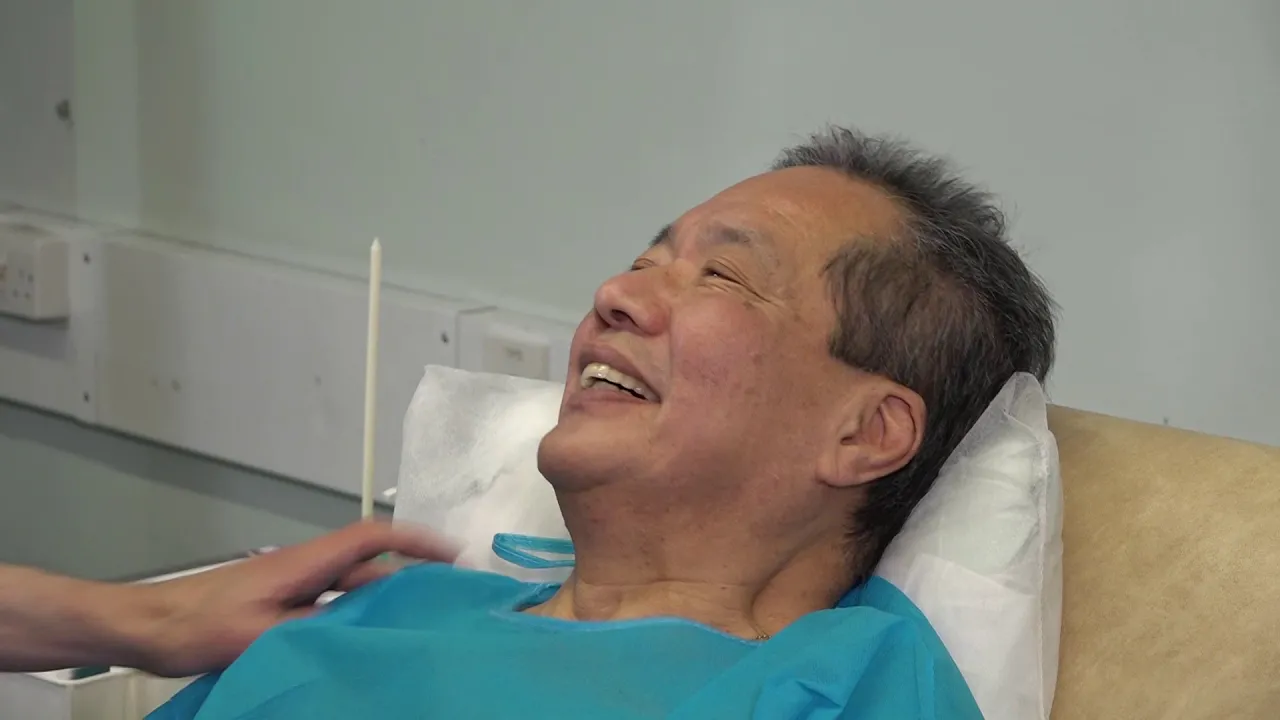
This video demonstrates clinical sensation testing for dermatomes L4 and C6, showing how to assess light touch sensation using tissue and sharp/dull discrimination using a dual-ended object, with the patient indicating sensations while eyes are closed to evaluate sensory nerve function.
Sensation Testing Guide: Light Touch & Sharp/Dull Exam
Added:Deep Dive
Basic neuroanatomy of the peripheral nervous system, specifically the structure and function of sensory nerves.
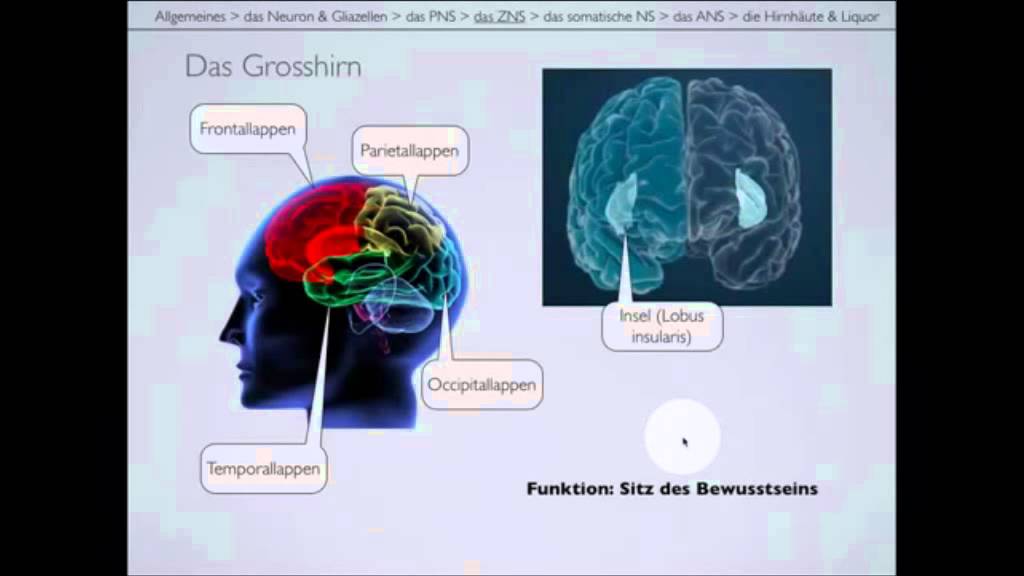
Langsame Version des Nervensystems Teil 2/2
126.6K
The peripheral nervous system consists of all nervous tissue except the brain and spinal cord, connecting the central nervous system to peripheral organs. It is divided into cranial nerves (12 pairs originating from the brain or brainstem) and spinal nerves (31 pairs originating from the spinal cord). Cranial nerves include specialized sensory nerves like the olfactory (smell) and optic (vision) nerves, which connect directly to the brain rather than the brainstem. Motor cranial nerves control eye movements, facial expressions, and other functions. The peripheral nervous system transmits efferent commands (motor output) and afferent information (sensory input) between the brain and body.
Click to watch →
Neuronas sensitivas | Neuronas sensoriales | Neuronas aferentes | Neurona sensitiva o aferente
8.1K
Sensory neurons have different structures depending on their location, whether in the peripheral or central nervous system. In the peripheral nervous system, pseudounipolar neurons are found. One prolongation extends away from the nervous system toward other regions of the organism, while the other directs toward the central nervous system (spinal cord or brainstem). The termination establishes contact with a new neuron to transport sensory information.
Click to watch →

ALHS 1090 - SRTC - Chapter 5
105
The peripheral nervous system is located outside or peripheral to the CNS and includes the nerves in the arms and legs. The PNS includes 31 pairs of spinal nerves, 21 cranial nerves, and the nerves in the arms and legs. Almost all nerves are part sensory and part motor. Some cranial nerves are all sensory and all motor. Sensory nerves gather information from the skin, muscles, and joints, including sensations such as temperature, touch, pressure, movement sense, position sense, and pain. These sensations allow us to distinguish between sharp and dull and to recognize different weights, shapes, and textures of objects. Central sensory information is sent to the brain, and the brain responds by sending messages back out to the body via motor nerves which control body movement. The message may be a conscious one that prompts action (such as putting on a coat if cold) or unconscious, causing reflexive responses (such as reflexively lifting your foot if you step on something sharp).
Click to watch →
![Sistema nervioso PERIFÉRICO [Vías sensitivas y motoras]](https://i.ytimg.com/vi/TkHV8SncmD4/maxresdefault.jpg)
Sistema nervioso PERIFÉRICO [Vías sensitivas y motoras]
100.9K
The peripheral nervous system consists of nerves and ganglia connecting the central nervous system to body tissues. Nerves are bundles of axons that transmit information bidirectionally: sensory nerves carry impulses to the CNS, while motor nerves carry responses from the CNS to effectors. Mixed nerves contain both sensory and motor neurons. The system is classified into cranial nerves (12 pairs connecting directly to the brain) and spinal nerves (communicating with the spinal cord). Cranial nerves include olfactory (smell), optic (vision), oculomotor (eye movement), trochlear (eye movement), trigeminal (face sensation and jaw movement), abducens (eye movement), facial (facial expression and taste), vestibulocochlear (hearing and balance), glossopharyngeal (swallowing and taste), vagus (visceral regulation), accessory (head and neck muscles), and hypoglossal (tongue movement).
Click to watch →

احياء الثالث متوسط 2024/ الفصل التاسع/ المحاضرة 2 / الجهاز العصبي المحيطي
90.1K
The peripheral nervous system consists of numerous nerves distributed throughout the body. Its primary function is to receive sensory stimuli from various sensory organs and transmit these signals to the central nervous system. For example, when you touch something hot, peripheral nerves in your hand detect the heat and pain, then send this information to the brain so you can react by moving away from the danger. The peripheral nervous system consists of two main types of nerves: spinal nerves (31 pairs emerging from the spinal cord connecting to muscles throughout the body) and cranial nerves (12 pairs emerging from the brain). Both types contain sensory fibers (for receiving sensations), motor fibers (for controlling movement), and mixed fibers (containing both sensory and motor functions).
Click to watch →
Deep Dive
The concept of dermatomes and how spinal nerve roots correspond to specific cutaneous regions of the body.
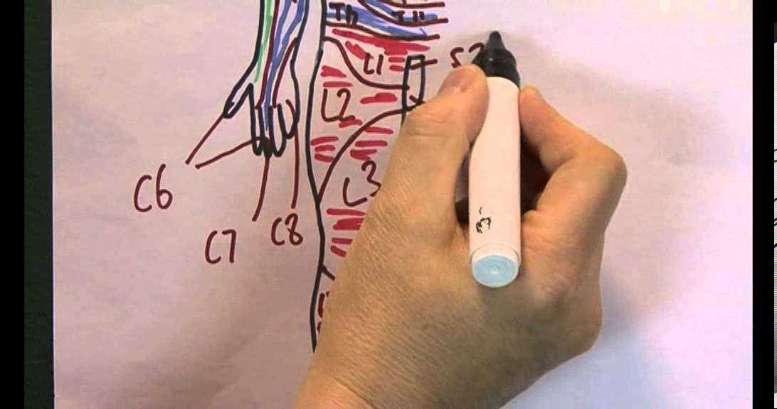
Skin 10, Dermatomes
43.1K
Dermatomes are specific areas of the skin surface supplied by particular spinal nerves, creating a systematic map where each spinal nerve root (C1-C8, T1-T12, L1-L5, S1-S5) innervates distinct regions of the body; this mapping allows clinicians to identify which spinal nerve is affected when a patient experiences pain, numbness, or paralysis in a specific body area.
Click to watch →

How to Control Your Sense of Pain & Pleasure
287.9K
The body surface is divided into dermatomes, which are territories innervated by specific spinal nerves. Each dermatome corresponds to a particular nerve root. When a rash or inflammation occurs, it often has a sharp boundary corresponding to the borders between dermatomes. For example, herpes simplex virus lives on the trigeminal nerve (fifth cranial nerve) and causes rashes with clear boundaries between affected and unaffected skin. Similarly, shingles rashes follow dermatome boundaries. This occurs because each dermatome is innervated by a single nerve root, and inflammation affects only one territory at a time.
Click to watch →
EMBRIOLOGÍA DEL SISTEMA MUSCULOESQUELÉTICO - Clase NMG
155
Dermatomes are skin regions controlled by specific spinal nerve roots. Each dermatome corresponds to a specific spinal level (C3-C8, T1-T12, L1-L5, S1-S5). This organization allows precise localization of sensory deficits and understanding of how spinal injuries affect different body regions.
Click to watch →

𝟎𝟕. 𝐋𝐨𝐰𝐞𝐫 𝐥𝐢𝐦𝐛 (𝐑𝐞𝐯𝐢𝐞𝐰 𝐨𝐟 𝐭𝐡𝐞 𝐜𝐮𝐭𝐚𝐧𝐞𝐨𝐮𝐬 𝐧𝐞𝐫𝐯𝐞𝐬 𝐚𝐧𝐝 𝐝𝐞𝐫𝐦𝐚𝐭𝐨𝐦𝐞𝐬)
24.7K
The lower limb's cutaneous nerves (sural, saphenous, femoral branch of genitofemoral, ilioinguinal, medial plantar, and lateral terminal branch of anterior tibial) provide sensory innervation to specific skin regions, while dermatomes represent the skin areas supplied by sensory fibers from individual spinal nerve roots (L1-L5 and S1), with key clinical correlations such as the saphenous nerve innervating the medial border of the dorsum of the foot at L3 level, and adjacent sensory loss on the big and second toes indicating injury to the lateral terminal branch of the anterior tibial nerve.
Click to watch →
Dermatome & Sensory Nerve Testing
8.6K
Dermatomes represent skin areas innervated by single nerve roots, while cutaneous nerve supply involves specific peripheral nerves. Cervical dermatomes C5-C8 produce distinct patterns: C6 affects forearm toward thumb, C7 involves middle finger, and C8 causes little finger tingling. The median nerve supplies thumb, index, middle, and half ring finger; ulnar nerve affects little finger and half ring finger. Lower limb dermatomes include L1-L5 and S1, with common pathology at L4-L5 and L5-S1 levels. Cutaneous nerves like axillary (regimental badge) and lateral femoral cutaneous (lateral thigh) can compress independently of spinal roots, causing isolated sensory symptoms without radicular pain.
Click to watch →
Deep Dive
The physiological difference between sensory pathways, specifically the spinothalamic tract (pain/temperature) and the dorsal column-medial lemniscal pathway (touch/vibration).

Sensory Pathways MADE EASY!!
145.4K
The spinothalamic tract transmits pain and temperature sensations through a different route than touch. Unlike the dorsal column pathway, it synapses and decussates immediately at the spinal cord level, bypassing the brainstem entirely. This fundamental difference in crossing points explains why spinal cord lesions produce distinct sensory deficits: contralateral loss of pain/temperature below injury, while ipsilateral fine touch is lost. Clinicians use separate touch and temperature tests to diagnose spinal cord injuries based on these pathway differences.
Click to watch →
Sensory Pathways | Touch vs. Pain and Temperature
71.7K
The human body uses two distinct ascending sensory pathways to transmit different types of sensory information to the brain: the dorsal column medial lemniscus pathway transmits fine touch, two-point discrimination, and conscious proprioception by ascending ipsilaterally through the spinal cord before decussating at the medulla, while the spinothalamic pathway transmits pain and temperature by decussating immediately at or near the level of entry into the spinal cord; this fundamental difference in decussation timing explains why damage to one side of the spinal cord affects different sensory modalities on opposite sides of the body.
Click to watch →
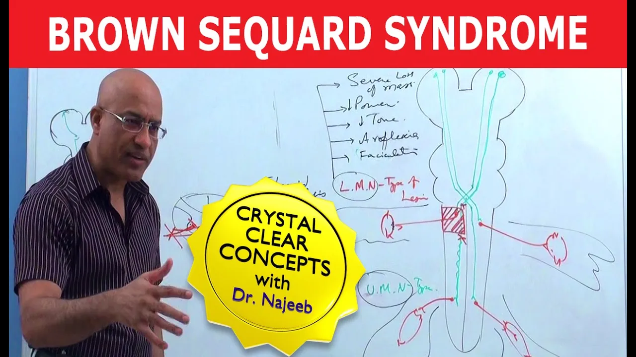
Brown Sequard Syndrome | Internal Medicine 💊
261K
The dorsal column system carries fine touch, vibration, proprioception, two-point discrimination, and stereognosis, ascending ipsilaterally within the spinal cord to gracile/cuneate nuclei before crossing to the thalamus. The spinothalamic tract carries pain, temperature, and crude touch, crossing contralaterally at the level of entry. This fundamental difference in crossing pattern explains why sensory loss patterns differ between the two systems. After hemisection, dorsal column fibers on the injured side are damaged, while spinothalamic fibers from the opposite side are damaged due to their contralateral crossing.
Click to watch →

Three Critical Vertical Pathways: Neuroanatomy Video Lab - Brain Dissections
54.4K
Two major sensory pathways transmit somatic information from the body to the brain. The spinothalamic tract carries pain, temperature, and crude touch, while the dorsal column-medial lemniscus pathway transmits proprioception, vibration, joint position sense, and fine touch. Both follow a three-neuron structure: first neuron in dorsal root ganglion, second neuron relaying in thalamus, third neuron projecting to postcentral gyrus. The key difference is decussation timing—the spinothalamic crosses in spinal cord, while the dorsal column crosses in caudal medulla. Both terminate in the ventral posterior lateral thalamic nucleus before reaching somatosensory cortex.
Click to watch →
¡VÍAS SENSITIVAS! RESUMEN PARA ESTUDIANTES
75.8K
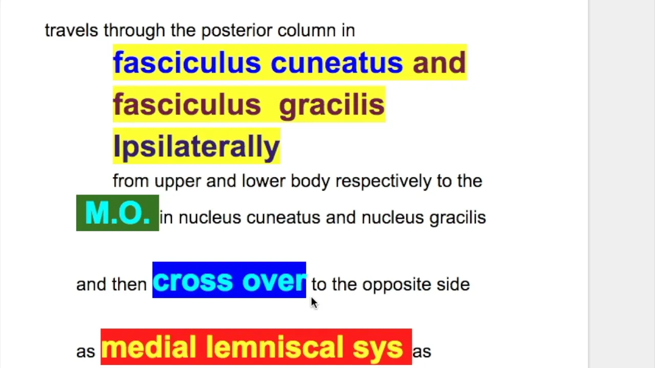
The somatosensory system consists of two main ascending pathways: the dorsal column-medial lemniscus pathway (lemniscus medial) responsible for fine touch, vibration, pressure, and proprioception, and the spinothalamic pathway (anterolateral) responsible for pain and temperature sensation. Both pathways use a three-neuron relay system where the first neuron's cell body is in the dorsal root ganglion, the second neuron's cell body is in the spinal cord or brainstem, and the third neuron's cell body is in the thalamus (ventral posterolateral nucleus). A key rule is that the second neuron always decussates (crosses to the opposite side) after synapsing. The dorsal column pathway carries information from the lower body through the fasciculus gracilis and from the upper body through the fasciculus cuneatus, while the spinothalamic pathway carries pain and temperature information through the anterior and lateral funiculi of the spinal cord.
Click to watch →
Deep Dive
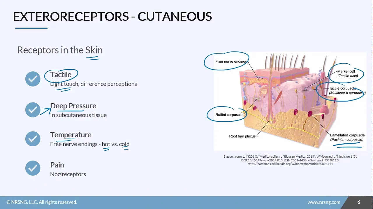
An understanding of basic cutaneous receptors, such as nociceptors for pain (sharp/dull) and mechanoreceptors for light touch.
Anatomy & Physiology Sensory Basics
117
Cutaneous receptors in the skin and mucous membranes detect four stimulus types: tactile receptors for light touch distinguishing sharp/dull/soft sensations; deep pressure receptors for heavy pressure; free nerve endings for temperature detection; and nociceptors for pain. Four pain types exist: superficial somatic pain in skin/tongue (cuts, burns); deep somatic pain in muscles/tendons/bones (fractures, tears); visceral pain from internal organs (stomach stretching, ischemia); and referred pain where signals from one area are misinterpreted as coming from another due to shared neural pathways, such as heart attacks felt as left arm pain.
Click to watch →

Repaso Histología CyD - ALDE
184
Cutaneous sensory receptors include free nerve endings (no capsule, respond to pain/temperature/light touch) and encapsulated receptors. Meissner's corpuscles (papillary dermis) detect light touch and low-frequency vibration. Pacinian corpuscles (reticular dermis/subcutaneous) detect deep pressure and high-frequency vibration. Ruffini endings detect skin stretch and sustained pressure. These receptors are mechanoreceptors, thermoreceptors, and nociceptors, providing comprehensive sensory information to the nervous system.
Click to watch →

المستقبلات - المستقبلات الحسية في الجلد || علوم بكالوريا سوريا
14.5K
The skin contains three main types of sensory receptors: mechanoreceptors (touch), thermoreceptors (temperature), and nociceptors (pain). Mechanoreceptors include Meissner's corpuscles (superficial dermis, fine touch on fingertips/palms/lips), Pacinian corpuscles (deep dermis, pressure/vibration), Ruffini endings (dermis and joints, heat/pressure/direction), and Krause endings (dermis, cold detection, concentrated in sole of foot). Thermoreceptors include Krause endings for cold and Ruffini endings for heat. Nociceptors are free nerve endings in the epidermis that detect pain. Encapsulated receptors (Meissner's, Pacinian, Ruffini, Krause) have low thresholds and respond to weak stimuli, while unencapsulated free nerve endings have high thresholds and respond only to strong stimuli.
Click to watch →

"Other Physical Senses" Open Neuroscience Initiative Chapter 8 (audio textbook)
327
Cutaneous receptors are classified into mechanoreceptors (pressure/stretch), thermoreceptors (temperature), and nociceptors (pain). Mechanoreceptors use mechanically gated ion channels that open upon physical distortion, causing depolarization. They are classified as slowly adapting (maintain firing during sustained stimulus) or rapidly adapting (respond only at stimulus onset/offset). Four mechanoreceptor types exist: Merkel's discs (pressure, edges, fingertips, 0.5mm resolution, slowly adapting), Pacinian corpuscles (high-frequency vibration, deep pressure, rapidly adapting, deepest receptors), Meissner's corpuscles (light touch, low-frequency vibration, rapidly adapting), and Ruffini endings (skin stretching, slowly adapting). Receptive field size varies - fingertips have ~10mm² fields while back areas have larger fields. Two-point discrimination tests measure receptive field size.
Click to watch →

Lab 5 - Cutaneous Receptors
3.1K
Cutaneous receptors in the skin include free nerve endings (detecting light touch, hot, cold, and pain), Merkel discs (sustained touch), Meissner's corpuscles (texture and vibration), Ruffini corpuscles (skin stretch), and Pacinian corpuscles (deep pressure and fast vibrations); cold receptors outnumber hot receptors and respond to temperatures between 8-28°C, while pain is transmitted through myelinated Aδ fibers for sharp pain and unmyelinated C fibers for dull persistent pain, with sensory information traveling through the spinal cord to the thalamus and ultimately to the somatosensory cortex, where receptive field size determines tactile acuity (measured by the two-point threshold test), and taste is detected by chemoreceptors called taste buds containing specialized epithelial cells that respond to five categories of taste (salty, sour, sweet, umami, and bitter) through different molecular mechanisms.
Click to watch →
Prerequisite Knowledge
- Concept 01Basic neuroanatomy of the peripheral nervous system, specifically the structure and function of sensory nerves.
- Concept 02The concept of dermatomes and how spinal nerve roots correspond to specific cutaneous regions of the body.
- Concept 03The physiological difference between sensory pathways, specifically the spinothalamic tract (pain/temperature) and the dorsal column-medial lemniscal pathway (touch/vibration).
- Concept 04An understanding of basic cutaneous receptors, such as nociceptors for pain (sharp/dull) and mechanoreceptors for light touch.
Subsequent Learning
- Step 01Interpretation of abnormal sensory findings, such as paresthesia, hypoesthesia, and anesthesia, to localize lesions.
- Step 02Performing advanced somatosensory tests, including temperature discrimination, vibration testing (using a tuning fork), and proprioception.
- Step 03Testing cortical sensory functions, such as stereognosis, graphesthesia, and two-point discrimination.
- Step 04Integrating sensory exam results with motor and reflex assessments to conduct a comprehensive neurological examination.
- Step 05Applying sensory testing in clinical pathology contexts, such as diagnosing peripheral neuropathy, radiculopathy, or spinal cord syndromes.
L4 Test
C6 Test
L4 Test
0:00Playing Section
- 1
Light touch applied with tissue on dermatome L4.
- 2
Patient reports sensation with eyes closed.
- 3
Result: seven out of eight intact.
Limitations of Manual Bedside Sensory Exams and the Shift to Quantitative Sensory Testing
While manual light touch and sharp/dull testing are clinical staples, they face significant criticism for their subjectivity and lack of standardization. Critics argue that these bedside tests suffer from poor inter-rater reliability, as the force applied by different examiners varies widely, and results rely heavily on subjective patient feedback. Furthermore, traditional exams are often binary and fail to capture subtle, early-stage neuropathies. As an alternative, many neurologists and researchers advocate for Quantitative Sensory Testing (QST) and electrodiagnostic studies (such as nerve conduction studies). QST utilizes calibrated, computerized stimuli to measure precise detection thresholds for thermal, mechanical, and vibration inputs. This provides reproducible, objective, and quantifiable data that can detect subclinical sensory deficits and track disease progression far more accurately than traditional manual exams.
Deep Dive
Interpretation of abnormal sensory findings, such as paresthesia, hypoesthesia, and anesthesia, to localize lesions.

طب المستقبل | المرحلة الثانية | PRACTICAL PHYSIOLOGY ( EXAMINATION OF THE SENSORY NERVOUS SYSTEM )
158
Common sensory symptoms include paresthesia (abnormal sensation), dysesthesia (unpleasant sensation), hypoesthesia (decreased sensation), anesthesia (complete loss), hyperesthesia (increased sensitivity), hyperalgesia (increased pain response), and allodynia (pain from non-painful stimuli). Lesion localization follows patterns: peripheral nerve lesions affect specific dermatomes, spinal cord lesions cause loss below lesion level, brainstem lesions cause contralateral loss, thalamic lesions cause contralateral loss, and parietal lobe lesions cause contralateral loss with cortical signs. Dissociated sensory loss (e.g., pain/temperature loss with preserved touch) indicates specific pathway damage. Systematic examination ensures comprehensive assessment and accurate lesion localization.
Click to watch →
SENSIBILIDADE superficial e profunda - Semiologia Neurológica | Prof. Felipe Barros
22.1K
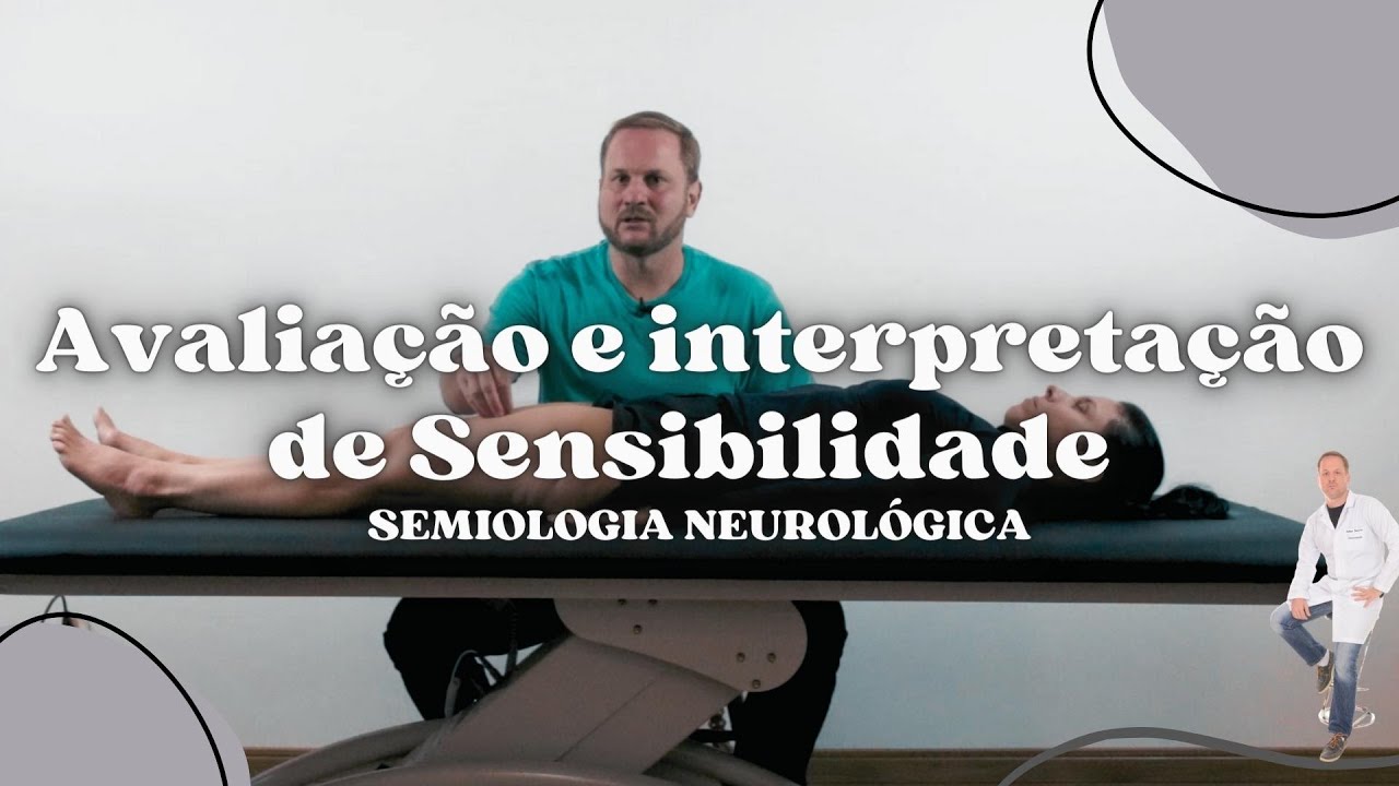
Abnormal sensory findings include: Disesthesia - difficulty recognizing stimuli (e.g., feeling a cotton ball but describing it as a shock); Hyperesthesia - increased sensitivity on one side compared to the other; Hypoesthesia - decreased sensitivity on one side; Anesthesia - complete absence of sensation. These findings help localize neurological lesions and indicate specific pathologies affecting sensory pathways.
Click to watch →

Examination of the sensory system
837
Sensory examination findings help diagnose neurological conditions: peripheral neuropathy (glove and stocking hypoesthesia); single nerve affection (radial nerve distribution); radicular affection (single root involvement); plexus lesions (one limb affected); spinal cord lesions (lower body affected); internal capsule lesions (one side plus face); polyneuritis multiplex (patches of different nerve affection). Spinal dermatomes: L1-L5 and S1 have specific lower limb distributions; thoracic dermatomes (D7-D12) follow costal margins; cervical dermatomes (C4-C8) follow upper limb patterns. These patterns localize lesions to specific anatomical regions.
Click to watch →

CÓRTEX: Áreas e Funções do LOBO PARIETAL (Anatomia em 3D) #MiniCurso 4/10
28.8K
Sensory information follows a pathway from peripheral receptors through the spinal cord to the thalamus, then to the postcentral gyrus (primary somatosensory area). Lesions here cause anesthesia (no sensation), hypoesthesia (reduced sensation), or paresthesia (abnormal sensations). The secondary somatosensory area in the superior parietal lobule interprets sensory information, and damage causes sensory agnosia—patients feel something but cannot identify what they're experiencing (e.g., unable to distinguish between pinching, caressing, or tapping). Understanding these pathways helps clinicians correlate neurological signs with specific lesion locations.
Click to watch →

Upper Limb Neuro Exam
358.5K
Specific terminology describes sensory abnormalities: Hypoesthesia - decreased sensitivity to light touch; Hyperesthesia - increased sensitivity to light touch; Paresthesias - pins-and-needles, numbness, or tingling sensations. Hypoalgesia - decreased sensitivity to pain; Hyperalgesia - increased sensitivity to pain. These terms are clinically significant for documenting neurological deficits and guiding diagnosis. When only discriminative sensations are impaired while elementary sensations remain intact, the lesion likely involves the sensory cortex rather than the posterior columns.
Click to watch →
Deep Dive
Performing advanced somatosensory tests, including temperature discrimination, vibration testing (using a tuning fork), and proprioception.

CNS Examination MBBS Practical - Part 3 | MBBS Practical Exam| | Free revision
239.5K
Sensory examination tests ascending pathways: spinothalamic tracts (pain/temperature) and dorsal columns (vibration/proprioception). Pain is tested with sterile pin after explaining sensation. Temperature uses cold/warm tubes. Proprioception tests joint position sense by moving joints and identifying direction. Vibration uses 128 Hz tuning fork, starting distally and progressing proximally. Key dermatome landmarks: C8 (middle finger), T5 (nipple), T10 (umbilicus), T12 (inguinal ligament). Abnormal proprioception requires testing proximal joints. Comparison between limbs is essential.
Click to watch →

The Examination of the Nervous System
337.9K
Sensory examination evaluates four modalities: pain (sharp objects like toothpick), temperature (cold preferred over hot), vibration (tuning fork 128 Hz placed on bone), and proprioception (joint position sense). Control testing establishes baseline sensation before examining limbs. Testing proceeds distally to proximally with eyes closed. Normal sensation shows consistent response throughout. Pathological patterns typically show distal impairment progressing proximally. Vibration testing compares patient's threshold to examiner's threshold. Proprioception testing holds the finger from the sides and moves it up and down while the patient identifies direction changes.
Click to watch →
The Neurologic Physical Examination
558.2K
The sensory exam evaluates four modalities through the spinothalamic and dorsal column-medial lemniscus pathways. Pain and temperature are tested using sharp objects like broken cotton swabs, starting distally and comparing left-to-right. Vibration uses a 128Hz or 256Hz tuning fork on bones, moving to different locations to assess distribution. Proprioception isolates the distal interphalangeal joint and moves it up/down while the patient identifies direction without visual cues. Testing is subjective and relies on patient honesty. Both vibration and proprioception can be tested interchangeably since they share the same pathway.
Click to watch →
Upper Limb Sensation Examination - Clinical Skills - Dr Gill
97.5K
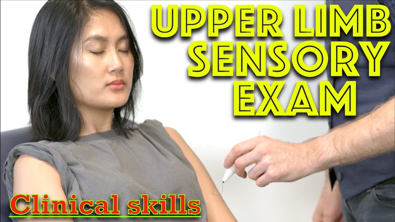
A comprehensive upper limb neurological examination assesses six sensory modalities: light touch (using neuroilament), fine touch (cotton wool), sharp-dull discrimination (sharpened point), vibration (tuning fork), proprioception (thumb positioning), and temperature (cold/warm stimuli), with normal findings indicating intact sensory function across all dermatomes.
Click to watch →
The Full Neurological Examination
1.1M
To test sensory function systematically: First test that the patient can feel the stimulus and understands what to do by testing on a part where sensation is known to be normal. If sensory loss appears glove or stocking distributed, start at the tips of fingers or toes and work upward until finding the sensory level. Test vibration sense using a C128 tuning fork on upper and lower limbs and trunk. Test proprioception (joint position sense) with eyes closed: separate the digit from adjacent digits, move the joint up and/or down, and ask the patient which direction the digit is being moved. Temperature testing is difficult in normal clinical settings but should be performed if abnormality is suspected.
Click to watch →
Deep Dive
Testing cortical sensory functions, such as stereognosis, graphesthesia, and two-point discrimination.
Astereognosis, stereognosis, two point discrimination, graphesthesia, Dejerine–Roussy syndrome
2.7K
Cortical sensory function is assessed through stereognosis (object recognition by touch with eyes closed), two-point discrimination (minimum distance to differentiate touch points, 3mm on fingertips), and graphesthesia (recognizing written numbers on palm with eyes closed); these tests depend on intact dorsal column-medial lemniscal pathway (first-order neurons in dorsal root ganglia → posterior columns → medulla nuclei → thalamus → sensory cortex), and lesions in the somatosensory cortex or thalamus (ventral posterior lateral/medial nuclei) cause contralateral deficits in discriminative touch, localization, proprioception, and stereognosis, with thalamic lesions potentially causing Dejerine-Roussy syndrome (severe post-stroke pain).
Click to watch →

Exame Neurológico
145K
Cortical sensory function is assessed by testing the parietal lobe and association areas. The graphesthesia test involves the patient closing their eyes while the examiner writes numbers on their palm, and the patient identifies the number. The stereognosis test involves the patient identifying objects placed in their hand while their eyes are closed. These tests assess higher-order sensory processing.
Click to watch →
Exame Neurológico - Função Sensitiva | Educação na Saúde
428
Cortical sensory functions require intact parietal lobe function. Stereognosis assesses object recognition by touch, asking patients to describe objects' shape, consistency, and material with eyes closed. Graphaesthesia evaluates recognition of symbols, letters, or numbers drawn on the skin with a blunt scalpel, demonstrated first with eyes open then tested with eyes closed. Two-point discrimination tests the ability to distinguish simultaneous tactile stimuli using a compass with adjustable points, with point distance varying by body area (smaller for fingertips, larger for other areas). These tests assess higher-order sensory processing and cortical integration.
Click to watch →

Pract: Neurological examination (sensory); Internal medicine
146
Cortical sensory examination performed only after superficial and deep pathways confirmed intact: (1) Two-point discrimination tests ability to distinguish two separate points, (2) Stereognosis tests ability to recognize familiar objects by touch, (3) Graphesthesia tests ability to recognize numbers/letters written on palm. These tests assess somatosensory cortex integrity. Normal thresholds vary by body location.
Click to watch →

Lobar Function Test - Parietal Lobe
103
Cortical sensation tests include tactile localization, two-point discrimination, stereognosis, and agraphesthesia. Before performing these tests, normal spinotheralamic sensation (touch, pain, temperature, position, vibration) must be confirmed. Tactile localization is tested by placing an object on the patient's body while their eyes are closed and asking them to identify the exact location. Two-point discrimination is tested by placing two points on the patient's body while their eyes are closed and asking if they feel one or two points. Normal values are: 5mm for finger tips (most sensitive), 4cm for dorsum of foot (less sensitive), and 5-7cm for trunk (least sensitive). Stereognosis is tested by placing familiar objects in the patient's hand while their eyes are closed and asking them to identify the object. Agraphia is tested by writing letters and numbers on the patient's forearm and palm while their eyes are closed and asking them to identify what was written.
Click to watch →
Deep Dive
Integrating sensory exam results with motor and reflex assessments to conduct a comprehensive neurological examination.

Sensory & Motor Examination | 4-hour Workshop on Neurological Examination for Neurology Residents
11.3K
The complete neurological examination integrates motor, sensory, and reflex testing within a systematic framework. Motor assessment evaluates muscle groups through resistance testing (finger abduction, fist making, shoulder shrugging, knee extension, straight leg raising) with appropriate force application—using full body weight for lower extremities while avoiding injury. Sensory testing examines two major pathways: posterior column-medial lemniscus (vibration, proprioception, fine touch) and spinothalamic tract (pain, temperature), with vibration testing being more sensitive and quicker. Reflex testing includes knee clonus (placing fingers above kneecap and wiggling while pushing down to observe rhythmic contraction-relaxation cycles). The brain organizes motor neurons in a U-shaped arrangement along the motor strip, representing functional movements rather than individual muscles. Proper technique requires pushing against the patient's own muscle group rather than using body weight or opposite limbs, with sensitivity taking precedence over specificity to detect early signs of conditions like transverse myelitis.
Click to watch →
Neurology: Clinical Skills - Motor, Sensory, & Reflex Neurological Exam #neurology #ubcmedicine
1.6M
This video demonstrates the complete neurological examination, covering motor assessment (inspection for muscle bulk and abnormal movements, pronator drift test for corticospinal lesions, tone assessment for rigidity/spasticity/flaccidity, and MRC power grading from 0-5), reflex testing using the deep tendon reflex scale (0-4) with weighted/unweighted hammers, and sensory evaluation of five modalities (temperature, pain, touch, vibration, proprioception) to assess the spinothalamic and posterior column-medial lemniscus tracts, with sensory deficits presenting in dermatomal or peripheral nerve patterns depending on lesion location.
Click to watch →

SZ & SMZ API-ICP PG MASTER CLASS 16th June
149
Systematic neurological examination includes: cranial nerves (vision, hearing, facial function, swallowing); motor system (muscle bulk, tone, power, reflexes); sensory system (pain, temperature, vibration, proprioception, cortical sensations); cerebellar function (finger-nose, heel-shin, Romberg's); and higher mental functions. Reflex levels (trapezius C2-C4, biceps C5-C6, knee C3-C4) help localize lesions. Hyperreflexia with clonus indicates upper motor neuron involvement. Sensory level helps localize the lesion.
Click to watch →
CNS Examination MBBS Practical - Part 3 | MBBS Practical Exam| | Free revision
239.5K
The neurological examination follows a systematic framework: (1) Higher mental functions, (2) Cranial nerves 1-12, (3) Motor system (bulk, tone, power, reflexes, coordination), (4) Sensory system, (5) Gait assessment. Motor examination includes inspection for wasting/hypertrophy, tone assessment (spasticity from corticospinal lesions, rigidity from extrapyramidal lesions), power grading (0-5 scale), and reflex testing (deep tendon: supinator, biceps, triceps, knee jerk, ankle jerk; superficial: abdominal, cremaster, plantar). Babinski's reflex indicates corticospinal tract lesion. Coordination tests cerebellar function (finger-to-nose, dysdiadochokinesia, knee-heel test). Sensory examination tests spinothalamic tracts (pain/temperature) and dorsal columns (vibration/proprioception). Cortical sensations include two-point discrimination, stereognosis, graphaesthesia. Gait assessment identifies patterns: festive (Parkinson's), circumduction (peripheral nerve), cerebellar (broad-based). Romberg's test distinguishes sensory ataxia from cerebral lesions.
Click to watch →

Neuro II, Motor, Sensory, Reflexes, Coordination
824
A comprehensive neurological examination systematically assesses motor function (muscle strength, tone, reflexes), sensory function (light touch, sharp/dull sensation, joint position sense), cerebellar coordination (finger-to-nose, heel-to-shin, rapid alternating movements), and gait/balance (Romberg test, heel-to-toe walking). The exam includes cranial nerve testing, deep tendon reflexes (biceps, triceps, brachioradialis, patellar, Achilles), and meningeal signs (Kernig's, Brudzinski's, nuchal rigidity) to evaluate neurological function and identify potential pathology.
Click to watch →
Deep Dive
Applying sensory testing in clinical pathology contexts, such as diagnosing peripheral neuropathy, radiculopathy, or spinal cord syndromes.
158. The Lower Limb Neuro Exam w/ Tom Jesson
2.6K
Test light touch first with cotton buds (not hands, which test thermal sensation), then pinprick for small fiber assessment. Compare affected to unaffected side or use abdomen/thigh for bilateral comparison. Test in loops around limbs (upper leg, lower leg, tops of toes) rather than strict dermatomes. In radiculopathy, sensory loss is typically fuzzy with overlapping nerve root function; in peripheral neuropathy, loss is clearly delineated. Pinprick often reveals additional sensory deficits not detected by light touch alone.
Click to watch →
The Sensory Exam Compilation
110.9K
Objectifying sensory exams involves having patients point to where they feel sensations and describe what they feel. For peripheral neuropathies, vibration testing uses tuning forks on bony prominences, with clinicians moving up the leg if ankle sensation is absent. Two-point discrimination tests tactile acuity using calipers. Diabetic foot sensation testing evaluates L4, L5, and S1 dermatomes on the lateral foot using a disposable pin. A positive test indicates loss of protective sensation. Clinical considerations include testing in quiet rooms, performing multiple trials per area (50/50 guessing chance), and recognizing that cortical lesions (neglect) may cause non-detection even with intact sensory tracts.
Click to watch →
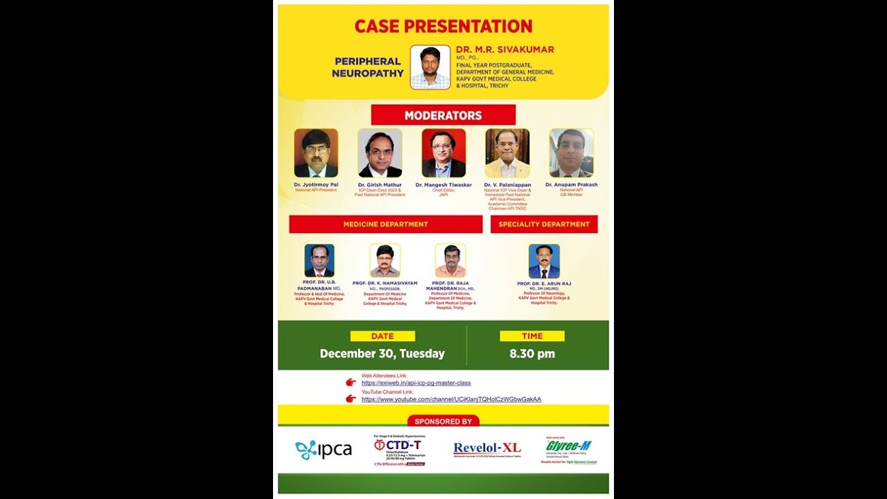
SZ SMZ API ICP Master Class Webinar 30 Dec 2025
238
This segment covers sensory examination in peripheral neuropathy. Sensory examination includes light touch, pain and temperature, vibration sense, joint position sense, and cortical sensation (two-point discrimination, tactile localization, stereognosis, graphesthesia). The examination should be performed systematically and note any asymmetry. The patient had impaired pain, touch, and temperature below knee and absent below ankles, impaired vibration sense below knee and absent at knee joint and ankle. These findings indicate large fiber involvement. The sensory loss pattern (below knee and ankle) indicates involvement of sensory tracts at the level of the peripheral nerves or spinal cord.
Click to watch →

Neuropathies périphériques. Dr kohil
10K
Sensory examination in peripheral neuropathy includes testing subjective symptoms (paresthesias, burning, pain) and objective deficits. Deep sensation (proprioception) is tested using a tuning fork placed on bony surfaces. Position sense is tested by moving toes/fingers with eyes closed. Temperature and pain sensation are tested using warm and cold objects. Diabetic neuropathy typically presents with 'glove and stocking' sensory loss, affecting feet and hands symmetrically.
Click to watch →

Neurological Examination of the Limbs - Explanation
3.6M
Sensory testing follows a dermatomal pattern validated against the sternum. For pin prick and light touch, the examiner tests L1 through S1 dermatomes systematically. If peripheral neuropathy is suspected, testing begins distally at toe tips and progresses proximally. Normal sensation is confirmed when the patient can feel sharpness or touch at all tested sites. The pattern helps localize spinal cord or peripheral nerve pathology.
Click to watch →
L4 Test
C6 Test
L4 Test
0:00- 1
Light touch applied with tissue on dermatome L4.
- 2
Patient reports sensation with eyes closed.
- 3
Result: seven out of eight intact.
Limitations of Manual Bedside Sensory Exams and the Shift to Quantitative Sensory Testing
While manual light touch and sharp/dull testing are clinical staples, they face significant criticism for their subjectivity and lack of standardization. Critics argue that these bedside tests suffer from poor inter-rater reliability, as the force applied by different examiners varies widely, and results rely heavily on subjective patient feedback. Furthermore, traditional exams are often binary and fail to capture subtle, early-stage neuropathies. As an alternative, many neurologists and researchers advocate for Quantitative Sensory Testing (QST) and electrodiagnostic studies (such as nerve conduction studies). QST utilizes calibrated, computerized stimuli to measure precise detection thresholds for thermal, mechanical, and vibration inputs. This provides reproducible, objective, and quantifiable data that can detect subclinical sensory deficits and track disease progression far more accurately than traditional manual exams.
Basic neuroanatomy of the peripheral nervous system, specifically the structure and function of sensory nerves.
The concept of dermatomes and how spinal nerve roots correspond to specific cutaneous regions of the body.
The physiological difference between sensory pathways, specifically the spinothalamic tract (pain/temperature) and the dorsal column-medial lemniscal pathway (touch/vibration).
An understanding of basic cutaneous receptors, such as nociceptors for pain (sharp/dull) and mechanoreceptors for light touch.
Interpretation of abnormal sensory findings, such as paresthesia, hypoesthesia, and anesthesia, to localize lesions.
Performing advanced somatosensory tests, including temperature discrimination, vibration testing (using a tuning fork), and proprioception.
Testing cortical sensory functions, such as stereognosis, graphesthesia, and two-point discrimination.
Integrating sensory exam results with motor and reflex assessments to conduct a comprehensive neurological examination.
Applying sensory testing in clinical pathology contexts, such as diagnosing peripheral neuropathy, radiculopathy, or spinal cord syndromes.
3K views19likes0:38@ChloeHopkins_PTAOriginal Release: 2024-11-27
for sensation testing of dermatome L4 we have light touch which I'm just using a tissue for this I'm just lightly touching him and then he's telling me whether he feels it or not with his eyes closed and here he has seven out of eight intact for sensation testing of C6 we'll do sharp and Dole I'm just slightly touching him with a sharp object and then on the end of it is a dole and he's just telling me how many he feels whether it's Sharp or Dole and for Sharp he has six out of six intact and for Dole he has six out of six intact
Up Next

5:05
Cerebellum Examination: Stanford Medicine 25 Bedside Skills
@StanfordMedicine25
1.2M views•2014-03-18

48:47
Graphic Medicine: Comics for Collaborative Healthcare Communication
@nationalpatientadvocate
189 views•2023-12-04

10:28
Neuroanatomy: Central and Peripheral Nervous System Divisions Explained
@AKLECTURES
136.2K views•2014-09-20

3:05
Stages of Labor and Vaginal Birth | Childbirth Animation
@nucleusmedicalmedia
52.1M views•2017-08-18
Related Study Plans & Knowledge Roadmaps
Structured learning paths in Medicine
medicine
Abnormal LFTs: Diagnostic Workup & Pathology
01Liver Anatomy and Basic Physiology
02Introduction to Liver Function Tests (LFTs)
03Hepatocellular vs. Cholestatic Patterns
+ 3 more learning modules
12h6 modules
Explore Roadmap
medicine
Analyzing Blood Smears: RBC, WBC & Platelets
01Introduction to Blood Components & Smear Preparation
02Normal Morphology of Blood Cells
03Erythrocyte Pathologies & Morphological Variants
+ 2 more learning modules
12h5 modules
Explore Roadmap
medicine
Mechanical Ventilation: Waveforms & Alarms
01Anatomy and Physiology of Breathing
02Introduction to Mechanical Ventilation & Core Modes
03Ventilator Settings and Waveform Interpretation
+ 2 more learning modules
12h5 modules
Explore Roadmap
medicine
Neuro Exam: Localization & Stroke Syndromes
01Foundations of Neuroanatomy and Vascular Supply
02The Neurological Examination: Step-by-Step
03Principles of Neurological Localization
+ 3 more learning modules
18h6 modules
Explore Roadmap