




![Thorax 8 - Trachée, bronches, poumons, plèvre [Descriptive anatomy of the respiratory tract]](https://i.ytimg.com/vi/fOG_CXzk_Xc/maxresdefault.jpg)


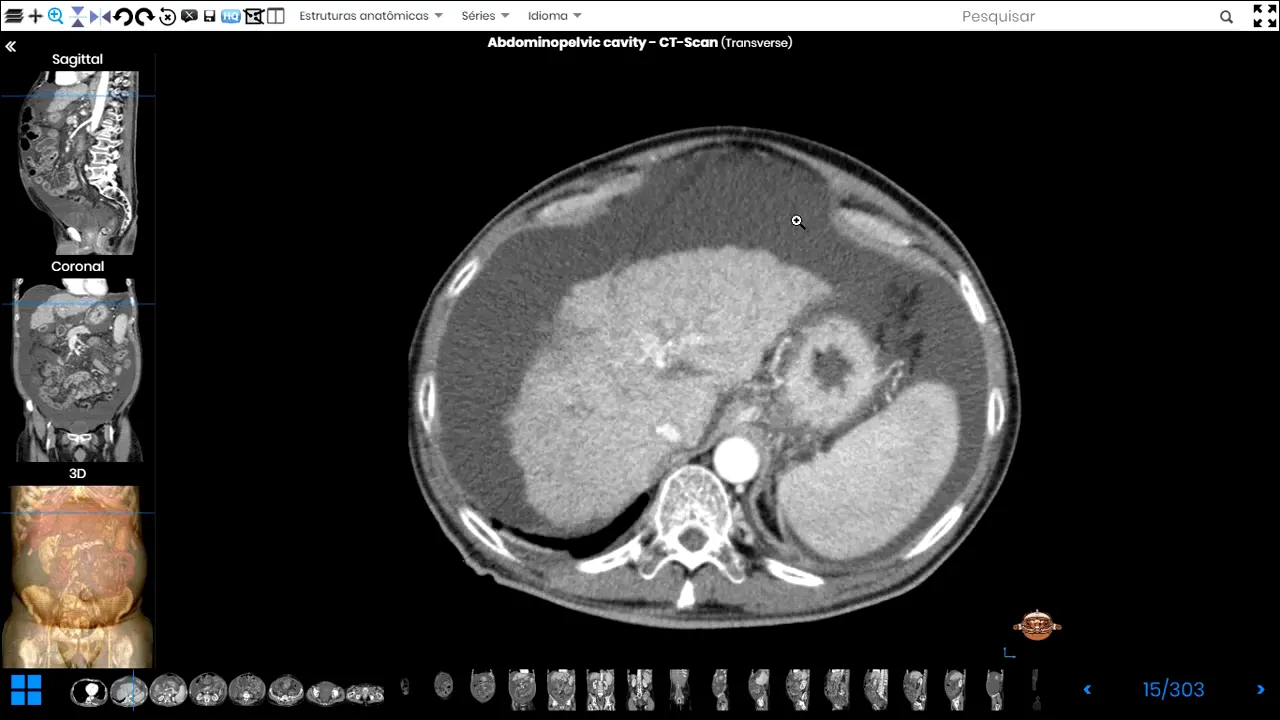









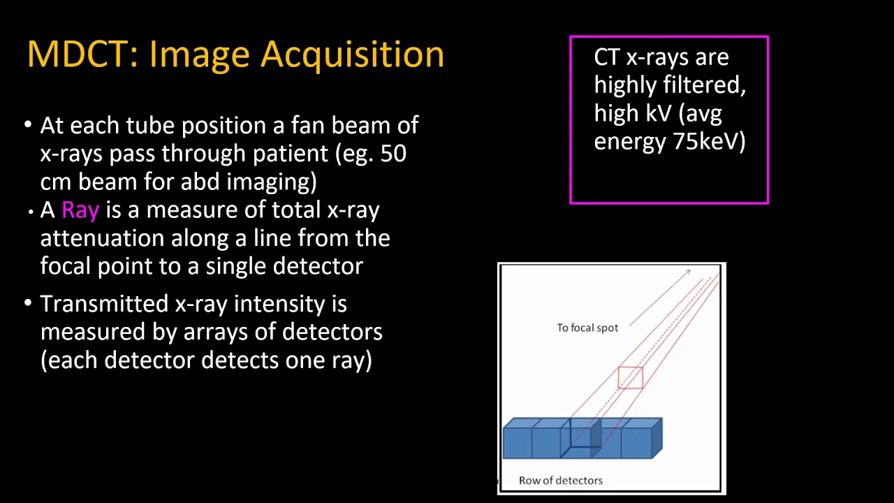


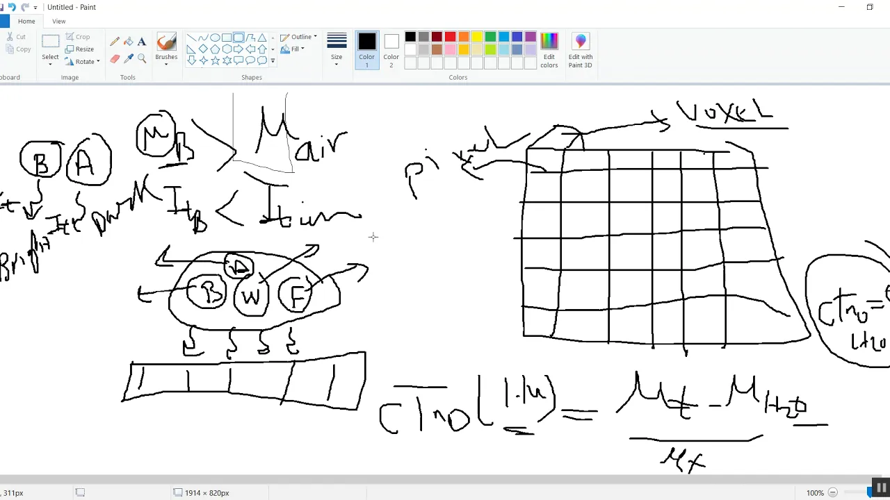



CT (Computed Tomography) produces 3D images by using multiple X-ray projections captured from different angles as the X-ray tube rotates around the patient, while X-ray produces only 2D images from a single projection. This fundamental difference allows CT to better localize structures, identify compression or invasion of adjacent tissues, and provide superior spatial resolution through multiple detectors and computer-processed matrix images.
RAD Crash Course Part 2 | CT Imaging & Clinical Applications
Added:Basic principles of X-ray physics, including tissue density attenuation (air, fat, water, bone) and how 2D projection imaging works.
Fundamental gross anatomy of the brain (cerebrum, ventricles, vasculature) and the thoracic cavity (pulmonary arteries, aorta).
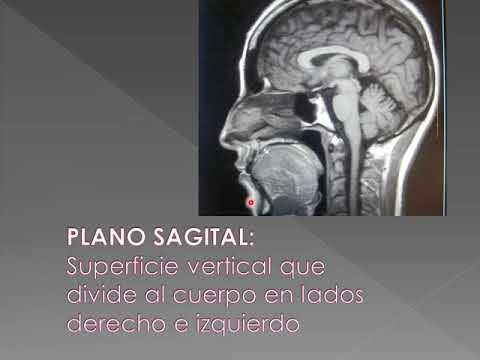
Understanding of cross-sectional anatomical planes (axial, sagittal, and coronal) used in medical imaging.
Basic concepts of intravenous contrast media and its role in enhancing blood vessels and organs in radiology.
Advanced CT modalities such as CT Angiography (CTA) and CT Perfusion (CTP) for assessing tissue viability in acute ischemic stroke.
Comparative imaging paradigms: Clinical criteria for choosing between CT and MRI based on diagnostic sensitivity, speed, and patient contraindications.
The physics of CT image reconstruction, artifact recognition (e.g., motion, beam hardening), and radiation safety principles (ALARA).
Integrating CT imaging findings into emergency clinical decision-making pathways, such as thrombolytic therapy protocols for stroke or anticoagulation for pulmonary embolism.
827 views7likes2:03:10Original Release: 2023-04-07
all right so what's the difference between X-ray and CT uh the difference is that an x-ray will give you a 2D image but um CQ will give you a 3D image right all right all right so you're going to be able to differentiate um uh structures better you're going to be able to localize them better right so if you look at this x-ray right here you can see that there's a mess right here right what you can tell is the mass is in the uh like in front of like anterior in the long or posterior and the lung you're not sure which lobe it's in you're not sure if it's compressing any any any problem any any uh vessels or any other major structures you simply can't tell because you only have a 2D image that is relatively poor in in contrast and in spatial resolution but if you look at the city of the same image of or the same patient you're going to be able to to to localize it specially exactly where it is and if it's compressing or invading into any other adjacent structures right um so yeah that's the main difference between CTS and x-rays that C2 will give you a 2D image while uh um sorry an extra will give you a 2D image while a CTO 3D image CTS use x-rays see same thing I said right the only thing is that x-rays will give you a single projection but a CT will give you multiple detection and multiple projections um to give you the 3D image same slide from yesterday all right so in a normal x-ray right so this is the X-ray tube right here right so in a normal x-ray you're going to have one detector and you're going to have one x-ray tube all right and it's true you have here one x-ray two but you have multiple detectors in a CT right so you're gonna have the x-rays coming what's done by projection um like a single production I mean like a single view think of a production as view right so you get one view when it comes to x-ray while the CT gets multiple views so um as I was saying you have an extra Europe as you can see here on X32 right just like in an extreme machine but the difference is you have multiple detectors right so you have here you have multiple detectors right here detecting the x-rays coming from the X-ray team and one more thing is that the X-ray Cube keeps moving all right so one second is here the other is here it just keeps um moving around the patient to give you multiple views all right now the multiple views come from the movement of the X-ray tube that's how you get the multiple multiple uh views or the multiple projections that you see in a CT scan the multiple detectors will give you better resolution right so the more detectors you have the better the resolution is because if you remember yesterday remember the thing about it I said about pixels right the more pixels you have the smaller they are the better it's the same concept right here if you have more detectors and each deductive is smaller in size they're better the resolution yes right this is just same thing right showing you that you get a view from here and then it moves and gives you gives you another view from here and then it moves and it gives you a third view from here right and then at the end you're going to end up with three views all right this is just an example in reality you get much more than just three uh views all right this is just an example so the end of the day you get three views right here here and then using a computer the using a computer what will happen is that a software will merge those pictures together depending on the attenuation all right so it will match each attenuation with each other all right and it will give you a matrix and a matrix is basically the end product or the end image that you see on a CD on a CT scan on on the computer screen the Matrix is is the final image right that's why it's called a computer tomography because the computer is the one that gives you the picture or gives you the view uh uh the result candidate all right so the multiple views you get from the X-ray tube as it keeps moving will be merged together to give you a matrix and this is basically what you see you will get this all right all right so how the image is formed it's it's formed by a software all right it's formed by a computer now if you go back here see this is this is the same x-ray tube where it's just moving so when the X-ray tube is right here it's going to give you a view that is like this this side of this patient all right and then when it goes to the middle it's going to give you this view of the patient and when it goes to the right it's going to give you this view of the patient and you will end up with three sets of views right or three sets of pictures that at some point are overlapping because you see this part right here this part is overlapping of this picture and this picture so this part is overlapping so if you get the pure raw pictures that the CT scan image or the CT scan machine gives you is going to be distorted because there's a lot of overlap going on but what happens is those three images or those three views are fed into a software or fed into a computer there's one x-ray tube and you have multiple detectors so if you go back here can you see those small cubes each one is a detector all right so you have one x-ray Cube but multiple detectors right so what happens is those three views are are fed into a software and a software yes the detectors move too so like they are always um uh the opposite side of the of the X-ray tube so if you can see the the the the detectors are all set on like a piece of metal or whatever right and they keep moving in relationship to the x-rays Loops or the X-ray app from my handle chart so they just keep moving moving together they they move together right they're not independent so if this goes to the left this will go to the right so it can detect all the things coming from the X-ray tube if you're welcome so as I'm saying you have three views that you got right so you have one two three you have three views right what happens is that those three used are then fed into a software or computer right and that computer will merge those views together all right they will merge them together eliminate any overlap in the picture all right improve the resolution to give you the final end product which is the Matrix and the Matrix is basically a combination of all the views that the CT scan or the X-ray tube took and this is the result that you get right and again that's why it's called computed tomography right because the computer it it's it's computed the computer is what gives you the the the final image all right sound like x-rays you know like you don't need to pass your X-ray image by a software or computer to give you the image right you can get it simply by film you don't even need that a computer to process this but in a CTE now uh uh do you guys have any questions about what I discussed uh up until now all right great now same concept as yesterday you have the spatial resolution you have the contrast resolution at the temporal resolution all right now before I skip this because I explained it yesterday does anyone want me to re-explain this because it's all right I can do it okay let's all right so just again uh did we do scan time no I'm gonna go through it scan time is is not that yes spatial resolution is is basically the pixels part of yesterday's lecture um if you remember that that's the picture and as you can see right here pixels right uh no we did not do scan time um I'm not sure if it's us explain Aslan um yeah it's not explained but scan time is is um I don't know how to explain it because because it's not a good thing scan time is not a good thing it's not anything you can focus on because um I'll talk about what you should focus on right now uh scan time is a bad thing that's basically it but the your professor didn't really explain it I'm not sure if I should it's going to be a waste of time but it's a bad thing you need to minimize can time that's basically what all you need to know right now what I want what is it about generally it causes Distortion in the image it causes um because anything that leads to an artifact and artifact is something that should not be there right and basically as can time um is is the longer you scan the patient all right the more possibility this patient is going to breathe the more the more frequently the patient is going to breathe and by breathing you move and when you're moving inside a CT scan that distorts the image so you want to minimize the scan time so you minimize the breathing artifact an artifact is a bad thing right it distorts the image you're welcome so what do I want you to focus on honestly like right here the only thing I want you to focus on is that you have one chest one you know one x-ray tube and you have multiple doctors and that's the difference between a CTN and x-ray you have multiple detectors all right and you need to know that the software is used to give you a CT image it's not something natural like an x-ray um that's basically what I needed to focus on from all those steps right now for spatial resolution as I mentioned yesterday those are pixels the smaller your pixels are the more pixels you have the better the spatial resolution all right I go in depth about this in yesterday's lecture if you want to go and watch it on my YouTube channel yep exactly they have the same thing you have General and characteristic radiation it's the exact same abbreviation you get from an x-ray right um so as you can see here a detector size um that is 7.5 millimeters would give you what you can see right here right but if it's smaller and as I said the smaller it is the more detectors you have the better the resolution is and you can see the difference here this has a big detector this has a small detector and you can see the difference in the images all right again the number of pixels as I mentioned the more pixels you have naturally the smaller they are which leads to a better picture all right now this is the different parts about about this lecture lecture this is what's different between uh CT and x-rays the attenuation all right and by attenuation I mean how differently different parts of the body absorb um absorbed the x-rays and then attenuation uh in CT we measured it by the house field units or the HQ units right that's you you can't say you it's because U is units right so your benchmark is water which is zero I choose right this is your benchmark this is where you start what's less than water yeah and air would be less than zero so it's going to be negative 500 negative 1000 depending on what type of air there is and then everything else is is is is just higher than zero they have muscles bones fat tissue whatever it's going to be always higher than zero right because the only thing that is zero is water uh this is just a slide here to like it's just a picture to show you the different um edges within different parts of the body um as you can see here uh along with beyond the negative 50 all right uh because it's filled with air all right um fat is is fat is a special fat is a special case for a special situation it's it's not really that negative but it is on the negative side all right so if you put it in contrast with air it's going to appear darker than here all right sorry water it will appear darker than water uh no you don't have to I'm just giving you an example of like how things absorb x-rays differently right uh all right contrast resolution how's this different from x-rays it's not different it's just we quantify it in in CT in CT we we Quantified we care how much x-rays um different types of the body absorb because later on I'm gonna talk about uh window and window is important because that's how that's where we use house House Field units in x-rays we don't care how much bone absorbs x-rays but here we do information depends on attenuation depends on different type parts of the body so like your bone will will absorb a lot of x-rays right so it will attenuate more and so on all right so it depends on the density of the tissue all right uh so a window sitting setting is is is um is the center okay so let's say you have a chest CT right and you don't care about the bones you don't care about the heart you care about the lung you're scanning this chest only for the lungs so what you do is you set the CT scan or the CT settings on a Long window and I'm going to explain to you what a lung window is and what different Windows mean right but basically um when you got a CT scan right you're going to have bones and bones are a thousand at shoes and you're going to have the lunge which is a negative 100 I choose and then you're gonna have the heart which is 200 at you so you have a wide range of tissues which with wide ranges of hu's right and you don't care about a lot of these you only care about the the lungs so what you do is you eliminate everything that is outside the house healed units of the lung so let's say the lung will appear between House Field units of of negative 50 and negative 200 all right anything less than negative 2 100 you eliminate and anything more than negative 50 you eliminate right so that you end up only looking at the lungs and that's what a when a window is right so let me show you this is just again same thing same thing all right this is a Long window right so if you notice guys look at the chest wall right you can't say you can't see anything here you can't like even if there was a pathology here you wouldn't be able to differentiate it from anything and so is the bones the bones yes of course of course right and that's why you okay so let's say you have normal lungs normal lungs on a lung window will all appear like this part right here right like they won't appear like they will all have the same color of this part right here because this looks healthy to me right if you have anything else inside the lungs that is distorting the lung or affecting the lung or inflaming the lung is going to give you a different attenuation so I want you to look at this part right here see see how this part is is brighter than than this part for example see this is telling you that there's there's something wrong right there's some sort of infiltration in the lungs and this is actually pneumonia all right so the lung would appear uniform in color in the lung window all right and and and to detect any abnormalities right and and let's say those are abnormalities are are very subtle they're small if you want to really see them you're going to have to set it to a Long window because in Long window everything else that is that does not have to do anything with the lung is eliminated all right so nothing is really distorting the view anymore it's just the lung and whatever is happening happening inside of it all right so for example here in the Long window we we usually put the ranges between negative 400 and 6 and and and negative 600 all right and that's how you view the Long window the the window width wasn't um it wouldn't distort your view you will still be able to see the lungs but if there's any subtle pathologies inside the lung there's a chance you will miss them because now you're not focused on the lungs you're focused on everything all right but when you're focused on the lungs any small or subtle changes in the lung would be more obvious all right you got it uh your professor didn't talk about the window width all right as far as I remember um so I'm not gonna really uh dig deep in that it's going to be a waste of time this is just another example of a media sign on window in a media style window you focus on the soft tissue structures inside the thorax or inside the mediastina this includes the pulmonary artery aorta the heart and stuff like this all right and here you want to eliminate everything right so what do you do is you should you put the level on level 50. remember what we said about the lung it's negative 200 to negative 600. when you put the level on 50 the lung is eliminated it's it's gone right because you put the level on 50. so negative 200 negative 600 all of that is eliminated it's gone so if you look at the lungs right here black darkness you can't see anything all right and you can see the bones right but if there's a fracture in the strip right here or a fracture here you're not going to be able to see it because the bone window is much higher than 50 and you eliminated that too all right so what you only can see is the mediastinum at all the media style structures and that's it foreign no you don't have to memorize the ranges it's just it's just to further explain to you how the window works but you don't have to memorize the windows you're welcome all right you have the liver window again same thing it's just to focus on the liver a bone window see how higher the bone window is it's on the 400 Hues right so everything else below 400 is eliminators all the soft tissue your brain your uh mucosal membranes everything this is a head CT everything is eliminated you can only see the bones and you can see details of the bones right I all donated contrast introduce contrast instruments the contrast resolution remember if you guys remember yesterday's lecture as I said you give contrast all right because the KV or the or the Kev it releases is way higher than everything else right so it's going to be starkling white it's just going to be very white so it's going to give you a perfect contrast with anything that is darker than it on on the side all right and you can see here like if you look this is the pulmonary trunk or the pulmonary artery right and if there's anything inside that pulmonary artery that is is distorting it like an embolus or something you're going to be easily able to see it because the the difference between this Sparkling White and that tiny small Speck of black is going to be way more obvious when you put contrast all right this is basically the same thing it's just telling you again that the photoelectric effect is increased with the less kvp if you guys remember from last night um but yeah again same thing the kvp has an exponential relation so if you double the dose of the kvp you quadruple uh the the energy and it tells you that the ma is a linear relationship so if you double the ma you you double the the energy and q1 dime so basically the longer the patient is is inside the tube of the CT scan the higher the dose that's basically it there's nothing else in the slide what is fov I'm not sure I'm not sure what fov is on Google let's Google it real quick field of view oh nice all right I believe that I feel to be okay so what they mean by field of view here is that do you guys remember the the slit I talked about that's basically I think that's what they're talking about that the the the the more surface area the patient is exposed to radiation all right the higher the dose so if you increase the area that the patient is exposed to by 10 or 10 10 centimeters you increase the dose by ten percent I believe that's what they mean Rock and that's the end of the structure all right that didn't take long 29 minutes not all bad you guys want to take a break before we go into the bigger lecture or uh is there oh yeah there is yeah two one time this is explained kvp is explained ma is explained I don't think fov is explained [Music] uh it's not important it's just an explanation um like if you yeah matter of fact if you introduced by tensor meters yeah see if you reduce it by 10 you reduce it by 10 if you increase it by 10 you increase by time the notes are there to explain right so if you understand what's going on you're good like it doesn't add anything really exactly exactly all right just focus on what you need to get a picture of don't don't you know diverge all right guys um do you want to take a five minute break before we go into the bigger lecture which is the clinical applications of CT or do you want to just dig into it all right all right let's take five minutes let's take five minutes right okay guys see you in five minutes all right um I have um all right so the CT applications right um it's kind of short the lecture that you guys took um I have another lecture I that I edit edited on and I added more clinical stuff um uh ways to better understand what you guys are seeing do you want to do that but it's going to be shway longer it's almost 50 slides what I do just want to stick to the lecture that the doctor gave you whatever you want I'm I'm chill with both all right um all right cool all right I hope it worked well I think it's this one no this is the one that the teacher that the professor gave you that's the one all right before I start CD the applications I need you guys to remember something and something that's very important if she's pregnant don't order CT or x-ray just don't all right and and if you and you if your only options are CT make sure that this patient has a shield on her abdomen okay so just keep that in mind it's going to be easier for you guys in the exam because um it helps you eliminate options it helps you eliminate choices all right this is the outline uh we will start by removing oh that's electric all right all right so keep that in mind okay now let's start um this is just more stuff okay so indications for CP scans um here in Saudi I'm not sure about the US really but here in South Saudi in trauma patients especially in the ER so like motor vehicle accidents and stuff like this we usually tend to order a pan CT scan for these patients all right so I scan for the whole body that's why it's called the pan CT scan right so keep in mind um trauma patients uh probably will get a CD scan all right now if you want to visualize the abdominal organs since most of them are are um soft tissue right and x-ray will not be that helpful like it could be helpful but it's not really that good so usually we do CT scan for abdominal organs for the long parenchyma so patients who have COPD lung fibrosis um sometimes people with lung cancer stuff like this they get a cp's count right and stroke patients they always get a CT scan always right if you suspect CT if you suspect the stroke get a CT scan and for uh CT and geography this is when you want to visualize the coronary arteries of the heart all right this is done uh when you want to maybe stent an archery for a patient who's having an MRI for example all right so keep that in mind now CT and Trauma as I said brain CT and spine uh just pelvis all right so for CT why do we get uh why do we get a CT scan and stroke like what what are you trying to see in a in a stroke patient when you get a CT scan all right you're not trying to see a stroke exactly right exactly it rules out hemorrhagic stroke you're not trying to see the infarct you're trying to see if there's bleeding or not right and that's the only reason you use brain CT in a stroke patient right you use it to to to rule out hemorrhagic transformation or hemorrhagic stroke uh in the patient all right that's why we use it without contrast because you don't need contrast if there's blood in the brain it's going to be obvious and you don't need contrast now we use a chest abdomen and pelvis get IV contrast because usually it gives you a better better view or of these organs right now clinical applications of the CT there so uh what about the pulse yeah and the pelvis school what about like the product for you the productive organs and the bladder so don't forget that bladder is also and you are um in your pelvis right so an acute conditions here a CT for the for any patient who has a signs of of of headache with an increased intracranial pressure what are the signs of increased intracranial pressure because not everyone who says I have a headache gets a CQ or gets an MRI no only patients who have increased signs of increased intracranial pressure so signs of increased intrapreneurial pressure would probably be something like papilledema right papilledema is when you do a fendoscopy or an examination of the eye and the optic disc becomes engorged and and and has poor demarcations all right vomiting is a sign of increased into cranial pressure nausea is a sign of increased intracranial pressure and so on all right so these patients get get a CT scan any patients who has a neurological deficit so suddenly he was sitting not doing anything and suddenly uh their right hand became weak right and they start slurring and they have um uh they have drooping of one of the eyelids right those patients get um a CT scan um what is DOB date of birth what does your date of birth have to do with anything oh difficulty breathing all right um difficulty breathing is not CT is not the first language or not the first line Imaging for a shortness of breath it's actually just x-ray but yeah you can get a CT for those especially for patients who have maybe a foreign foreign body um maybe if they have a tumor that's compressing on their bronchus stuff like this all right for abdominal pain a CT is done for any signs of ischemia of the colon or the small bowel it's done also for about obstruction and obviously trauma as I mentioned all right and then everything else is non-acute all right as I mentioned contraindications with contrast it depends on the it depends on the situation so for example an abdominal pains you would get you would get depth contrast right if you um and neurological diseases like strokes and stuff like this you probably won't get a seat you will probably won't give a contrast for that you're welcome all right so as I mentioned in contraindications uh pregnancy um is is the number one contraindication for for CT scans right if the patient has allergy to IV contrast then you can't give IB contrast um and then it depends on your experience as a radiologist if you want to do it without CT without IV contrast or just not do the CT at all that is up to you and doing an impairment again because of the IV contrast if you guys remember from last lecture one of the problems with ID contrast is that it's it's excreted by the kidneys right so the patient has been an impairment um you might not want to give the patient IV contrast all right window what window is this guys it's a bone window perfect yes see everything else is eliminated you can't see the brain you can't see mucosal structures you can't see anything at all you don't you can only see one thing and one thing only and that is the bone right so that is a bone window why do we get this why would you need a CT scan with bone window exactly for for skull fractures perfect what window is this this is we didn't talk about this by the time so if you get it I'll be impressed I call it brain window sure it is it is technically brain window but this is what we call a soft tissue window right so keep that in mind this is a soft issue window all right uh so nothing really here to show you except that these are different levels of a CT Imaging what else would be soft tissue in the brain um cranial nerves would be soft tissue but it's really hard to see cranial nerves um generally would be anything that is actually soft tissue so your heart is soft tissue remember the mediastinal window we talked about that is also called soft tissue window all right just in the media sign and that's why it's called the media center window it's just personally at least I don't know if this is something but I've never heard of something called a brain window maybe it exists I just never heard of it all right now this slide just tells you that we have different levels especially when it comes in our brain CT all right so this is this is the the this is uh the highest all right uh out of all those three and then you go to the middle and you hear you can see the brain stem right here you can see the the Pawns of megala and whatever and then this is the level of the of the eye you can see the cerebellum right here all right so keep in mind that when you see the eye you're relatively low in the brain and the Brain uh structures all right when you see you and you can see the eyes you're at the level of the cerebellum right here right and this is the fourth ventricle right so keep that in mind like if you if you look here you can see the lateral ventricles right you can see the lateral ventricles and that's how you know your up in the brain yep just see fourth ventricle right here this is the brain stem yep nothing really to talk about uh what what is happening here guys yeah perfect um so you can see here that this is there's no edema and there's no Hemorrhage no um all right there's this training so see that's the thing if it's Hemorrhage is going to be right keep that in mind right if there's Hemorrhage it's going to be bright it's going to be White Edition color not not dark what happens What's Happening Here is that there is atrophy of the brain tissue all right so the ventricles and the and the and the uh gyri and salt Kai are are deeper right so they accommodate more CSF that's why you think there's there's increased CSF here but there's no increase CSF no it's just the brain tissue has shrinked down so there's more space for dcsf right and this happens in two peoples generally elderly and who can tell me the second type of people who get brain atrophy other than elderly alcoholics right known as schizophrenia alcoholics so people who have chronic history of alcoholism they will have brain atrophy um Alzheimer's elderly right but yes you're right okay um I saw a question uh how how will we see edema edema will be more uh localized more focused about in one area all right it's not going to be diffused like you're seeing it right here all right and it's going to be like if you look closely you will be able to differentiate between the white matter and the dark matter when it comes to uh edema that differentiation between the White and the gray matter is gone all right that's how you know you're dealing with edema and there's going to be a shifting of the midline so you can see that the midline is in the midline right here right but if you have a demo like let's say on the right side you will see that that the brain is moving to the left side even though something normal hydrocephalus in terms of being hydrocephalus is not edema though hydrocephalus means there's increased in there's increased tsf fluid in the ventricles so there's no illum please have this point after yes of course I'll give it to you guys all right so this is one of the slides you're welcome now one of the those are one of the slides I didn't that I added all right this just gives you um gives you different presentation like clinical presentations of each types of brain pathologies right so for example in an epidural Hemorrhage these people usually are trauma patients all right and these people will probably present with something like loss of consciousness headache and ipsilateraliasis right so um not I don't really want to go through the whole thing right it's just for your own sake right uh if you want to you know learn more about this um and I and I bolded the things that are are special to each case so if you have sub argument hammerage for example you're gonna have something called a Thunderclap addict so these patients will tell you oh this is the worst headache of my life okay and these patients usually have a ruptured aneurysm right and they could be trauma but structured aneurysm once AMS AMS is um uh altered mental status all right so if they're going to be drowsy confused you know their mental status is decreased okay um but yeah that's basically uh Etienne okay all right so the types of hemorrhages we have four types we have epidural subdural subarachnoid and enter cerebral subdural is the most superficial one right it's it's above the dura mater and as we know that urometer is the outer layer of the meninges it's the third one the one on the top right so anything above it is a urinal hematoma if it's under the dura but above the arachnoid it's called a subdural hematoma if it's under the arachnoid it's a subarachnoid hemorrhage and if it's inside the brain tissue it's called an intracerebral or intraper and common temperature right who can tell me what what's this diagnosis uh it's written right here can we hide this all right we can't write this it is subaruenoid and and the way you can tell it's subarachnoid is because you can see the grooves of the brain right because because the arachnoid covers the brain entirely all right so that the grooves in the brain will be filled with blood and you're going to get a sign called the Star Sign light right exactly the star shape perfect you're going to get the star shaped right here all right it's going to be white in color or bright in color and bright indicates blood all right and that's how you know there's a subarachnoid hemorrhage these patients will probably tell you I have severe headache and this is the worst headache of my life they might have decreased levels of consciousness uh they might have a history of of of subargenoid hemorrhage in their family because one of the more common causes of sublichinoid hemorrhages is berries aneurysm which is which which could be related to familial diseases okay all right another another picture can someone tell me what's going on here no there's no idea this is another subaracoid Hemorrhage right if you look at if you look at the exactly it's another subarachnoid hemorrhage you can see the grooves of the brain it's just it's probably it's probably um artifact patient moved during the CT or something all right don't worry about this but like if you look at the grooves in the brain right you can see that they are brighter right you can see that they are bright and this indicates bleeding and probably some PowerPoint because it it moves along alongside the grooves of the brain right see as I said B is not severeign B what's B where's B oh no B is not sub-archy B is the norm so this is just a comparison so let's see if we can talk but we want them very much so we assume fluid if you this is an edema testing all right yes if you if you can't have it if you can't differentiate between the gray and white matter you assume you assume that there's a demon right but not not bleeding a demon where is the star sign and name right it's incomplete star assignment like here it's a complete star sign all right you can see it completely right but here it's incomplete right you can see that some of the groups are filled with blood but not all of the groups not all of the brain groups are filled with blood so you get kind of like an incomplete filling you don't have to get the Star Sign every time all right you're welcome all right so this is an epidural and a sub dealer all right uh one of the ways I remember is that the one that looks like an eye all right like like when you look at an eye from the side right you look at someone's eye from the side it looks like this right because it's like bulging right just like that's how I know this this is this is this is a subdurate looks like a moon all right because it takes the the concave Lemon versus banana that's another way yeah that works Lemon versus a banana yeah so the the one that is convex the concave is a subut the one that is complex is is is is is is a is an epidural hematoma all right and one more thing I want you guys to remember you will never see an epidural hematoma that is from here till here all right you'll never see that why because the brain has sutures all right so like at some points there's like an attachment of the brain to the skull all right so like right here there's an attachment right here there's an attachment and so on and if you have an epidural bleeding right the bleeding is going to be contained between two sutures right it's not going to cross suture lines all right you'll never see that the blood will start seeping right here right never in an epidural hematoma because it's it's confined to the to the suture lines but with a subur hematoma you can see that the hematoma start term here and ends all the way here why because it does not respect suture lines it crosses suture lines all right so keep that in mind who can tell me what causes epidural hematoma like what is torn in an epidural hematoma what vessel is bleeding in an epidural hematoma the middle meningeal artery yes right and since you said an artery this means it's a high velocity bleed right so these patients will present with an acute onset right so it's going to be very acute like hours maybe minutes maybe it's going to be on the same day right they're going to die if it's more than a day they're dead all right the point is it's it's an arterial bleed so when you see an arterial bleed think of Rapid deterioration of this patient all right in a subdural hematoma What's storm and veins don't have high pressure so they bleed slowly right so these patients can present to you after weeks they can present that from Once all right and this is a very common presentation and as the daily patients who fall all right and then four weeks after that they present to the ER with altered mental status confusion dizziness uh um their wobbly right and you do a CT scan and you can see this all right so so keep that in mind in subdural hematomas you can have a chronic suburah hematoma all right houses of prognosis uh epidural is way worse but uh but you need to keep something in mind that brain herniation and midline shift is more common with subdural hematomas so keep that in mind um but yeah so in epidural in some Dural hematomas I said you can have acute and you have chronic right and when the blood is fresh it's it's bright right you can see that it's bright right here because this is fresh blood it's new blood this is acute epidural hematoma and this is acute subura hematoma if you get a chronic subdue hematoma this line is going to be dark right because old blood appears dark all right so keep that in mind thank you all right what can you guys see here yeah this is ischemia of the middle uh cerebral artery territory all right this is the anterior cerebral artery territory no it's not ideal this is an infarctica all right and I'm going to tell you how to differentiate between a scheme and an unfortunate so this is ischemia of the middle meningitial artery territory this is this is probably not acute because you can see it on CT usually you won't be able to see it on CT if it happened like an hour ago or or 30 minutes ago Ninja right so if it's acute you usually need between three to four point five hours um actually more no no wait three to four point five hours is the cutoff for treating stroke but with the CT scan usually it starts appearing by six hours all right so if you can see it on CT it's probably uh uh too late a little bit right but basically this shows you a scheming right here right uh and you can see that it's it's in the middle initial middle uh cellular R3 territory this is the anterior cerebral artery territory and this is the posterior cerebral r33 okay you can see here that Q is chemical stroke right see here a middle aged woman with few weeks of headache nausea and vomiting what do you guys think this is okay this is this is a brain tumor all right because because of one thing and how can I tell it's a brain tumor um if you guys look at this part right here right you can see that it's it's it's not not about the infiltration it's about the contrast this is a non-contrast city and this is a contrast city and what you can see here is that the CT is being taken up by this Mass right here and the mass that takes up uh contrast means that this Mass has vessels and it has arteries and veins in it and that indicates tumors okay because apps Seas only contain fluid and debris they don't contain vessels right and that's how you know that this could be a potential tumor all right foreign contrast here yes the added contrast to this picture right here this is without and this is with and you can even tell the contrast because if you look at the CSF fluid right here the CSF looks dark great but right here it appears a little bit whitish all right so these are the types of Edema why can't you see the other vessels what other vessels because they're not because they they are not appearing on this cut of the CT scan if you go down a little bit you're going to be start because most of the vessels are at the at the at the base of the of the of the brain right and then they start branching so these are the different types of the edema all right you have two types you have cytotoxic and you have vasogenic all right now cytotoxic leads to leads to uh loss of the of the of the of the uh white matter gray matter differentiation so if you guys take a look here right can you show me where does the white matter start and where does the gray matter start probably can right because well I can't but like if you look right here you can see that this is the white matter and you can see it starts becoming darker right so this is the gray matter right here so here you can see the differentiation but here you can see the differentiation and this is what we call loss of the white matter gray matter differentiation or the or the value differentiation and this is is characteristic for cytotoxic edema all right now for vasogenic edema the the differentiation is still there you can see it right here all right why is it hypodense because it's water all right so edema is basically a collection of water and water is dark like you can even compare to the CSF CSF is also mostly water right so you can also see that it's dark to look at the ventricles all right honestly this I feel like this is too advanced for you guys um but the CT basically show you uh this is a spine CT all right and it's showing you factors of of of of the of the um of the C spine you can see that right here there are fractures right you can see that this vertebra right here is also fractured make sure I haven't seen it sometimes yes of course so to differentiate between vasogenic and cytotoxic edema all right there you need to look at one thing can you differentiate the white matter from the dark matter all right or the gray matter if you can all right you can see the difference clearly then this is vasogenic if you can't see the differentiation then that's a cytotoxic so take a look at this part right here and show me where is the white matter and where's the gray matter you can't all right so this means that this is a cytotoxic edema right this is cytotoxic because you lost the differentiation between the White and the the grammatic but if you look here all right you can tell that this is the white matter and this is the gray matter you can still see the differentiations right you can still see it so if that is still there then this is uh basogenic you work all right so as I said guys this is just I think it's too uh Advanced but anyways this just shows you as a fracture of the c-spine you can see that this vertebra right here is literally shattered in the middle right here right I can show you another fracture right here in the spinous processes of the vertebra but there's nothing really that I can comment on more uh the cervical spine the spine in your neck all right what is this window Oh my days it keeps showing up yes this is the Long window because as you can see everything else is white except the lungs and you can see in details everything about the one you can see all the structures inside the lung everything is is very clear when it comes to the lung structure and that's how you know this is a Long window what window is this media style window yes soft tissue window perfect yeah what what long is this what lung what window is this what can you see perfectly what is what is very obvious on the CP scan like yep you can it's the bone window thank you this is the bone window all right the only thing you can see here really are the bones right you can't really see the monks you can't really see the heart I mean you can tell this is the heart because that's its location right but you there's something wrong with the heart you won't be able to tell to to tell that there's something so this is the bone window why are you guys saying yeah it is bone window okay uh so this is just different views this is the soft tissue or the media style window this is the Long window and this another Long window just in a different name what is this view called guys axial Coronel uh what do you guys think this is what is this called this is a coronal window C is a coronal window and these are axial uh sorry chronic views this is a coronal view as you view right uh now there's another uh another table I made all right for different diseases of the chest that you guys can go on and read you have the aortic dissection and one of the one of the uh characteristics of Arctic dissection is an asymmetrical uh blood pressure let me bold that for you guys you can also have a tearing chest pain that radiates the back all right this is also characteristics right of aortic dissection right aortic dissection is also very common in Mars fan syndrome and actually the most common cause of death in Marfan syndrome is is important dissection right you have pulmonary embolism very common in in hyper coagulable States like malignancy and and pregnancy those are hypercoagulable states so pulmonary embolism from dvts are very common right pleuratic chest pain is is very common in these patients all right pneumonia um obviously there's going to be fever all right and then you have pulmonary fibrosis uh especially uh something very specific about it is non-productive cuff all right so it's dry cough is very common um but yeah you can go through this this uh this uh table on your own if you want to learn more about the presentation of each diseases you know sometimes I don't even need to look at the at the Imaging thing I don't even need to look at the X-ray or the CT like if I read the question that's it I know the answer I don't even need to go through the CT scan yes pleuretic chest pain is just pain when you breathe so when you take a deep breath pain increases okay all right moving on middle-aged woman with shortness of breath for a few hours what do you guys think is going on here pulmonary what embolism yeah all right so you guys if you guys look right here right this pulmonary artery and this pulmonary artery right it's so clear because of the contrast and then you got this little black thing inside the pulmonary artery and this is a pulmonary embolism right again another picture of a pulmonary embolism this is a clear pulmonary artery but then you look at this right here you can see that there's this black thing inside the pulmonary artery right all right what do you guys think is going on here pneumonia right someone has fever it's probably pneumonia right but this is not just only pneumonia this is something called empyema right and there's so this is the lungs and you can see that it's compressed it's it's it's compressed from both sides by these black things and these black things are what we call emphahima this is just a collection of fuss and fluid and bacteria uh in the in the flu in the plural cavity it gets it got so big to the point where it started compressing the lung tissue all right how to write that empyema is it mentioned on the next slide maybe it is yeah right there we go see empaema and ammonia right so this is the lung this is the collapse right lung and this is the Empire sorry demo middle aged man with upper back pain and dizziness where you guys think is going on I ordered this section that's great as I said this is the pulmonary trunk right or the pulmonary artery all right and then you have the what what is this this is this is the aorta right and if you look at the aorta you can see this this small line right here and by the way you can get you can get a embolism in the in the order right like it's not going to be like this right so this is the dissection especially in a the presentation is an upper back pain all right back pain think of a base action right here and since this middle this at this man is in middle age man what do you think uh the cause of the dissection is uh it depends on the dissection all right because there are two types of dissection you have type A and type B uh um type A involves involves the ascending aorta and if the dissection involves the ascending aorta this is a surgical emergency right and this patient needs to go to the emergency right now but if it if it's type B and it only involves the the descending aorta or the aortic Arch you can treat it medically but the presentation itself it can it can be prolonged like it can be like two three three days ago this patient started having this chest pain all right but it's going to get keep get keep on getting worse till the patient knows that he or she needs to go to the um to the ER and this is always looks like this on CT yeah different variations obviously but like yes you're going to find something in that you work done all right you're gonna find two lumens in the aorta that's how you say that this is dissection you're gonna that that the aorta is split into two lumens right so it's not like it's like two structures now it's not only one aorta it's like divided into two so you can see you have here the true Lumen and the false movement right here right uh how do I use some hypertension it's a middle-aged manner you think I have hypertension see exactly it's probably more fan sorry a young patient middle age patient is probably something that this patient had had been demand or like grew up with right so it's probably more fan syndrome or any other connective tissue disease like maybe uh Genesis imperfecta or stuff like this but it's probably more functional right but keep in mind aortic dissection is a disease with the elderly it's most commonly in the Elder patient who has hypertension all right so keep that in mind yeah it works in that section all right 55 year old male with the shortness of breath for six months why do you guys think this patient has all right this patient probably has fibrosis of his lungs all right CF as in cystic fibrosis well uh is this a no no it's not cystic fibrosis it's the patient is too old to have cystic fibrosis all right cystic fibrosis patients are usually children they're not elderly um and how do I know that this is probably fibrosis because of the duration of the presentation you can see that this patient having shortness of breath for six months right you can't have pneumonia for six months is the Bulge in the middle normal this bulge right here this is what you're talking about uh they're near the order yeah this is the ortic knob it's normal yeah it's the aortic knob this is a normal structure you're welcome right so the so the length of presentation is very long for for for pneumonia it's very long for any inflammatory conditions it's very long for cancer even right six months is very long uh so this patient probably has has has uh pulmonary fibrosis and you can look like throughout the lung field you can feel that the whole lot both lungs the whole lung is affected you can see that there's Distortion of the lung parenchyma in all the lungs on both sides all right usually if the patient has pneumonia it's probably going to be on one side it's going to be localized to one part of the of the of the lung and so on right but this is a diffuse Condition it's a diffuse ethology and it's taking way too long for it to be anything but fibrosis and if you do a CT scan this is a normal lung all right you can see it's perfectly healthy right but then when you look at the that patient's long you can see these changes and I'm going to talk about the changes that you can look at right so these are the these are the changes you can look at all right you can see there are three things you need to look at you can look at honeycombing you can look at bronch cactuses and you can look at reticulations right now what what is bronchiatasis now we have you have bronchus right you have a bronchus right if the bronchus becomes dilated that's what we call Brown cactuses all right and if you look here right here can you see this this one right here this is a very dilated bronchus and this is this is a sign of brown cactuses all right this is just dilated bronchus you can see it right here too right a very dilated bronchus that's bronchaknesses what is Honey honeycombing honeycombing is literally the formation of of small pockets of air right and and you can see it right here can I zoom in right can you guys see this small pockets of air next to each other that resemble honeycomb all right and you can see it right here too it's even better here right this is honeycombing of the lungs and honeycombing is very specific to fibrosis whether it's idiopathic fibrosis or fibrosis because of an inflammatory condition when you see honeycombing it's probably fibrosis now what are articulations reticulations are thin bands of fibrosis all right so you will see them wait right here can you see those bands of fibrosis like if you compare it to the normal lung right here you can't see those bands of fibrosis anywhere those are bronchosis by the way these are terminal bronchioles all right but if you look right here these are bands of of of um of fibrosis all right and these these this is what we call reticulations it's just showing you the fibrosis itself can you repeat honeycombing honeycombing is the formation of small of small pockets of air right they're small small pockets of air next to each other all right and and you can see them right here see this is this is one this is another this is another this is another and they're all next to each other to the point where they look like honeycomb I don't know if you guys have seen unicorn before get them from the process of fibrosis so the fibrosis is going to start separating the the lung tissue all right until it starts forming those small pockets of air this is what honeycomb look at right let me see if there's better Imaging look at this this is this is amazing picture of honeycomb as a now Bank can you guys see this this is a perfect look uh perfect example of honeycombing all right is honeycombing clear now okay great so that's what you see on on pulmonary fibrosis okay uh now this is a splinic laceration right and you're gonna see just part of the spleen is is darker than the other part of the spleen and these you expect them in in um in trauma patients all right so trauma patients will have this okay okay so this this slides basically tells you what you should take into consideration to choose the right uh Imaging modality all right should we should I do an x-ray should I do a CT scan should I do an MRI whatever all right and this this depends on on on the level of evidence available all right to answer that clinical question so for example you get a patient that is that is um unconscious right but you know that this patient was involved in a car crash or a car accident right and you also take this patient's chest right and there is no uh there is no air entry on the right side what is the first thing you're going to order and this patient is unstable hemodynamically is unstable he is unconscious is hypotensive yeah we'll order an X-ray right why wouldn't you order a CT scan although a CT scan is a correct answer it will show you exactly the timing exactly it takes time and you can't bring the CT machine to you you can't bring the CT machine to the to the emergency department while we have portable x-ray machines all right so it depends on your clinical suspicion it depends on on what's available to you right so keep that in mind that um but yeah okay selecting appropriate so I'll Define a clinical problem okay so you need to know what the patient has what what is the patient complaining of or what's abnormal in this patient all right and then form a list of differential diagnoses right and then choose the appropriate uh appropriate um modality for this patient so for example if the patient has um okay I have a question for you guys if the same case I gave you Manchester was a pregnant lady would you still get an x-ray yeah you would still get an x-ray no you you would get an x-ray because right now the patient is dying the patient is is dying right and it doesn't matter if the baby is going to get a little bit of ionizing radiation who cares right I need to know if this patient has a pneumothorax or not the benefits outweigh the risks yes thank you Lee that's that's why you would get you'd still get an x-ray okay so keep that in mind you need to to use your clinical suspicion all right but let's say that a pregnant lady came to you she's stable but she's been complaining of of um two days of right uh lower quadrant pain and you do a physical exam and this patient has rebound and guarding would you get a city or would you get an ultrasound you'd get an ultrasound right because yes it's true the number one modality to diagnose Apprentice is a CT scan but she's pregnant right and she's stable if she's pregnant and she's stable you don't need to expose her to CT scan so you can do an ultrasound perfect see so you gotta also always outweigh the risks right so you need to know what do you have what you can do what you can't do so you can choose the best Imaging modality okay and now I have some cases for you guys we're just going to go through them real quick all right so case number one is an 18 year old male came to the ER with abdominal pain all right temperature is 37.9 heart rate is 80. the patient has right lower quadrant tenderness what is the appropriate radiological examination I would get I would get a CD scan for this patient the patient is is a male right so he's not going to be pregnant and he is stable he is clinically stable the only thing he's complaining of is the right lower quadrant tenderness so what you would think of is appendicitis right and if you think of appendicitis exactly the number one Imaging modality for appendicitis is a city uh probably with contrast for appendicitis you would get it for with contrast all right you're welcome so yeah you will get a CT scan with contrast and you can see it right here this inflamed appendix and how do you know it's inflamed because you will see that there's edema around the appendix itself like if you just zoom in a little bit can you see this whitish hue right next to the appendix this is signs of Edema right and that's how we know that the appendix has opened all right so the clinical problem is abdominal pain it could be appendicitis gastroenteritis or inflammatory ball disease either ways all of them will be diagnosed with the CT okay now do you have guys have any questions about case number one great uh case number two is a 36 year old male presents to the ER with the right lower quadrant pain that radiates the groin the temperature is 30 you know does inflammation here look like that in the brain uh do you mean the appendix is that what you're talking about fluid will always the fluid will appear dark all right and if you look compared to the compared to the walls of the appendix right the water or the edema is darker than the walls of the appendix so it's darker all right but again wait a second yeah the edema is outside it's around it's it's specifically in the fat surrounding the the the the appendix Iraq so for case two it's a 36 year old male who presents to the ER with the right lower quadrant abdominal pain that radiates to the groin the temperature is 36.8 heart rate is 80 CBC is normal and urine test shows microscopically maturity what is the radiological examination CTU dot contrast what do you guys think the patient has yeah the patient has tones all right what what gave away that it's a stone because it's radiating to the groin all right so I don't know if you guys took that but a stone has a presentation that we call um uh from loin to groin right so there's going to be pain that starts in the loin or the side of the body and then it goes and radiates to the groin okay so that's how I know it sets um you don't need contrast in a kidney stone because the kidney stone is inside the the the ureters and you don't need contrast for that okay so either ways even if the patient has good GFR and good kidney function test you don't need IV contrast anyways so you will go ahead and do a CT scan without contrast all right you're welcome and there we go someone will appear bright so contrast there you go another reason why we don't do um contrast okay and you guys you can see that you're the the the stone right here all right demo renal colic co-stone urethric Stone yep we're good any questions about case number two perfect case number three is a 28 year old female who presents with the yellow abdominal pain her temperature is normal her heart rate is normal and she has right lower quadrant tendons what would you order okay all right usually in the ER you would get a pregnancy test first okay you get a pregnancy test first because you don't know she could be pregnant all right so what you do is you gotta you get a pregnancy test first and then you choose a modality what if you don't have um what if you don't have a pregnancy test what would you get foreign yeah okay what else can you get let's say you did an ultrasound and it didn't work or like you couldn't see anything sure you can't Shield her but like when I say a shield by the way like there's still chance that the baby is exposed to radiation rate like a shield will decrease the amount of radiation but there could still be exposure yes it would get an MRI all right an MRI is a good option it's a it's a good substitute for for uh for a CT right y MRI MRI is better than CT scans but one it takes way longer we're into an MRI yeah it would take longer but you don't care if it takes longer because she is hemodynamically stable right like she is she's not she's not her life is not why is it safer because it does not use x-rays MRI does not use X-rays at all right yeah use magnets all right you're welcome so what you can get is an ultrasound wait is this our case yeah I don't know why she said CT with contrast but ultrasound is the answer okay yeah I have a question about the case about the female so if it's appendicitis don't be uh I don't I don't really afraid that the appendix can ruptured if you wait longer uh yeah of course that can happen um the appendix can rupture a few weeks longer but what's happening right now the patient that the patient is stable right obviously the first thing you're going to get is a pregnancy test and if it doesn't work or you don't have a pregnancy test you got an ultrasound the the problem arises is that when the ultrasound doesn't show anything the next best modality for a pregnant lady would be an MRI all right that is the best this patient has a chance of becoming emergent in in any time okay but right now she's hemodynamically stable and you deal with her as a hemodynamically stable patient if she becomes hemodynamically unstable that's a different story all right you would get an ultrasound if it doesn't show anything then you might consider CT scan with shielding or you can go straight to surgery right all right thank you you're welcome uh seating contrast what teams for appendicitis yes CT scan with contrast is the Imaging modality for um for appendcitis it's the number one modality I agree with you it's just the point is you still haven't figured out if this patient is pregnant or not all right if you haven't then ultrasound is your first Imaging modality not CDs yeah okay and actually she shows you here an ultrasound there's an ultrasound now I don't have a normal one okay but when you compare it to a normal one this is the appendix right and this wall this is the wall of the appendix and it's way thickened this is a very thick appendicular wall all right and a thick wall indicates inflammation so keep that in mind okay so you can see here that this wall is thickened that's how you know it's inflamed do we have any questions about the third case foreign and that's a baby by the way um and if you guys look right here okay you can see that this is the wall of the appendix right here and you can see that there's fluid collection around it right there's this Hue around it and this is edema okay this is edema around the appendix so that's how an MRI looks like case four is a 36 year old slim and tall male slim and tall male what do you guys think when I say a slim Antonio what do you start thinking of yeah okay I know it's cliche and you shouldn't do that but like in a question that says a slim and tall male like there's no reason they would mention the participation is Slim and Tall unless they're trying to tell you something okay and they're trying to tell you that this patient might have more fan syndrome right but anyways um one thing I need you guys to remember in a patient who's slim and Tall even if they don't have more fan syndrome they have a higher chance of getting spontaneous uh pneumothorax okay so keep that in mind that's just a piece of information that I want you guys to remember now a 36 year old slim and tall male presents to the ER following a multi-vehicle accident with headache so the patient has a headache several episodes of loss of consciousness followed by a regaining of Consciousness and chest pain temperature is 37.2 heart rate is 100 wait yeah 120 and BP is 140 over 98. a patient has right matriasis and the absent right lung sounds what is the best Imaging modality this neem said full City and I agree with you I think you should get a full CT what do you think this patient has what is mcdriasis majorities is dilation of the pupils so the patients will have one pupil that is blown out or very dilated and the other pupil is normal or constricted right and that that is majoriasis meiosis is the opposite it's very very small um you're welcome um an x-ray sure but if you lose if you look here this patient has two problems right nutrition doesn't have one he has two he has loss of consciousness he has a headache all right and he has regaining of Consciousness so he loses Consciousness and then regains Consciousness again right this patient also has chest pain all right so he has problems in two systems his his head and his chest he has signs and symptoms of of of of of a stroke and he has the signs and symptoms of a pneumothorax not aortic dissection absent right lung sound so the lung is affected and in a motor vehicle accident you think of of what what what uh lung pathology you would think of in a motor vehicle accident that gives you an absent right lung sound attention is your number one hemothorax is another answer pneumonia is not going to happen in a car crash so so keep that in mind guys um this patient probably has it with works and the patient also probably has has a bleeding in his brain so what you will get is a pan CT scan what is a pan CT scan I think one of you guys mentioned I think it was this name right a full CT scanner fancy he's kind of the whole body head chest abdomen pelvis everything if it were hemophorex would you hear clink clink no you would not hear crinklings you will hear absent breath sounds there will no there will be no breast sounds but how do you differentiate between the pneumothorax and pneumothorax that's when you percuss uh pneumothorax would be hyper resonant because there's air inside so you'll have hyper resonant percussion but with with an uh hemothorax you will have dull percussions because there's blood inside you're welcome okay Lena says this is an epidural Hemorrhage what do you guys think okay yeah this is an epidural Hemorrhage because it's a lemon or I from the side right so it's bulging inside this is a human this is this is an epidural helmetone okay exactly it's complex and what is this what is this right here this is a number for accuracy this is very dark indicating air and you can see that the lung is compressed all right the lung is pushed down by this air right here so this is a tension number 4X what do you guys think this is this is dissection if you look at the aorta you can see that there's there's two lumens inside the overtime and if you guys remember the uh the type A and type B new uh dissections that I talked about remember this is a type A aortic dissection why because it involves uh it involves the ascending and the descending uh aortic uh aorta all right so this this is a straight to surgery kind of case then this is a type A aortic dissection right so you can see that this patient has here an epidural hematoma this patient has the section this patient has pneumothorax and uh yeah so a pan CT is the right answer for this patient type P is only descending or the aortic Arch what differentiate between Type A and type B is the involvement of the ascending aorta if it involves the ascending it's type A and this is surgery if it involves the descending or the aortic Arch this could be managed with medications you're welcome all right so we're done with this lecture do you guys have any questions all right uh do you want to take a five minute break okay see you guys in five minutes all right guys uh we technically have one lecture to go um because as I said the last lecture is not there isn't really a lot of things I can talk about but I'm just going to go through it real quick in case you guys have any questions right so let's start talking about the ultrasounds this is the ultrasound launcher yeah this is the ultra sunlight all right okay so as the name implies ultrasound uses sound waves all right to create an image and I'm going to show you how that that happens right and I need you guys to remember that the wavelength of of ultrasounds are way um way longer than X-rays and and CT scans because an x-rays they are on the other side of the of the spectrum of the uh of the Waves right they are uh high in frequency and low in wavelength or magnitude all right um and in ultrasounds it's on the other end of the spectrum where they have a low frequency and and and larger or bigger wavelances okay so this is just an explanation this is a high frequency weight all right and this is a low frequency wave and you put and when you put them actually let me show you a picture uh Spectrum why are these not open or not you can see it right here see see where x3s are X-rays will be right here and you can see that they have a higher frequency all right and shorter wavelengthses while ultrasound are on uh ultrasounds are on this side of the of the of the spectrum they have shorter frequencies uh or lower frequencies and they have a larger or bigger or wider wavelances okay and and the higher the frequency is or the uh the faster the frequency is the more ionizing and damaging radiation that is for the patient so you can see that the gamma rays are right here and gamma rays are what you can see in uranium and stuff like this this is very damaging to the human being now the the lower you go the better it is you can see the microwaves are here okay so yeah that's basically what this says no the velocity is constant for specific waves honestly I feel like this is all just gibberish I don't think you're going to get tested on this it just it's a basic formula um that tells you that the speed of the sound all right is equal equal to the wavelength multiplied by the frequency all right so you just um the the frequency like okay so you take this right here you can see that the frequency is 2 multiplied by the wavelength or the the distance between two waves and this is the the the the speed of the sound okay um honestly I don't think you need to remember anything about this it's just there again the higher the frequency the shorter the wavelength the lower the frequency the longer the wavelength um that the sound that humans can hear are are um generally uh that ranges of 20 khcs all right while ultrasound are way higher than this it's the opposite is supposed to be characteristics yeah it's just characteristics of alternate okay um while ultrasounds are in the megahertz right so we can't hear them as humans you can't see you can't hear the ultrasounds okay so uh ultrasounds are are less than the audible sound okay so in the wavelength and the wavelength the ultrasounds are uh shorter than the audible sound but the energy is higher than the other sound because they are um they are the frequency of ultrasounds are higher than than what we can hear okay again same thing no difference okay now this is actually how how the image is formed I need you guys to know ultrasound sonography yeah it's the same thing when you see when you hear sonography or ultrasounds they're synonymous they're the same thing okay there's this one picture I want you guys to see yeah this picture right here okay now this is the transducer of an ultrasound or the handle of an ultrasound and the most important part of it is the piezoelectric Crystal this Crystal right here and one of the one of the characteristics of this Crystal is when you give it um electricity okay it's it vibrates and how are sound waves mate or how our sound waves created sound waves are created by vibration right so when you vibrate you get sound waves and that's how ultrasounds work when you give a current or electricity to this piezoelectric Crystal it starts vibrating really quickly which gives you ultrasounds or sound waves okay now when those sound waves hit the organs and then they are reflected back they are hitting the piezoelectric Crystal and one of the other characteristics of piezoelectric crystals is that when they vibrate they give you electricity back okay so it's it's it's a it's a two-way street if you give it electricity it gives you vibrations if you give it vibrations it gives you electricity okay and that's how an image is is is is is is is reflected back on the crystal right so the sound waves that can you please repeat that the ultrasound okay so the you have the crystal right and you gave this Crystal electricity how will ultrasounds formed when you give it electricity it starts vibrating the the crystal starts vibrating and it starts vibrating it gives you ultrasounds now those ultrasounds are going to travel and they're going to hit the organs inside the human body when they hit the organs inside the human body they are reflected back okay so they hit them they hit them and then they reflect back when they are reflected back they hit what the crystal they hit the piezoelectric Crystal and as I said when the sound waves hit them piezoelectric Crystal they make it vibrate again and when it vibrates it gives you electricity okay and this electricity is taking to a computer and a computer reads this electricity as an image okay was that clear in front of them so you put the transducer right on the organ that you want to um visualize so if you want you want to visualize the liver you put it on the right upper quadrant right so it's immediately on the right upper quadrant now at the beginning you're going to see the skin you're going to see the soft the the the the added post tissue but right under the other postage you're going to start seeing the lungs uh sorry the the liver right and that's how you see that's the so you want it no the connective tissue will be visible right but they're going to be on the top because the top is the skin then the connective tissue then the the subcutaneous fat and then the organ that you want to see okay so if you look right here right see those this will be like the skin and then you're gonna have the fat and then these are the the connective tissue and then you're gonna see the organ you want here so it hits the organs then back to the piezoelectric Crystal and when it hits when you hit sound waves or you hit vibrations with the piezoelectric crystals it vibrates right when it vibrates it gives you electricity so it has a dual dual function if you give it electricity it gives you vibrations if you give it vibrations it gives you electricity you get a point and then the electricity formed will be sent to the computer and the computer will give you an image all right so that's the transducer all right um you're wrong that's basically the same explanation right it's it's a it's a two-way street it's a two-way mechanism all right that's basically what it explains here um yep same thing acoustic impedance is basically uh tells you how different parts of the body absorb or reflect back ultrasounds differently right that that's basically how how um what acoustic impedance is or how how ultrasounds travels through this organ specifically all right so for example uh it's not gonna travel through a bones right it's going to be all reflected back to the uh back to the uh probe right when it hits bones that's why it's going to appear very wide because all of the all of the ultrasounds are back to the to the transducer but if it hits the liver which is a soft tissue some of the ultrasounds are going to pass through the liver and and not all of them are going to be reflected back so the liver is going to be darker because not all of the ultrasounds went back some of them just went through or got absorbed that's basically what this saliva tells now we have propagation of the ultrasound in the media okay so when the ultrasounds leave the probe um things can happen all right one of them is reflection the other is refraction and then the third one is scatter okay now if you can guys some of them will be absorbed right here right they will they will never make it back to the transducer right they're just absorbed reflection is that when it hits the organ it goes back straight to the transducer all right it's just hits it and goes back to the transducer nothing else happens it doesn't get absorbed it doesn't get refracted it doesn't get scattered it's just reflected back and this is what happens with uh with bones and needles and stuff like this all right or they can scatter all right and by scatter they they they they they go you guys remember last yesterday's lecture they just go right and left right and left right and they they don't contribute to the formation Advantage they're just scattered and then the last one is refraction so like if you point it directly at the liver right the angle changes because the light or the sound waves got refracted okay if you can look it right here see it goes in a straight line it's the organ and then it's refracted okay it just changes direction or changes the angle um when it hits the organ uh two movies okay so attenuation depends on the loss of energy so as I said yeah what about refraction it gives you a different angle right so like it's not going to be a straight image of what you're seeing right in front of the probe okay so it's it's just a different angle you're going to have to manipulate the probe more to get the angle right okay exactly like light and water it's exactly the same thing not distorted it's just in a different angle so like if you want to get a part of the explain it um okay so this is this is the liver you're trying to see okay and if you point it directly like this and there is refraction what you're gonna see what you're gonna see is is gonna be this angle you're going to see this part right or whatever is next to the liver you're not going to be able to see the liver or whatever you want you're trying to see in the liver so to get the anger right you're gonna have to manipulate the transducer or manipulate the the probe to eliminate the reflection all right Iraq attenuation is loss of energy as I said okay so there's no loss of energy when it hits the bone because everything that hits the bone will be reflected back to the transducer so this would probably be bone okay now the attenuation differs when you lose the energy of the sound waves and you lose the energy of the sound waves when they pass through organs now they can't pass through bones right so they are reflected all back so you don't lose energy but when they pass through something like the liver some of them are absorbed some of them are stopped right and that's how you start losing energy in sound waves and that's what that that what gives you different attenuations in the image all right again reflection flexion increases with air and Bone nothing really to talk about here you know it's the same thing again reflection this by the way shows you a needle if you can look right here you can see a needle right here all right okay so an air um how can I explain it air think of it as a vacuum all right or think of it of like the the ultrasound won't have anything to bounce off in air right so it's going to be completely dark all right so like there's nothing for the for the for the for the ultrasounds to bounce off what is the needle for um it's probably a diagnostic or or a treating process so for example a fine needle aspiration you know this is an ultrasound guided biopsy an ultrasound guided um procedure all right that's when we use needles foreign so for refraction my timers comes back into later honestly I don't know what this slide's trying to say it's it's basically the same thing it it it gives you something that is not true or or the light is not like it's it's what you pointed your probe at is not what you're looking at okay something else because the light is giving you a different angle that's that's basically what the slide's trying to tell you again transducer okay so we have a contrast resolution spatial resolution and temporal resolution again um now here uh it's basically the same thing right but there are some things that I need you guys to to focus on all right and by the way uh here we measure it by decibels all right so the the the attenuation um measurement unit is is decibels if you guys remember in CT it was um it was household units here it's decibels okay all right so in the axial it's the you have you have to you have two types of resolution when it comes to spatial resolution you have the axial resolution we have the last resolution okay the axial resolution relates to the wavelength of the of the of the ultrasound itself and the shorter the wavelength is the higher the spatial resolution so if you want to get better spatial resolution what you can do is you increase um the the frequency of of the of the ultrasound so you increase how frequent they are you know and when you increase the frequency what do you decrease you decrease the wavelength exactly well it's it is technically opposite to x-rays yes but they are two different you know um that so don't try to um mix them together but yes technically yes they are different to each other so in axial resolution so for an axial resolution if you uh uh have a shorter wavelength AKA higher frequency you get a better spatial resolution so you need to remember this for the lateral resolution itself uh does not have it doesn't have anything to do with sound waves um but it depends on what type of piezoelectric Crystal you use there are different types and there are higher quality ones and lower quality ones this is more technical issue you don't really know need to know what you need you know um but the one you need to focus on is the axiot resolution and you need to remember that the the shorter the wavelength is the higher the frequency the better your spatial resolution is come on again it's the same thing the axial resolution increases with the frequency of the transducer you increase the frequency you increase the axis resolution this is just an example if you look this is a four uh melee uh why did I stutter all right this is for MHz right and this is six and you can see that the one with six you can see better quality or better better uh differentiation of structures when when you increase uh when you increase the frequency okay so we increase the frequency you can see better you can see the structures better you can differentiate between them better right here not so much you can still see a lot of things but the differentiation between structures is portray come on all right now we're talking about conditions or the clinical application of some of these stuff do you guys have any questions in the physics the most important thing you need to remember is the piezoelectric Crystal and how it works and you need to remember that the spatial resolution is better with a shorter wavelengths and higher frequencies so that's what you need to remember from the physics basically any you're welcome now we're talking about trade clinical ultrasound you have hemangiomas hemangiomis is is basically a benign lesion that is made up of uh blood spaces or spaces that is filled with blood okay it's it's proliferation abnormal proliferation of blood vessels okay and they're collecting them about into our balls all right and this is how it looks like all right and although it's filled with blood I don't know uh it will it will it will look um it will look ISO echoic more to look just like the structures near it because you're gonna notice that there's Distortion you're gonna notice that there's a mass right here okay you guys know what posterior enhancement is or posterior posterior acoustic enhancement test acoustic enhancement is if you have a fluid or if you have an apps for example that is filled with fluid when you put light through it when light goes through it on this side it's going to come on the other side brighter so you're going to have a shadow that is brighter than than than the one that was sent so if you guys look right here okay look at this one right here you can see that behind it it got brighter so this tells you that this area right here is filled with fluid you know so what it enhances when it becomes brighter that's how you know that this cavity right here or this Mass right here has fluid inside it was this clear okay and this is what we call posterior enhancement okay hemangiom was our collection of blood vessels that have grown uh randomly or or chaotically uh and and they appear echogenic or they appear as the same or or almost similar to the normal tissue okay so they appear almost similar to the normal tissue you have something called anechoic okay and then you have something called hyper record hyperechoic is one when this mass is way brighter than the surrounding tissue and any coink means that that that that this structure is black compared to the 2D surrounding structure and then you have hypoechoric hypoechoic means it appears darker darker doesn't mean black just means that it looks darker so you have hyperechoic looks brighter you have any you have Echo uh you have isoechoic it means it looks the same color and then you have hypoicoid means it looks darker but then you have any means it's black and hemangiomas appear either isoechoic or slightly hyper record amen and why would they appear slightly hyper record because they are filled with blood and blood is fluent and fluid enhances the sound waves they make them brighter come on you're welcome sorry blood does not not not fluid now let's talk about the gallbladder Shreya amen what is the most common cause of an ultrasound like why would you order an ultrasound for a gallbladder what's the most common reason cholelithiasis right or gallbladder stones that is probably the most common cause causal status is also another cause and if you look at a normal gallbladder the the two ways you can assess a gallbladder is by the presence of Edema around the gallbladder and the thickness of the of the gallbladder wall this is a normal thickness of a gallbladder wall right here in cholecystitis or incology thiasis the gallbladder wall will appear to thickened okay anytime there is a organ and that organ has a covering and that covering is thickened there is inflammation so in cholecystitis the gallbladder wall is going to be thickened and you're going to have edema around it also you're gonna find a stone inside the gallbladder and this is a stone inside the gallbladder right here how can you tell this is a stone is edema dark edema is is fluids right sure we don't use ultrasound to to um see the thing is and no edema is a collection of of um of what it's a collection of fluids some and fluids appear dark but it's not edema it's not Frank so it's like it's not a collection of of of of of fluids only right because if it was a collection of fluids only it's going to appear absolutely dark it's fluids within the tissue so it's going to appear hypo echoic it's going to appear darker than the normal tissue but it's not going to be black you're welcome now this is a gallbladder stone because yes it is bright but also because something we call um okay so you have two things you have a posterior uh enhancement okay and then you have a posterior shadow you have acoustic enhancement and an acoustic shadow acoustic enhancement means that this is filled with fluid the fluid made it brighter so it became enhanced so this is posterior enhancement but if it's a solid if it's a if it's a stone it's not going to allow for the floor for the wavelengths or the ultrasounds to pass through it so you're going to get a posterior Shadow and this is what the posterior Shadow is look at this dark shadow right here this is because the ultrasounds did not pass through the stone okay and this is what we call a posterior Shadow that's how we know this is a stone okay and you can look here there's multiple Stones here and how do you know that these are Stones because no ultrasound passed behind them right here see it's all dark all right again this is a normal ultrasound of the kidney and if you look right here there's a stone right here and you can see posterior enhancement right here posterior enhancements right under behind the stone it's just dark and this is posterior enhancement huh this is a this is a case of hydronephrosis guys what is hydronephrosis kidney big basically yeah uh yeah sorry I mean a shadow there's a there's a posterior Shadow now if you know how the kidney is is connected to aorticer right if the ureter is blocked and the the urine starts backing back into the to the kidney the kidney becomes dilated okay and that's what that's what you see the calyces and the pyramids of the of the of the kidney becomes very dilated this is what you see here in in this ultrasound you can see that these caleces are very compare them to this one you can see they are very small see small finger like pyramids and caleces and pelvis right then compared to this one it's very dilated very dilated okay and this is how we know this is hydronephrosis any questions about hydronephrosis uh renal stones and gallbladder stones great house and we'll stay with me almost done all right so the thyroid thyroid is like probably the hardest thing to visualize on an ultrasound I'm not really sure what she's trying to tell you here but this is how a normal ultrasound looks like I guess um honestly I don't know what to explain here like okay how do you know that this is assist this one right here how do you know this is a system this is a cyst how do you know exactly the enhancement behind the SC it got brighter right here here like compared to this area to this area right here see this is this is brighter that's how you know it's just it's it's a it's a because it's filled with fluid and the fluid enhanced the ultrasound you know now how yeah this is yep you guys can read this but yeah and yeah I highly doubt she's gonna any um dig deep into thyroid ultrasound but this this is a very good uh summary of what this picture shows you know now how do you know that this is uh uh this is an audio how do you know this is an audio or cancer basically it's ISO echoic yeah yes that's a very good explanation it's it's ISO it's ISO echoic and if you go behind it compare this area to this area there is no difference there's no there is no posterior enhancement or posterior uh Shadow okay and that's how you know that this is a mess this is this is a tissue you know this is not a stone and this is not fluid this is this is a mass piece of mass of tissue and that's why we start thinking about thyroid cancer in this case there is no shadowing so if it's it's if it's if it's a part of the body if it's a tissue okay so like let's say it's uh thyroid cancer or liver cancer or whatever it doesn't contain uh fluids but it also doesn't contain something solid like like a stone so you won't have posterior enhancement and you won't have a posterior Shadow you're just gonna have a normal background it's not going to enhance and it's not going to be shadowed and that's what you see here if you compare this area right here which is right behind not right behind this and then you compare this area right here there is no difference and that's how you can tell there is no shadowing and also there is no uh enhancement of The Shadow was this clear normal organs do look this way exactly right and and if you didn't see that there is a if you didn't see that there's like something like a capsule or or like a differentiating line between the normal structure and the cancer you would think that this is normal but because you can see that this is obviously a mass right this is obviously something that is not does not belong to this area you should start suspecting that this is cancer but there's because there's no enhancement and no Shadow you can cancel out access uh or assist and you can also cancel out stones we're good unless you're very good okay so capsule plus the five uh acts like normal tissue yes it looks like normal pressure doesn't act like it looks like normal tissue you know and but it also obviously looks like a mess it looks like it's something that doesn't belong there so uh this is this is just the adrenals adrenals sit on top of the of the kidneys uh they have an inverted y shape right so so like this is a y they are they're like this I I don't know how to explain it it's like wait let me just show you a picture I'm pretty sure you guys saw one foreign s right here so so if this is a y this is an inverted y um so this is this this is what you see on adrenal um uh you can see that you can see that this is the ultra this is the kidneys right here and it sits on top of it right here this is this is the kidneys you can see here the pelvis the the calyces of the kidney all right you can see the this is the pelvis right here looks huge here actually yeah I'm not sure what what is going on here but this looks like a very huge adrenal band with you know where's the baby I have no idea what to do but like look this is the liver guys this is this is the liver so you can see that this is the kidney and this is the liver right here because the liver and the kidney they sit on top of each other and you can see that this is the adrenal glands this thing right here is the adrenal band come on and we're done any questions about the ultrasounds why is it black I'm not sure honestly no not really sure sorry about that let's read the can see liver and kidney and adrenal glands in between the kid usually has higher probe frequency you'll see all details interior and posterior yeah it doesn't really shame I'm sorry all right uh then we have the last lecture let's get this over with guys honestly um do you want me to go through it honestly it's it's just straightforward it tells you what the patient when the patient starts fasting um before a procedure so for example if you go here right where is it I call a colonograph colonography in a colonography it tells you that the patient should start fasting um uh I mean two days prior to the exam the patient has took me on liquid diet so only juices clear soup water stuff like this right and then the day prior to the exam the patient has to take gastrographen at 18 uh hour and then drink two two to three liters of water honestly we didn't take this like it wasn't in our curriculum back then this is the first time I see this lecture by the way like even the last two years I did not see a structure so I'm not sure if you should remember all of this but I don't know honestly you know the problem is there's nothing for me to explain that's the problem like there's nothing for me to explain it's just pure memorization that's the problem with this lecture it's not in the previous batch yeah see because I I gave the crash course for the past three years I would know if this was there but like it wasn't um so I'm not really sure if I should give it to you guys if you go through it it's literally just memorization like GI fluoroscopy see the patient should not have anything to eat or drink prior to the midnight okay what am I supposed to explain and then you go to small bowel series the patient should not have anything after midnight okay like sure so uh yeah it's just memorization um if you guys um if you guys want to email me this is my email right let me type it in the chats you know what app store chats okay this is my email if you oh wait I'll face up no not on Gmail at a facebook.edu okay this is my email in case you guys have any questions regarding this lecture or any other lectures anything that is not clear please do email me uh whenever you want um it's also my phone numbers this is my phone number two all right uh so you want to email me text me call me whatever I don't I'm always available all right I have I have chronic insomnia so you can call me at 5am I'll be up and yeah uh thank you so much for attending this lecture and I hope it was beneficial and I hope you do amazing on your Radiology midterm and Final you are very welcome of course I'm glad I'm glad to hear that okay guys uh take care and yeah please don't hesitate to contact me
Up Next
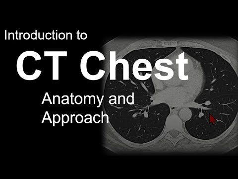
36:53
CT Chest Introduction: Anatomy & Reading Approach
@navigatingradiology
471.2K views•2022-03-26

48:47
Graphic Medicine: Comics for Collaborative Healthcare Communication
@nationalpatientadvocate
189 views•2023-12-04

10:28
Neuroanatomy: Central and Peripheral Nervous System Divisions Explained
@AKLECTURES
136.2K views•2014-09-20

3:05
Stages of Labor and Vaginal Birth | Childbirth Animation
@nucleusmedicalmedia
52.1M views•2017-08-18
Related Study Plans & Knowledge Roadmaps
Structured learning paths in Medicine
medicine
Abnormal LFTs: Diagnostic Workup & Pathology
01Liver Anatomy and Basic Physiology
02Introduction to Liver Function Tests (LFTs)
03Hepatocellular vs. Cholestatic Patterns
+ 3 more learning modules
12h6 modules
Explore Roadmap
medicine
Analyzing Blood Smears: RBC, WBC & Platelets
01Introduction to Blood Components & Smear Preparation
02Normal Morphology of Blood Cells
03Erythrocyte Pathologies & Morphological Variants
+ 2 more learning modules
12h5 modules
Explore Roadmap
medicine
Mechanical Ventilation: Waveforms & Alarms
01Anatomy and Physiology of Breathing
02Introduction to Mechanical Ventilation & Core Modes
03Ventilator Settings and Waveform Interpretation
+ 2 more learning modules
12h5 modules
Explore Roadmap
medicine
Neuro Exam: Localization & Stroke Syndromes
01Foundations of Neuroanatomy and Vascular Supply
02The Neurological Examination: Step-by-Step
03Principles of Neurological Localization
+ 3 more learning modules
18h6 modules
Explore Roadmap