












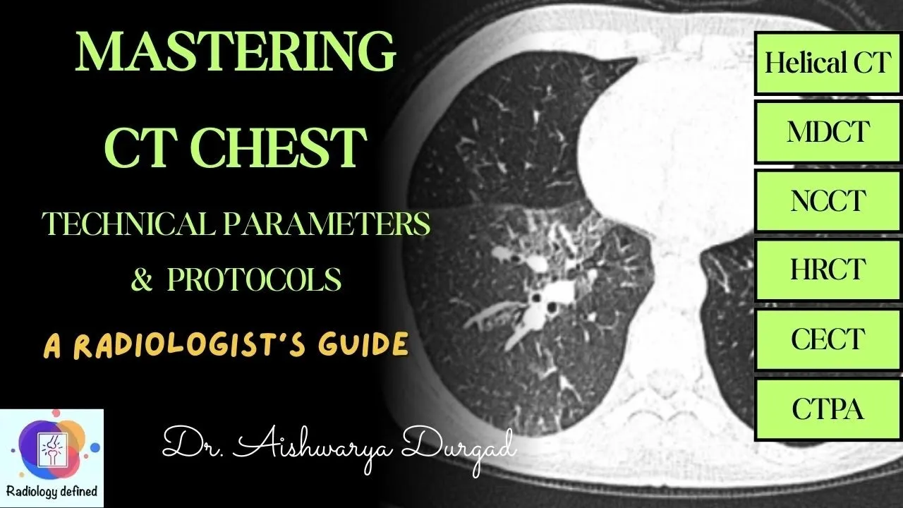







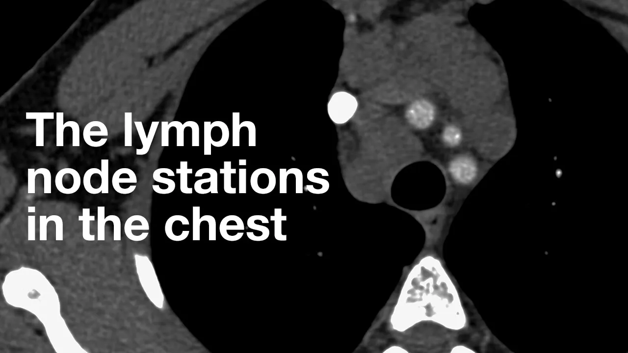




This video introduces the fundamental anatomy of the chest on CT scans and provides a systematic approach to reading chest CT images. The thoracic cavity contains the lungs, heart, and mediastinum, which is divided into four clinically important compartments: superior (above pericardium), anterior (fatty tissue/thymus), middle (heart and great vessels), and posterior (spine/esophagus). The heart has four chambers with specific anatomical features, while the lungs have distinct lobar and segmental anatomy. The systematic reading approach involves starting with the scout image, then evaluating soft tissue windows for mediastinal structures, vessels, and lymph nodes, followed by lung windows for parenchymal pathology, and finally bone windows for skeletal assessment.
CT Chest Introduction: Anatomy & Reading Approach
Added:this interactive video series is designed to get you comfortable reading ct scans of the chest by interactive i mean that you'll be able to scroll through full ct cases on your phone or computer as you follow along in this first video we'll cover basic anatomy and an approach to reading ct of the chest in future videos we'll cover how to read a ct pulmonary angiogram or ctpa including how to diagnose acute and chronic pulmonary emboli and then cover basic patterns of lung disease like consolidation ground glass nodules septal thickening etc and the associated differentials that you need to know to open up the ct chest that we're looking at you can use your phone camera to scan the qr code on the screen and then scroll through the case on your phone while you follow along or if you're on a computer you can click the link in the description and open up the case in a separate browser alongside the video the cases will look something like this and you can access the different series on the left or if you're on your phone you'll see an icon in the top left that you can click to bring up the various series before watching this i strongly suggest watching our video a practical introduction to ct also if you want to support the channel join our patreon at patreon.com navigating or become a member of our youtube channel by clicking join in the description here you'll have access to case-based courses that we are building including an abdominal and pelvic mri course and other scrollable ct and mri teaching cases all right so let's get started so this is a ct scan of the chest and again if you'd like to follow along you can open up the case via qr code or the link in the description so this ct scan of the chest was performed with iv contrast with a routine protocol but there are a number of ways that you can image the chest with ct so there are multiple protocols and we won't get into too much detail but for now you should understand the basics of contrast timing and how that affects what the images look like so you can have a non-contrast ct chest i.e no iv contrast was administered notice that there's no contrast here in the heart or vessels if we give iv contrast how long we wait after injection practically speaking is going to determine the phase of contrast after we inject contrast into a vein it travels to the right heart and then into the pulmonary arteries so if we image really really early at that stage about 15 seconds or so after injection the pulmonary arteries would be filled with contrast and look very very bright this would be a ctpa or ct pulmonary angiogram protocol the scan that we want when we're looking for pulmonary emboli if we waited a little bit longer about 20 seconds after injection or so contrast moves from the pulmonary arteries into the lungs back to the the pulmonary veins in the left heart and then into the aorta and other arteries if we image then it would be a cta or a ct angiogram of the chest with the aorta and other arteries very bright we use this to assess the arterial vasculature for example when ruling out an aortic dissection if we waited even longer the contrast would distribute further and we'll have more optimal enhancement of soft tissues around 60 to 70 seconds or so this is a routine contrast enhanced ct of the chest now we've intentionally oversimplified things here for now and left out protocols like low dose ct chest and high resolution ct chest but that's the basic principle that you want to understand for now and we'll get to the rest later for now let's get back to this ct and start looking at the anatomy in detail so when we scroll through these axial images of the chest most of what we're interested in here is within the thoracic cavity and the thoracic cavity is this entire cavity that is surrounded by chest wall which includes the bones and muscles that surround the cavity the thoracic cavity extends all the way up to this opening at the top of the thoracic cavity called the thoracic inlet basically it's the hole at the top bordered by the first ribs which are here and the manubrium and the thoracic cavity extends all the way down to the thoracic outlet which is closed by the diaphragm and borders the inferior aspects of the ribcage and to the bottom of the sternum in the thoracic cavity we have the right lung and the left lung and between the lungs is the mediastinum which we'll cover first the mediastinum is split up into compartments which are very important to know clinically because different processes happen in each of the compartments and so the differential diagnosis for things in each of the compartments is going to be different there are a bunch of ways to split up the mediastinum but the easiest and most logical way is to use the fibrous pericardium as your landmark so it's worth knowing a little bit about the pericardium so the pericardium is the sac that the heart sits in with the fibrous component being the outermost layer which you can actually see here on the ct it inserts into the diaphragm inferiorly here and up all the way to the level of the roots of the great vessels here is the fibrous pericardium on the sagittal images so everything superior to the pericardium and below the thoracic inlet is considered the superior mediastinum the superior mediastinum contains trachea esophagus and a bunch of vasculature that we'll talk about later the anterior mediastinum is anterior to the pericardium so this space right here in adult patients it contains mostly fatty attenuation notice the attenuation here is similar to the subcutaneous tissues but in younger patients it can contain the thymus it can also contain lymph nodes and sometimes thyroid which is up here if it extends inferiorly so that's why when you see an anterior mediastinal mass the classic differential includes thymic lesions like thymoma thyroid lesions and lymphoma amongst other things like teratoma and other germ cell tumors the middle mediastinum is within the pericardium mostly the heart and the vessels joining the heart including part of the ascending aorta and the pulmonary trunk the svc it also contains the tracheal bifurcation and some lymph nodes and lastly the posterior mediastinum is posterior to the pericardium the posterior mediastinum of course contains the spine the paraspinal nerves the vasculature back here and the esophagus the differential for a posterior mediastinal mass is logical if you know the anatomy with neurogenic tumors being the most common let's look through this anatomy in a bit more detail starting with the middle mediastinum and the heart so this here is the right atrium which is where the major systemic veins drain namely the inferior vena cava or ivc which defines the right atrium this is coming from the lower body and the superior vena cava from above this here is the right atrial appendage and you can see this structure here as well called the crista terminalis which is a muscular ridge running from the superior vena cava down to the ivc it's a normal structure and not a clot this is the right ventricle notice that normally it is pretty thin walled not so important for this introductory talk but important in general the right ventricle is defined by the moderator band which is this band coming from the interventricular septum here and extending towards the margin of the right ventricle also known as the septomarginal trabecula you may not see it very well on routine ct and for now don't worry about it too much the right ventricle pumps blood into the pulmonary artery so this is the pulmonary trunk and the right and left pulmonary arteries from here blood goes into the lungs and then drains into the pulmonary veins these are pulmonary veins that i'm pointing out the pulmonary veins drain into the left atrium which sits posteriorly here this is the left atrial appendage the left atrial appendage is a common location for blood clots to form it's also a common area where you can see mixing artifact the left atrium is normally less than four centimeters or so in ap dimension lastly this is the left ventricle here notice the relatively thick wall compared to the right ventricle you'll also notice the papillary muscles here the left ventricle pumps blood into the aorta so this is the aortic valve and the ascending aorta okay and that's a nice segue to move from the heart to the vascular anatomy so starting with the arteries again this is the aorta with the aortic valve ascending aorta aortic arch and descending aorta important arterial branches to know include the coronary arteries so this is the left coronary artery here with the left anterior descending that extends down the interventricular septum and the circumflex as well as the right coronary artery which arises from the right coronary cusp the three major branches of the aorta are first the brachiocephalic trunk which splits into the right subclavian and the right common carotid artery that i'm following now the second major branch is the left common carotid artery and the third major branch is the left subclavian artery other clinically important arteries to be aware of two arising from the subclavian vein are first the vertebral artery which i'm following now and second is the internal thoracic artery here it is on the left extending inferiorly along the anterior chest wall lastly the intercostal arteries are important to be aware of clinically they travel along the under surface of the ribs even if you don't see them that well they're important to know clinically for example if there is trauma and there's active extravasation or bleeding coming from this location you know it's arising from an intercostal artery all right moving along we briefly talked about the pulmonary artery here including the pulmonary trunk right and left pulmonary arteries we'll cover the rest of the pulmonary artery branches all the way to the segmental level in a future video on ct pulmonary angiogram major veins to know include the left jugular vein which joins the left subclavian vein here to become the left brachiocephalic vein similar on the right the right subclavian vein and right jugular vein joined to make the right brachiocephalic vein the two brachiocephalic veins joined to become the superior vena cava which joins the right atrium the arch of the azages here joins the superior vena cava just above the level of the right main stem bronchus so this is the right main stem bronchus here just above it is this arch of azagus joining into the superior vena cava the azages itself comes from inferiorly here and extends up to the level of the arch and it runs along the right side of the aorta and the posterior mediastinum we mentioned the inferior vena cava that joins the red atrium inferiorly one other vein i didn't mention earlier is the largest cardiac vein also known as the coronary vein here that runs along the posterior heart and drains into the right atrium as well moving along to the airways this is the trachea that bifurcates into the right main stem bronchus and the left main stem bronchus the tracheal bifurcation is also known as the carina notice that the right main stem bronchus here is a lot shorter than the left also know that the right main stem bronchus usually has more of an inferior angulation to it compared to the left which is why foreign bodies and aspiration tend to go down the right side more than the left this here posteriorly is the esophagus extending all the way down to the level of the ge junction and stomach before we go on to the lungs one last important thing to touch on here are the lymph node stations and we'll cover the most clinically important ones so starting with the supraclavicular lymph nodes as well as the low cervical lymph nodes which are low in the neck the prevascular nodes anterior to the great vessels here the aortal pulmonary zone or ap window which is just inferior to the aorta and above the pulmonary artery hence a for aorta p for pulmonary artery window this space here we have paratracheal nodes which are surrounding the trachea and that includes left paratracheal nodes you can see a normal appearing lymph node here on the left for example and right paratracheal nodes the dividing line between left paratracheal and right paratracheal is the left lateral border of the trachea so left paratracheal nodes are out here and right paratracheal nodes are anterior and to the right as we scroll down here past the carina we have the subcorinal station this is a normal subcorina lymph node and then more inferiorly and posteriorly we have para esophageal nodes if they happen to be present we also have bilateral hilar nodes adjacent to the main stem bronchi and then we have interlobar lobar segmental and subsegmental nodes as you go outwards but don't worry about those for now just understand the naming convention in the mediastinal nodes for now other important lymph nodes to be aware of in the chest include axillary nodes bilaterally internal thoracic nodes along the internal thoracic chain and cardiophrenic nodes down here okay so let's move on to the lungs themselves so we'll start with the right lung which is separated into three lobes by the fissures so here on the sagittal images you can see the oblique fissure here and the horizontal fissure here which separates the lungs into the right upper lobe right middle lobe and right lower lobe when i scroll through the axial images we are in the upper lobe here i can see the oblique fissure here and so all this posterior to the oblique fissure just like on the sagittal image as you can see are part of the lower lobe here as i scroll down you'll start to see the horizontal fissure which is this line here and so now we have the right upper lobe this is above the horizontal fissure right middle lobe between the horizontal and oblique fissure down here and this is all right lower lobe on the left we have only the oblique fissure separating the left upper lobe from the left lower lobe posteriorly and inferiorly on the axials here again you can see the oblique fissure this is all upper lobe and posteriorly and inferiorly this is all lower lobe the inferior part of the left upper lobe is called the lingula here near the heart now if you're going into radiology you'll probably need to know the segmental anatomy of the lungs as well so we'll cover that in detail in the ctpa video but let's briefly go through it now if this is your first time hearing this stuff don't worry if it's over your head you'll probably need to review this on your own and it just takes time and you'll get it eventually so the right lung is split into ten segments and the left into eight on the right starting with the upper lobe the upper lobe is split into three so we have the apical segment we're going to follow the bronchi to see where we're going so this is the apical segment here we have the anterior segment and we have the posterior segment in the right middle lobe again following the bronchi to indicate where i'm going in the right middle lobe we have a medial segment and a lateral segment notice that it splits into a medial and lateral bronchi here and the right lower lobe is split into five segments so this is right lower lobe here this posteriorly and superiorly is the superior segment of the right lower lobe pretty high in the chest and then as i scroll down it splits into four basal segments the anterior the medial the posterior and the lateral basal segments so again three in the red upper lobe apical anterior and posterior two in the right middle lobe medial and lateral and five in the right lower lobe superior and then the four basal logically anterior medial posterior lateral the left is split into eight segments with a few differences from the right so the left upper lobe basically you're just combining the apical and posterior segments so you have the apico posterior segment and then you also have an anterior segment so anterior is here and apico posterior is all this the lingula which is down here is instead of medial and lateral like the right middle lobe split into the superior you'll see a bronchus coming off here so the superior and then inferior segments so we're at four so far and then the left lower lobe again has a superior segment up here and then we have basal segments and on the left we only have three instead of four the anterior and medial segments are combined and then we have our lateral and posterior basal segments so the major differences between the two lungs are that we have the apico posterior combined in the left upper lobe and the anteromedial in the lower lobe which gets rid of the two segments making it eight on the left and ten on the right the other difference is that the lingular are separated into superior and inferior instead of the medial and lateral in the right middle lobe again if you're new to that and this terminology is new that's likely way too much for now don't worry it's not that important at this point and we'll go through it again when we talk about pulmonary emboli okay so surrounding each of the lungs is a double layer of pleura you don't actually see them when you have normal pleura on ct but the space between these layers is called the pleural space so you're going to be looking for things like pleural effusions that will fill that space dependently and when it's filled with gas and pneumothorax amongst other pathology all right so that's the meat and potatoes of the anatomy that i wanted to cover a few other things to briefly mention we can see the lower neck here as well on cts of the chest including the thyroid gland notice how bright it is we also have the entire chest wall which includes subcutaneous fat musculature and bones starting with the bones we have our ribs starting from the first rib all the way down to the 12th rib we have our spine posteriorly here we have our manubrium and sternum anteriorly here we have our bilateral clavicles the acromioclavicular joints up here can sometimes be seen this is the scapula back here and then we can often see part of the humerus on sagittal images the spine and the sternum there are many muscles that you can see on ct of the chest that are beyond the scope of this talk including pectoralis major here and pectoralis minor bilaterally rotator cuff muscles back here paraspinal muscles we can also see quite a bit of the upper abdomen here and we covered the anatomy here in detail in our ct abdomen and pelvis talk okay so that's it for the anatomy let's move on to a brief approach so usually i start by looking at the scout image that's because the scout image often covers more anatomy than does the ct itself looking for any incidental findings then i start by looking at the soft tissue window first looking for any medical devices things like pacemakers lines tubes staying on soft tissue window i then look at the chest wall including all the way up to the lower neck which also includes the thyroid looking for incidental thyroid nodules or thyroid masses as part of my scan of the chest wall on soft tissue windows i'll quickly look down the spinal canal for any incidental findings and then start looking at the thoracic cavity itself starting with the heart and mediastinum so generally when i'm looking at the heart itself i'm looking for specific chamber enlargement or other pathology like clot or tumor i'm looking in the pericardial space here posterior to the fibrous pericardium for any evidence of pleural effusions pleural thickening pleural calcification as i'm scrolling up here i look for coronary calcifications on non-gated routine cts it's difficult to assess the coronary arteries much more than looking for calcifications most of the time so knowing the anatomy is important to pick up on what is coronary calcifications or stents i then look at the mediastinal vessels so specifically looking at the aorta and pulmonary artery to start starting with the pulmonary artery i usually measure the pulmonary artery here in this axis the pulmonary trunk is considered enlarged if it's over three centimeters which would also indicate pulmonary hypertension i also then follow the pulmonary arteries themselves when we give an iv contrast even if it's a routine ct like this because we'll often pick up on incidental pulmonary emboli i also measure the diameter of the ascending aorta only if it looks big to me and your eye will get and your eyes will get used to what is too big and if it's over four centimeters that's considered aneurysmal for the ascending aorta i'm gonna follow the aortic arch and down to the descending aorta looking for any other aortic pathology as well as briefly following the major branches of the aorta i'm looking at the esophagus in the posterior mediastinum and as i'm looking at all these things i'm also looking for any obvious mediastinal masses in each of the compartments that we mentioned in the anatomy section i then look for lymph nodes i'm looking through all of the chains that we mentioned so starting with the lower neck bilaterally and looking closely for supraclavicular lymph nodes in the mediastinum the prevascular chain ap window paratracheal nodes sub subcorinal nodes paraesophageal nodes looking for hilar nodes along the internal thoracic bilaterally cardiophrenic then on soft tissue window i'm looking at the pleura for any pleural effusions dependently pleural thickening calcification masses and then switch to lung window to look for pneumothorax first in the setting of trauma you're going to pick up little tiny pneumothoracies looking anteriorly and medial here or posterior medial here this is also non-dependent for this part of the lung while i'm still on lung window i'm looking at the airways and then i start to look at the lungs and so everyone has their own search pattern for how they look through the lungs i tend to start by doing a quick overview of both lungs to look for any obvious pathology i usually split it into halves on the axial cuts so i'm looking right anterior first and then right posterior and then the same on the left left anterior and left posterior i then take my time and look at the lungs in more detail looking for smaller lung nodules i tend to use the mip images if they're available mip images are maximum intensity projections with thicker slices so as you can see the vessels look very elongated and linear that is contrary to what a long nodule would look like so for example there is a lung nodule it doesn't look linear like these vessels so lung nodules will pop up as dots amongst all of these connecting linear structures that are the vessels so when i'm looking at the mips depending on your plane i actually split the chronos into three so i'm looking superiorly here first from front to back middle and then inferiorly scrolling relatively slowly here's another nodule for example and then do the same on the left if you happen to identify a nodule on the mips the next step is to then cross correlate and find that same nodule on your normal axial cuts i then switch to bone window to look at the bones looking at the ribs i generally split them into posterior and anterior so i'm looking posteriorly on the right here and then anteriorly or anterior laterally here on the right same thing on the left repeat looking at both clavicles here the manubrium and sternum i tend to look at the axial images for the spine as well and the scapulae as well as the humeral heads here and then i do pull up the sagittal images as well to look at the spine and sternum again lastly on soft tissue window i'm looking at the upper abdomen for incidental findings and we went through an approach on the abdomen and pelvis talk but again looking at the liver or what i can see of it the gallbladder pancreas spleen adrenals and kidneys remember lung cancers like to metastasize to the adrenals so you're specifically looking for adrenal nodules when looking at cases of lung cancer i also follow the stomach to the duodenum or what i can and have a look at the bowel as well as for any incidental lymph nodes in the upper abdomen but again watch the ect adamant and pelvis talk and this will take you a very short amount of time to look through all right so that's the end of this video in the next video we'll cover how to look at ct pulmonary angiogram with a more detailed look at segmental anatomy and how to diagnose acute and chronic pulmonary emboli thanks a lot
Up Next
Lung Nodule Stratification with Fleischner Guidelines 2017
@internationalcancerimaging159
42.6K views•2020-03-30

Graphic Medicine: Comics for Collaborative Healthcare Communication
@nationalpatientadvocate
189 views•2023-12-04

Neuroanatomy: Central and Peripheral Nervous System Divisions Explained
@AKLECTURES
136.2K views•2014-09-20

Stages of Labor and Vaginal Birth | Childbirth Animation
@nucleusmedicalmedia
52.1M views•2017-08-18
Related Study Plans & Knowledge Roadmaps
Structured learning paths in Medicine