The T4 bacteriophage infects E. coli through a sequential process: long tail fibers recognize host cell surface receptors, triggering base plate conformational changes that unravel short tail fibers for irreversible binding; subsequent tail sheath contraction activates GP5 protein to puncture the outer membrane and degrade the periplasmic peptidoglycan layer, allowing viral DNA to enter the host cell; after completing the lytic cycle, the host cell lyses to release newly assembled phages.
Bacteriophage T4 Life Cycle: 3D Animation of E. coli Infection
Added:Deep Dive
Basic structural components of a bacteriophage (capsid, sheath, tail fibers) and its viral genome.
Bacteriophage | Structure and Replication of Bacteriophage | B.sc Botany 1st Semester | Paper | LU
3.2K
All viruses have a basic structure consisting of two main components: (1) Nucleic acid (genetic material) which can be either DNA or RNA, but never both at the same time, and (2) Protein coat (capsid) which protects the genetic material. The capsid is made up of protein subunits called capsomeres. Bacteriophages specifically have a characteristic structure consisting of three main parts: (1) Head region - contains the genetic material, (2) Neck region (also called collar), and (3) Tail region. The head contains the nucleic acid genome, while the tail is used for attachment and injection of genetic material into the host bacterium. Bacteriophages infect bacteria through a specific mechanism: (1) The phage attaches to the bacterial surface using its tail fibers, (2) The tail region folds and injects the viral genome into the bacterial cell, (3) The viral genome enters the bacterial cytoplasm and takes over the host's cellular machinery, (4) The host cell becomes hijacked and starts producing viral components instead of its own.
Click to watch →
Bacteriophage
432
Bacteriophages consist of two main structural components: (1) A head (capsid) made of protein that contains the genetic material (DNA or RNA), and (2) A tail structure with a base plate and tail fibers. The head is hexagonal in shape and houses the viral genome. The tail serves as a delivery mechanism for injecting genetic material into bacterial cells. The tail fibers are crucial for recognizing and attaching to specific bacterial receptors.
Click to watch →

Virus | Structure and Classification | Biology | Extraclass #bacteriophage
106.8K
Bacteriophages (bacterial viruses) have a distinctive structure with five main parts: (1) Capsid head - where genetic material is packed into a protein structure; (2) Collar - connects the sheath to a thin disc at the head end and to a base plate at the tip end; (3) Sheath - an external contractile envelope wrapping the tail tube in some viruses; (4) Baseplate - a multi-protein molecular machine that controls host cell recognition; (5) Tail fiber - a rod-like structure approximately 1400 angstroms in contour length with a total mass of about 600,000 daltons.
Click to watch →

شرح الدرس الأول في DNA بكل ملاحظاته | البيولوجيا الجزيئية | أحياء ثانوية عامة 2026
36.3K
This segment covers bacteriophage structure and life cycle. Bacteriophages are viruses that infect bacteria, containing DNA as genetic material. Structure includes: protein head (capsid) containing DNA, protein tail (sheath), and tail fibers for attachment. The life cycle follows a precise timeline: (1) Attachment (0 min) - bacteriophage attaches to bacterial cell using tail fibers, (2) Injection (4 min) - bacteriophage injects DNA into bacterial cell, (3) DNA Replication (15 min) - bacteriophage DNA is replicated, (4) Protein Synthesis (20 min) - bacterial cell produces bacteriophage proteins, (5) Assembly (28 min) - new bacteriophages are assembled, (6) Lysis (32 min) - bacterial cell bursts, releasing new bacteriophages. The bacteriophage's DNA directs all production, while the protein coat remains outside.
Click to watch →

जीव जगत का वर्गीकरण NEET 2024 Biology TOP 20,000 Questions Score 360/360 Doctor Sahab Series ✅
76K
A bacteriophage (bacterial virus) has a specific structure with labeled parts: A is the head (capsid), B is the collar, C is the tail sheath, and D is the tail fibers. The head contains the genetic material, the collar connects the head to the tail, the tail sheath is a contractile structure, and the tail fibers help the bacteriophage attach to bacterial cells. Understanding this structure is essential for understanding viral infection mechanisms.
Click to watch →
Deep Dive
The cellular anatomy of Gram-negative bacteria (such as E. coli), focusing on the outer membrane, cell wall, and inner membrane.

Lec 2 ||Bacterial cell structure 🦠🦠
297
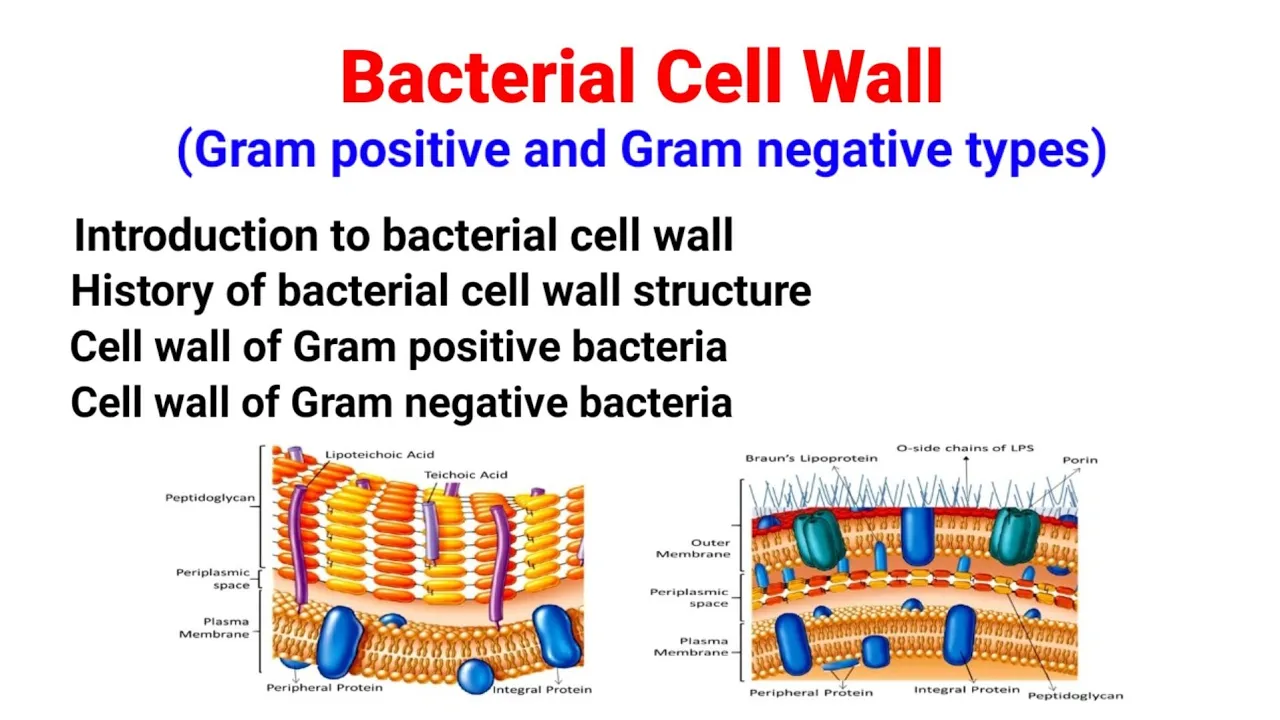
Gram-negative bacteria have a more complex cell wall structure consisting of: (1) An outer membrane containing lipopolysaccharide (LPS) and lipids; (2) A periplasmic space containing peptidoglycan (only 1-2 layers, much thinner than Gram-positive); (3) The inner cytoplasmic membrane. The outer membrane contains lipopolysaccharide (LPS), which includes a lipid component (endotoxin) and a polysaccharide component (O-antigen).
Click to watch →
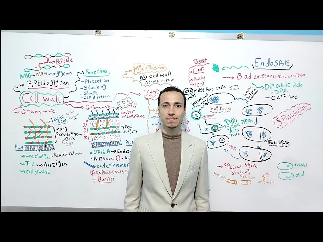
Bacterial Structure part 2 | Dr. Mohamed El Kot
2.9K
Gram-negative bacteria have a thin cell wall with only 1-2 layers of peptidoglycan, insufficient to retain Gram stain (appearing pink/red). The outer membrane contains lipopolysaccharides (LPS), lipoproteins, and phospholipids. LPS consists of lipid A (hydrophobic anchor), core polysaccharide, and O-antigen. The outer membrane acts as a barrier against phagocytosis, complement-mediated lysis, and antibiotics. The periplasmic space between inner and outer membranes contains enzymes and transport proteins.
Click to watch →
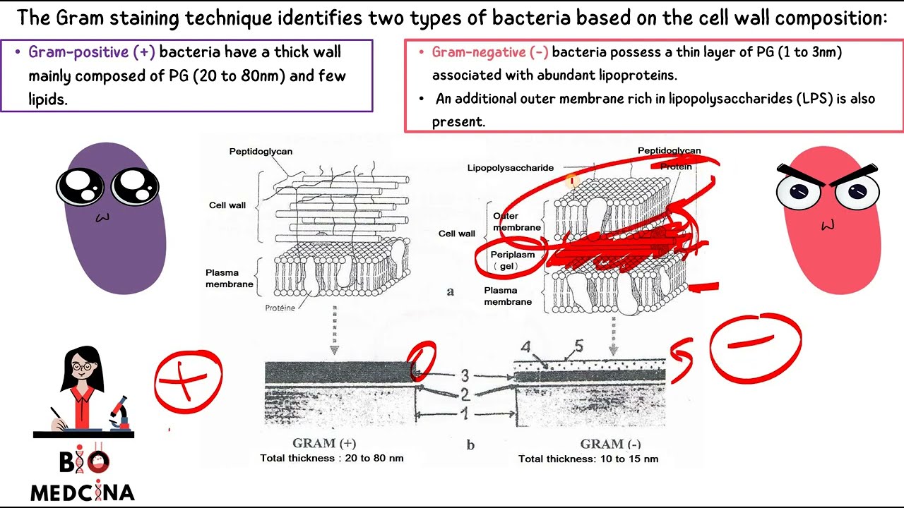
PROKARYOTIC CELL 2 باللغة العربية biologie cellulaire S1 English
21.7K
Gram-negative bacteria have a complex cell wall structure consisting of multiple layers: (1) An inner cytoplasmic membrane; (2) A thin peptidoglycan layer; (3) An outer membrane containing lipopolysaccharides (LPS) and proteins; (4) A periplasmic space between the outer membrane and peptidoglycan layer. The outer membrane provides additional protection and acts as a selective barrier. This layered structure distinguishes Gram-negative bacteria from Gram-positive types and affects their susceptibility to antibiotics.
Click to watch →

Bacterial characteristics - Gram staining | Cells | MCAT | Khan Academy
474.3K
Gram-negative bacteria have a more complex cell wall structure with multiple layers: an inner plasma membrane, a thin peptidoglycan layer, an outer membrane containing lipids, a lipopolysaccharide (LPS) layer (composed of lipid regions at the bottom and polysaccharide chains at the top), and an outer capsule. Between the inner and outer membranes lies the periplasmic space, which contains enzymes and other molecules. The thin peptidoglycan layer and presence of LPS contribute to the unique properties of Gram-negative bacteria.
Click to watch →
Bacterial Cell Wall | Gram positive & Gram negative bacteria cell wall| Structure|Salient features|
63.7K
Gram-positive bacterial cell walls contain peripheral and integral proteins in the plasma membrane following the fluid mosaic model, a thick peptidoglycan layer made of NAG-NAM disaccharides with tetrapeptide chains and peptide interbridges, teichoic acids providing negative charge and linking to peptidoglycan and plasma membrane, and surface proteins like M proteins functioning as virulence factors. Gram-negative bacterial cell walls include peripheral and integral proteins, a thin peptidoglycan layer, an outer membrane composed of lipopolysaccharide (LPS) with lipid A (endotoxin), core polysaccharide, and O-side chain (antigen); brown lipoproteins anchoring the outer membrane; and porin proteins enabling nutrient transport and contributing to drug resistance. The periplasm contains hydrolytic enzymes and transport proteins for nutrient acquisition. The outer membrane in gram-negative bacteria enhances virulence, explaining why most gram-negative bacteria are pathogenic compared to gram-positive bacteria.
Click to watch →
Deep Dive
The Central Dogma of molecular biology, including how DNA is replicated, transcribed into mRNA, and translated into proteins.

Central Dogma Lec 01: Molecular Biology l Lifesciences l For all Bio Classes l By Neha Bajpai
198
The Central Dogma of Molecular Biology describes the flow of genetic information: DNA replication copies DNA during cell division using DNA polymerase; transcription converts specific DNA segments (genes) into RNA using RNA polymerase; translation converts RNA information into proteins. These three processes form the foundation of molecular biology and explain how genetic information is expressed and transmitted across generations of cells.
Click to watch →

TRANSLATION | PROTEIN SYNTHESIS EXPLAINED | MOLECULAR BIOLOGY & GENETICS~EXAM QUESTIONS
843
The central dogma of molecular biology describes the flow of genetic information in cells: DNA replicates to make copies of itself, DNA is transcribed into mRNA, and mRNA is translated into proteins. The three processes are: replication (DNA to DNA), transcription (DNA to mRNA), and translation (mRNA to proteins).
Click to watch →

DNA Replication, RNA Transcription & Translation | Genetics
3.1M
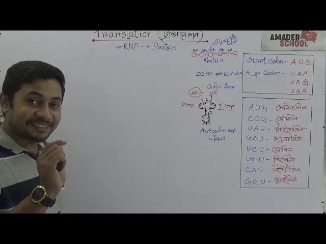
The central dogma describes the flow of genetic information in cells: DNA is replicated to ensure heredity, transcribed into mRNA to carry genetic instructions out of the nucleus, and translated into proteins that perform cellular functions. DNA replication uses complementary base pairing (A-T, G-C) to create identical copies. Transcription produces mRNA using uracil instead of thymine. Translation decodes mRNA codons (groups of three bases) into amino acid sequences using tRNA molecules. With 64 possible codons specifying 20 amino acids, the genetic code is redundant and nearly universal. Proteins, composed of amino acid chains, carry out most cellular functions including enzymatic reactions, structural support, and transport.
Click to watch →
DNA Translation | mRNA to Protein | ডি এন এ ট্রান্সলেশন । HSC Biology || Fahad Sir
192.2K
The central dogma of molecular biology describes the flow of genetic information from DNA to RNA to protein. DNA replication copies genetic material, transcription converts DNA into messenger RNA (mRNA), and translation synthesizes proteins from mRNA templates. This fundamental process underlies all cellular functions and gene expression.
Click to watch →

Molecular Biology Review Video
15.5K
The central dogma describes the flow of genetic information: DNA replicates itself (DNA to DNA), DNA is transcribed into RNA (DNA to RNA), and RNA is translated into protein (RNA to protein). Replication is the copying of genetic material. Transcription is the process of copying DNA information into RNA. Translation is the process of converting RNA information into protein sequences. This directional flow of information is fundamental to all cellular life.
Click to watch →
Deep Dive
The concept of receptor-ligand specificity, which explains how viruses recognize and bind to specific host cells.

Replicación viral/ inmunología y virología SESIÓN IX
1.9K
Each virus species has evolved to recognize and bind to specific receptor proteins on particular host cell types. This receptor specificity determines which cells a virus can infect and replicate within. For example, HIV requires CD4 receptors on T helper cells, while the coronavirus requires ACE2 receptors on respiratory epithelial cells. This principle explains why different viruses cause different diseases and why some viruses can infect multiple cell types while others are highly specific. The specificity is determined by the molecular compatibility between viral attachment proteins and host cell receptors.
Click to watch →

Chapter 5- Virology
114.6K
Viral infection begins with attachment to specific receptors on host cell surfaces, determined by receptor-ligand interactions. Both enveloped and naked viruses use specific attachment sites—spike proteins for enveloped viruses and corner fibers for icosahedral viruses. Entry occurs through receptor-mediated endocytosis (engulfment in vesicles) or membrane fusion (direct merging of viral and host membranes). Host range determines which cells and species a virus can infect, governed by receptor specificity. Viruses range from narrow host ranges (infecting only one cell type in one species, like Hepatitis B) to broad host ranges (infecting multiple species and cell types, like rabies). Mutations in host cell receptors can confer natural resistance, as seen in HIV-resistant individuals lacking functional CCR5 receptors.
Click to watch →

【総集編】身近で危険な恐ろしい感染症まとめ|人食いバクテリア・手足口病・マールブルグ病など
65.1K
Viruses can only infect cells that have specific receptor molecules matching the virus's surface proteins. This 'lock and key' mechanism determines which species and cell types a virus can infect. This specificity explains why some viruses can jump between species while others are restricted to particular hosts.
Click to watch →

Biochemistry ICMR JRF Life Science 2023 PYQS | MPQS Series | IFAS | L1
30.8K
Receptor-ligand interactions are highly specific, meaning one receptor typically binds only one specific ligand. This specificity is determined by the complementary shape and chemical properties between the receptor binding site and the ligand. While some receptors can bind multiple ligands with similar structures, the general principle is that receptor-ligand interactions are highly selective, ensuring precise cellular communication.
Click to watch →

অণুজীব hsc (2nd part) | Nano Biology Course 200/200 | #nanobiology | onujib hsc
420
Viruses are obligate parasites with highly specific host cells. Each virus can only infect specific host cells because they require specific receptors on the host cell membrane to attach and enter. The virus binds to these receptors through a chemical reaction, allowing it to firmly attach to the cell surface. Only after this attachment can the virus enter the host cell. For example, chickenpox virus only infects skin cells, cold viruses only infect respiratory tract cells, and hepatitis viruses only infect liver cells. A virus can only infect a host cell if that cell has the specific receptor the virus needs.
Click to watch →
Prerequisite Knowledge
- Concept 01Basic structural components of a bacteriophage (capsid, sheath, tail fibers) and its viral genome.
- Concept 02The cellular anatomy of Gram-negative bacteria (such as E. coli), focusing on the outer membrane, cell wall, and inner membrane.
- Concept 03The Central Dogma of molecular biology, including how DNA is replicated, transcribed into mRNA, and translated into proteins.
- Concept 04The concept of receptor-ligand specificity, which explains how viruses recognize and bind to specific host cells.
Subsequent Learning
- Step 01The lysogenic (temperate) life cycle of bacteriophages (such as Lambda phage) and how it differs from the obligately lytic T4 cycle.
- Step 02Bacterial defense mechanisms against viral invasion, specifically restriction-modification systems and the CRISPR-Cas adaptive immune system.
- Step 03Phage Therapy, including the clinical application of bacteriophages as an alternative to antibiotics for treating multidrug-resistant infections.
- Step 04The role of bacteriophages in horizontal gene transfer, specifically the processes of generalized and specialized transduction.
Attachment
Signal relay
Firm binding
Membrane puncture
Cell wall breach
DNA entry
Cell lysis
Attachment
0:01Playing Section
- 1
T4 phage recognizes host receptors via long tail fibers.
- 2
This initial contact triggers the infection process.
The Lysogenic Cycle and Chronic Phage Infections
While the 3D animation of bacteriophage T4 illustrates the destructive lytic cycle, it represents only one pathway of viral-bacterial interaction. An important alternative paradigm is the lysogenic cycle, exhibited by temperate phages like Lambda. Instead of lysing the host, these phages integrate their DNA into the host genome as a prophage, replicating harmlessly alongside the bacterium for generations. Furthermore, some viruses engage in chronic infections (such as filamentous phages) where progeny are continuously released without killing the host cell, or enter 'pseudolysogeny' during nutrient starvation. These alternative life cycles demonstrate that phage-host relationships are not always predatory and lethal, but can be mutualistic, dormant, or cooperative, fundamentally shifting our understanding of microbial ecology.
Deep Dive
The lysogenic (temperate) life cycle of bacteriophages (such as Lambda phage) and how it differs from the obligately lytic T4 cycle.

Semester-1 Core Paper | Details of Viruses |Virus life cycle| Bacteriophage lytic & lysogenic cycle|
260
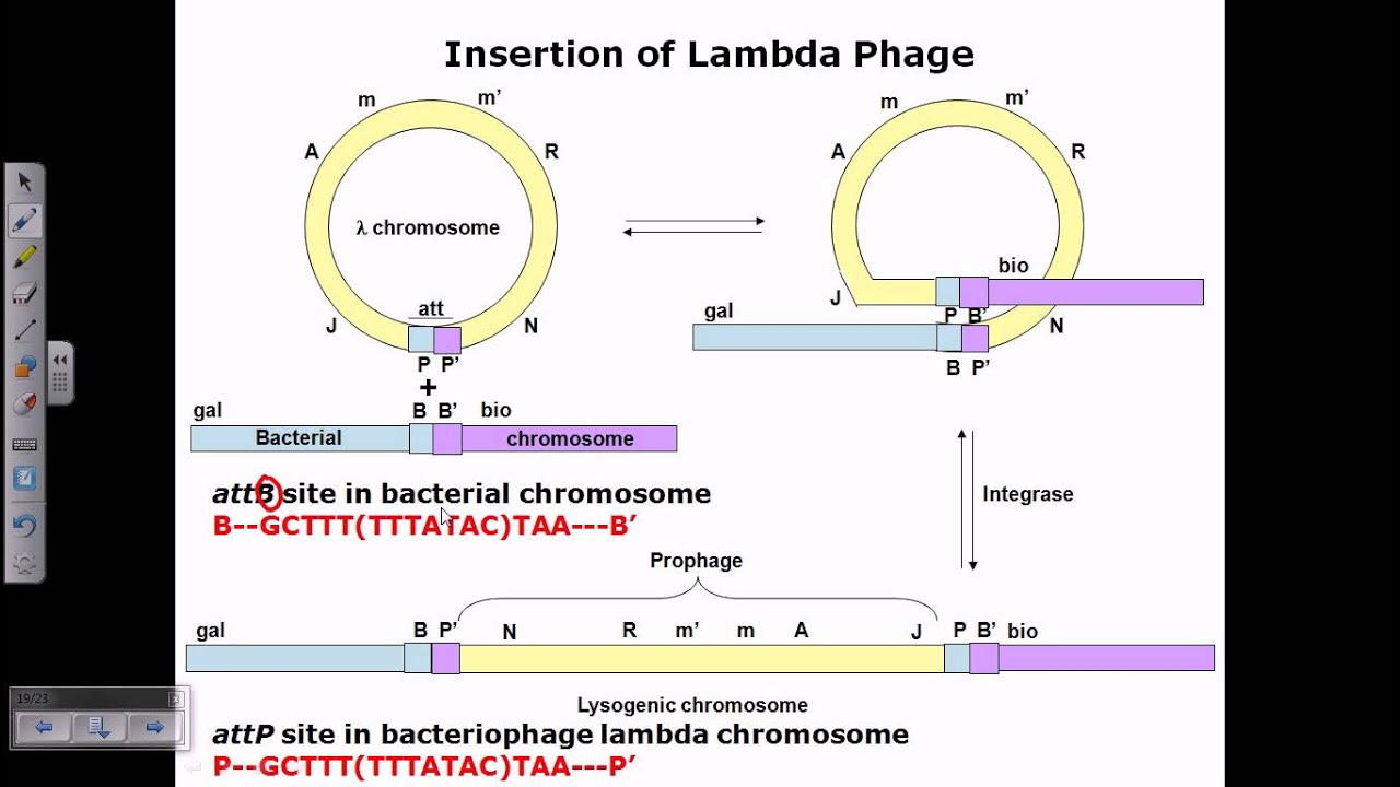
The lysogenic cycle (Lambda phage) differs from lytic at step 3: phage DNA integrates into host chromosome as prophage, replicating passively with host DNA during cell division. Under stress (UV damage), prophage excises and enters lytic cycle. This allows silent phage multiplication without killing host, demonstrating viral persistence strategies.
Click to watch →

Lytic vs Lysogenic Cycle Switch I Regulation Of Phages I Control Of Gene Expression I Lambda Phage
112.2K
The lambda bacteriophage uses a sophisticated transcriptional switch mechanism to decide between lysogenic (dormant integration) and lytic (productive replication) cycles, controlled by two competing regulatory proteins: CI repressor (which promotes lysogeny by binding to operators and blocking transcription from PR and PL promoters) and Cro protein (which promotes lysis by blocking CI and activating late gene expression); the decision depends on the relative synthesis rates of these proteins and environmental conditions, with CI being more stable and favored under good bacterial growth conditions while Cro dominates under poor conditions, ultimately determining whether the phage integrates into the bacterial chromosome or proceeds to replicate and lyse the host cell.
Click to watch →
Lysogenic cycle of lambda phage
64.5K
The lambda phage lysogenic cycle is regulated by the lambda repressor protein, which binds with higher affinity to operator regions O1, O2, and O3 in sequence; initially, low repressor concentration binds only O1 to block lytic cycle transcription, while higher concentrations bind O2 cooperatively to enhance lysogenic transcription via interaction with RNA polymerase and C1/C2 proteins, promoting integration (int gene) and excision (xis gene); however, very high repressor concentrations bind O3 to repress both directions, and prophage induction occurs when environmental stress triggers RecA-mediated degradation of the repressor, allowing the phage to switch back to the lytic cycle.
Click to watch →

Weekly Revision (1) - قصر العيني 2026
11.3K
The lytic cycle is the reproductive cycle of virulent bacteriophages: attachment, injection of genetic material, synthesis of viral components, assembly of new viruses, and lysis (bursting) of the bacterial cell to release new viruses. The lysogenic cycle is the reproductive cycle of temperate bacteriophages: attachment, injection of genetic material, integration of viral DNA into bacterial chromosome (becoming a prophage), and replication with the bacterial chromosome. The prophage can remain dormant for many generations and can be induced to enter the lytic cycle under certain conditions.
Click to watch →

Lysogenic cycle of bacteriophage | Lysogenic cycle in Hindi
7.4K
The lysogenic cycle is one of two life strategies used by temperate bacteriophages like Lambda. In this cycle, the virus attaches to a bacterial host and injects its circular DNA, which then integrates into the bacterial chromosome becoming a prophage. The prophage remains dormant and replicates passively with every bacterial cell division. When environmental conditions become favorable, the prophage excises itself and enters the lytic cycle, producing new viral particles that lyse the host cell. This dual-cycle strategy allows bacteriophages to persist in bacterial populations while maintaining the potential for rapid propagation when conditions permit.
Click to watch →
Deep Dive
Bacterial defense mechanisms against viral invasion, specifically restriction-modification systems and the CRISPR-Cas adaptive immune system.
Chapter 9 Bacteria and Viruses
215
Bacteria evolved defense mechanisms against viral infection including receptor reduction, polysaccharide coat secretion, and restriction-modification systems that cleave foreign DNA. The CRISPR-Cas system provides adaptive immunity: during adaptation, foreign DNA is cleaved into protospacers and incorporated into the CRISPR array; during interference, these spacers guide Cas proteins to recognize and cleave matching viral DNA. This system has been adapted for gene editing technology.
Click to watch →
ГЕНЕТИЧЕСКОЕ ИЗМЕНЕНИЕ ЛЮДЕЙ
18K
Bacterial restriction-modification systems protect against viral DNA by methylating bacterial DNA and cutting unmethylated viral DNA. CRISPR spacers are acquired from viral DNA upon first infection, creating a memory of encountered viruses. This adaptive immunity allows bacteria to recognize and destroy specific viruses upon re-infection, analogous to vertebrate adaptive immunity.
Click to watch →

What are restriction enzymes
235.4K
Restriction enzymes evolved as bacterial defense mechanisms against viral invaders (phages). When phages infect bacteria, they deliver their genomes to degrade the bacterial chromosome. In response, bacteria produce restriction enzymes that identify and cleave foreign DNA. However, bacteria face a dilemma: these same enzymes could accidentally destroy the bacterial genome. To solve this, bacteria developed a complementary system using methyltransferases that modify their own DNA with methyl groups, creating molecular tags that signal restriction enzymes not to cleave self-DNA. This restriction-modification system provides bacteria with adaptive immunity against viral infections while protecting their own genetic material.
Click to watch →

Гонка вооружений. Разнообразие бактериальных систем противовирусного иммунитета. Артём Исаев
1K
Bacteria employ multiple defense strategies against phage infection. Prevention of adsorption includes receptor modification, biofilm formation, capsule secretion, and phase variation. Upon successful infection, bacteria activate restriction-modification systems that cleave foreign DNA, CRISPR-Cas systems that provide adaptive immunity, and abortive immune systems that kill infected cells to prevent phage propagation. These systems work at different stages of infection and provide layered protection. The restriction-modification system is the most common, while CRISPR-Cas provides adaptive immunity that can be inherited by daughter cells.
Click to watch →

Rotem Sorek talk at the 22nd HFSP Awardees Meeting
764
Bacteria face constant viral infection pressure from bacteriophages, which are the most abundant entities on Earth (10 times more than bacteria). Bacteria have evolved two main immune arms: CRISPR-Cas provides adaptive immunity by memorizing past infections through stored viral DNA fragments that block future infections; restriction-modification systems provide innate immunity using restriction enzymes that cleave foreign DNA while modification enzymes protect bacterial DNA. These systems represent fundamental strategies that bacteria use to survive in environments teeming with viruses.
Click to watch →
Deep Dive
Phage Therapy, including the clinical application of bacteriophages as an alternative to antibiotics for treating multidrug-resistant infections.

MICROBIOSCIENCE 2026, DÍA 1.
548
Phage therapy uses bacteriophages as alternatives to antibiotics for treating bacterial infections, particularly antibiotic-resistant bacteria. Phages have high specificity for their target bacteria, minimizing effects on beneficial microbiota. Current applications include treatment of multidrug-resistant infections, with ongoing clinical trials for cystic fibrosis and multidrug-resistant urinary tract infections. A documented case involved a patient with multidrug-resistant Pseudomonas infection who was cured using phage therapy after failing all antibiotics. Phage therapy represents an emerging approach to combat antibiotic resistance, though it carries risks due to viral mutation potential and requires careful monitoring.
Click to watch →
Utility of Bacteriophages to Combat Drug Resistant Bacteria
145
Bacteriophages are viruses that specifically target and kill bacteria by attaching to bacterial cells, injecting their DNA, replicating within the bacteria, and lysing the cell every 22 minutes; unlike broad-spectrum antibiotics that disrupt the entire human microbiome, phages are highly species-specific and can effectively combat antibiotic-resistant bacteria such as Pseudomonas and MRSA, with clinical trials showing significant reductions in bacterial loads (up to two-log reductions) in patients with cystic fibrosis and complicated bacteremia, suggesting phage therapy could serve as either a standalone or complementary treatment to antibiotics for severe resistant infections.
Click to watch →

Phage Treatment Saves A Life
69.7K
Bacteriophages are viruses that specifically target and destroy certain bacteria; this experimental therapy has shown promise for treating multidrug-resistant bacterial infections when conventional antibiotics fail, as demonstrated by a successful case at UC San Diego where an international team identified and deployed phages against a patient's unique bacterial isolate, leading to rapid recovery.
Click to watch →
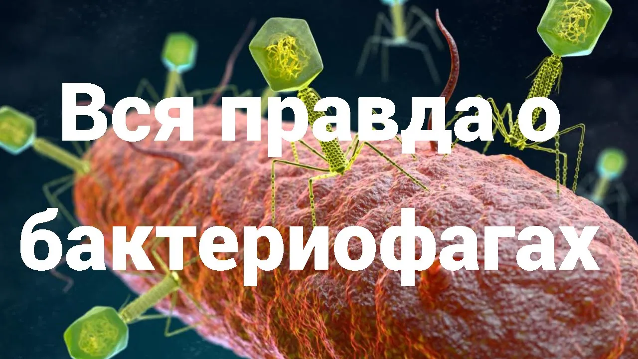
Бактериофаги - «идеальные хищники»
4.8K
Antibiotic resistance is a global health crisis, with approximately 700,000 deaths annually from antibiotic-resistant infections. This has prompted renewed interest in phage therapy as an alternative treatment. In 2016, the first successful intravenous phage therapy case in the United States was reported, treating a patient with a severe multidrug-resistant infection. In 2018, a 15-year-old cystic fibrosis patient was successfully treated with a combination of natural and genetically modified phages. Western medical communities, particularly American infectious disease specialists, are increasingly recognizing phage therapy as an important alternative or complement to antibiotics. The high specificity of phages for their bacterial hosts allows precise targeting without disrupting the rest of the microbiome, though this specificity also creates challenges for developing broad-spectrum treatments.
Click to watch →
How phages may help combat drug-resistant infections
4.8K
Phage therapy uses viruses that specifically infect and destroy bacteria to treat antibiotic-resistant infections; unlike broad-spectrum antibiotics, phages are highly targeted, making them promising alternatives for patients who have exhausted conventional treatments and face life-threatening drug-resistant bacterial infections.
Click to watch →
Deep Dive
The role of bacteriophages in horizontal gene transfer, specifically the processes of generalized and specialized transduction.
![Bacterial Transduction [Generalized & Specialized] – Horizontal Gene Transfer Part – III](https://i.ytimg.com/vi_webp/M3qPV0soD5Q/maxresdefault.webp)
Bacterial Transduction [Generalized & Specialized] – Horizontal Gene Transfer Part – III
77.9K
Bacterial transduction is a horizontal gene transfer mechanism where bacteriophages (viruses that infect bacteria) transfer bacterial genes between cells. It occurs through two mechanisms: generalized transduction during the lytic cycle, where random bacterial DNA fragments are accidentally packaged into viral particles and transferred to new hosts; and specialized transduction during the lysogenic cycle, where improper excision of the viral genome carries specific bacterial DNA sequences near the integration site. A key distinction from transformation is that transduction always transfers double-stranded DNA, whereas transformation involves single-stranded DNA uptake.
Click to watch →

Generalized transduction vs Specialized Transduction
315.4K
Generalized transduction involves lytic phages packaging random bacterial DNA fragments during viral particle assembly, allowing any gene segment to be transferred between bacteria; specialized transduction occurs when lysogenic phages accidentally package bacterial DNA adjacent to their integrated viral genome during the switch from lysogenic to lytic cycle, transferring specific bacterial gene fragments along with viral DNA.
Click to watch →
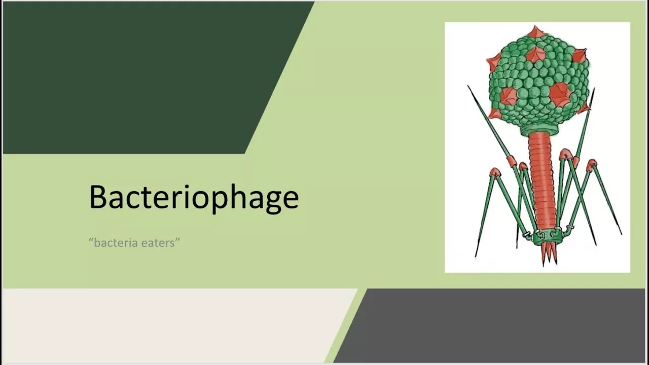
Bacteriophage - "bacteria eaters"
3.2K
During the lytic cycle, bacterial DNA fragmentation creates opportunities for horizontal gene transfer. General transduction occurs when random host DNA fragments become packaged into phage capsids, allowing transfer to new hosts—any genomic region can be transferred. Specialized transduction, associated with the lysogenic cycle, transfers only host DNA flanking the prophage integration site. Both mechanisms enable genetic material exchange between bacteria, contributing to bacterial evolution and antibiotic resistance spread.
Click to watch →

GENETICA BACTERIANA (2/2)
15.3K
Transduction is a mechanism of horizontal gene transfer mediated by bacteriophages (viruses that infect bacteria). There are two types: generalized transduction occurs when a phage accidentally packages bacterial DNA instead of its own viral DNA during the lytic cycle, and when these phages infect new bacteria, they inject bacterial DNA from the previous host. Specialized transduction occurs when a phage integrates into the bacterial chromosome (lysogenic cycle) and, upon excision, carries adjacent bacterial genes along with its own genome. Both mechanisms allow bacteria to acquire new genetic material and potentially new traits, contributing to bacterial evolution and adaptation.
Click to watch →

Bacteriophage
131
Bacteriophages facilitate horizontal gene transfer between bacteria through transduction. Generalized transduction (using lytic phages like P22) occurs when bacterial DNA fragments are accidentally packaged into phage particles. When these particles infect new hosts, the bacterial DNA can recombine with the recipient's chromosome. This process is rare (1 in 100,000-1,000,000 particles) but was historically important for bacterial genetic mapping. Specialized transduction (using temperate phages) transfers only genes adjacent to the prophage integration site. This mechanism allows non-pathogenic bacteria to acquire virulence factors from pathogenic bacteria, potentially converting them into pathogens.
Click to watch →
Attachment
Signal relay
Firm binding
Membrane puncture
Cell wall breach
DNA entry
Cell lysis
Attachment
0:01- 1
T4 phage recognizes host receptors via long tail fibers.
- 2
This initial contact triggers the infection process.
The Lysogenic Cycle and Chronic Phage Infections
While the 3D animation of bacteriophage T4 illustrates the destructive lytic cycle, it represents only one pathway of viral-bacterial interaction. An important alternative paradigm is the lysogenic cycle, exhibited by temperate phages like Lambda. Instead of lysing the host, these phages integrate their DNA into the host genome as a prophage, replicating harmlessly alongside the bacterium for generations. Furthermore, some viruses engage in chronic infections (such as filamentous phages) where progeny are continuously released without killing the host cell, or enter 'pseudolysogeny' during nutrient starvation. These alternative life cycles demonstrate that phage-host relationships are not always predatory and lethal, but can be mutualistic, dormant, or cooperative, fundamentally shifting our understanding of microbial ecology.
Basic structural components of a bacteriophage (capsid, sheath, tail fibers) and its viral genome.
The cellular anatomy of Gram-negative bacteria (such as E. coli), focusing on the outer membrane, cell wall, and inner membrane.
The Central Dogma of molecular biology, including how DNA is replicated, transcribed into mRNA, and translated into proteins.
The concept of receptor-ligand specificity, which explains how viruses recognize and bind to specific host cells.
The lysogenic (temperate) life cycle of bacteriophages (such as Lambda phage) and how it differs from the obligately lytic T4 cycle.
Bacterial defense mechanisms against viral invasion, specifically restriction-modification systems and the CRISPR-Cas adaptive immune system.
Phage Therapy, including the clinical application of bacteriophages as an alternative to antibiotics for treating multidrug-resistant infections.
The role of bacteriophages in horizontal gene transfer, specifically the processes of generalized and specialized transduction.
688.9K views4.6Klikes1:21@AmhausOriginal Release: 2012-03-12
The T4 phagee initiates infection of an E.coli bacterium by recognizing cell surface receptors of the host with its longtail fibers.
A recognition signal is sent through the longtail fibers to the base plate.
This unravels the short tail fibers that bind irreversibly to the E.coli cell surface. The base plate changes confirmation and the tail sheath contracts causing GP5 at the end of the tail tube to puncture the outer membrane of the cell.
The loome domain of GP5 is activated and degrades the paraplasmic pepidocan layer.
The remaining part of the membrane is degraded and DNA from the head of the phagee can travel through the tail tube and enter the e.coli.
[Music] After the live cycle is complete, the host cell bests open and ejects the newly built viruses into the environment, destroying the host cell.
[Music]
Up Next

23:11
Bacteriophages: Nature's Most Abundant Viruses | Graham Hatfull
@scicommlab
36.1K views•2011-04-23

50:12
Bacterial Communication: Quorum Sensing and Biofilm Formation Explained
@JHUAAP
12.6K views•2011-06-28

5:35
Enteric Nervous System Explained: The Gut's Brain | Neurobiology Lecture
@alumniu6029
438 views•2018-09-12

7:08
Bacteriophages: Earth's Deadliest Killers and Future Antibiotics
@kurzgesagt
34.6M views•2018-05-13
Related Study Plans & Knowledge Roadmaps
Structured learning paths in Biology
biology
RNAi: Biogenesis, RISC & Gene Silencing
01Molecular Biology Foundations: DNA to Protein
02Introduction to RNA Interference (RNAi)
03Molecular Biogenesis of miRNA and siRNA
+ 2 more learning modules
10h5 modules
Explore Roadmap
biology
Mycorrhizae: Signaling, Biophysics & Ecology
01Foundations of Mycorrhizal Symbiosis
02Molecular Signaling and Root Colonization
03Biophysics and Bioenergetics of Nutrient Exchange
+ 1 more learning modules
11h4 modules
Explore Roadmap
biology
Circadian Clocks: TTFL, SCN & Periphery
01Foundations of Molecular Biology & Gene Expression
02Introduction to Chronobiology and Actograms
03The Core TTFL: Molecular Architecture of the Clock
+ 3 more learning modules
14h6 modules
Explore Roadmap
biology
Population Genetics: Math, Drift & Selection
01Genetic & Probability Foundations
02The Hardy-Weinberg Principle and Baseline Models
03Mathematical Modeling of Natural Selection
+ 3 more learning modules
18h6 modules
Explore Roadmap