











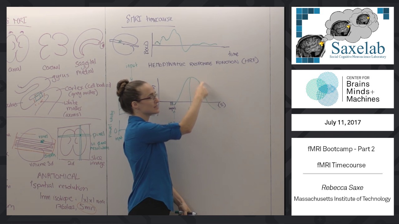



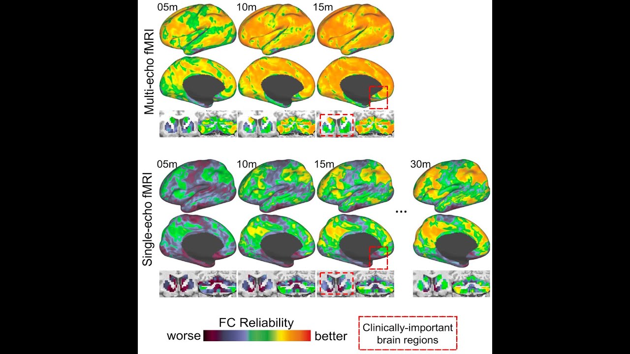


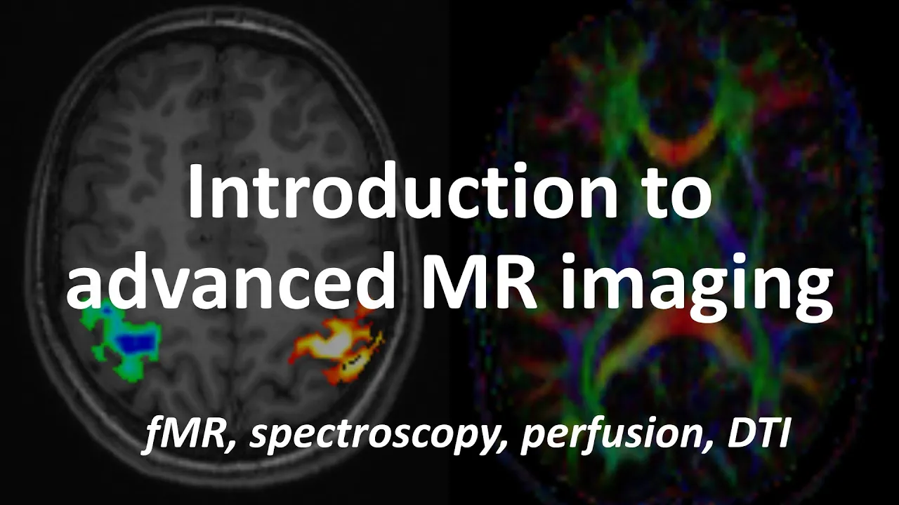





fMRI (functional Magnetic Resonance Imaging) measures brain activity by detecting changes in blood oxygenation levels using magnetic fields; when neurons become active during tasks, they consume more oxygen and glucose, causing nearby blood vessels to dilate and increase blood flow to those areas, creating a measurable Blood Oxygen Level Dependent (BOLD) signal that fMRI can capture approximately every two seconds to reveal which brain regions are more active during specific activities.
Functional MRI (fMRI) Explained: How Brain Imaging Works
Added:Basic neuroanatomy, including the major lobes of the brain and the concept of functional localization.
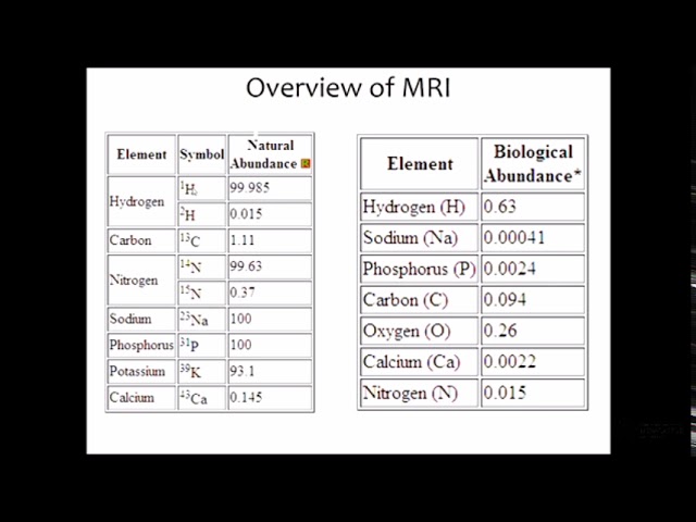
The fundamental physics of standard Magnetic Resonance Imaging (MRI), specifically how magnetic fields align hydrogen protons in the body.
Basic circulatory system physiology, particularly the transport of oxygen by hemoglobin and the difference between arterial and venous blood.
The concept of metabolic demand in cells, specifically how active neurons require increased glucose and oxygen.
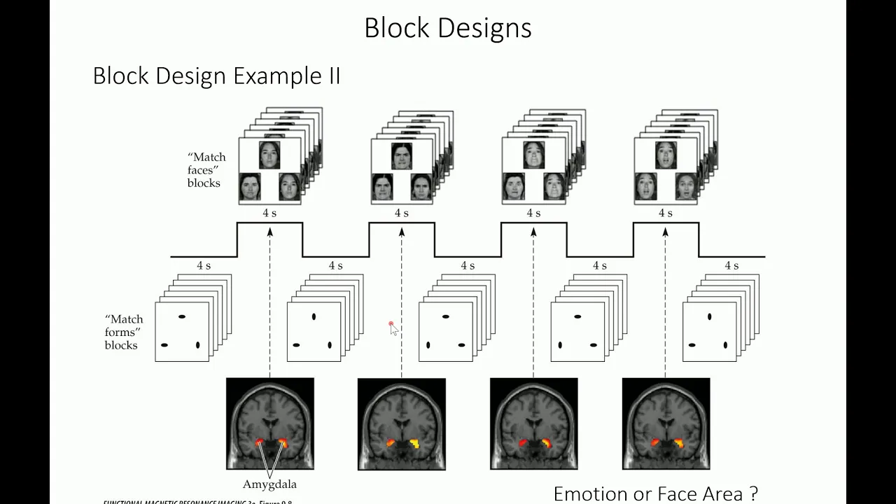
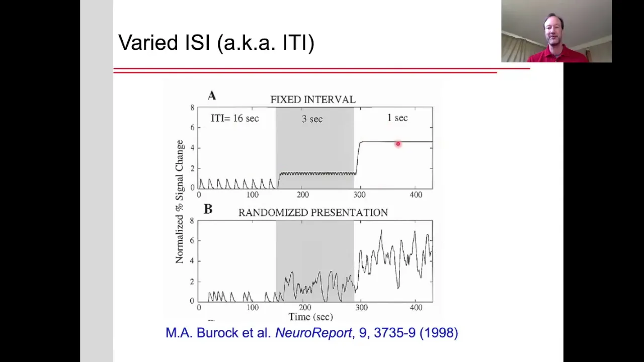
fMRI experimental methodologies, including the difference between block designs and event-related designs.
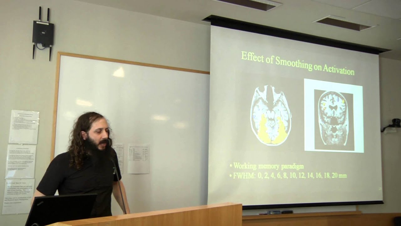
Data preprocessing and statistical analysis techniques for brain scans, such as motion correction, spatial smoothing, and general linear modeling (GLM).
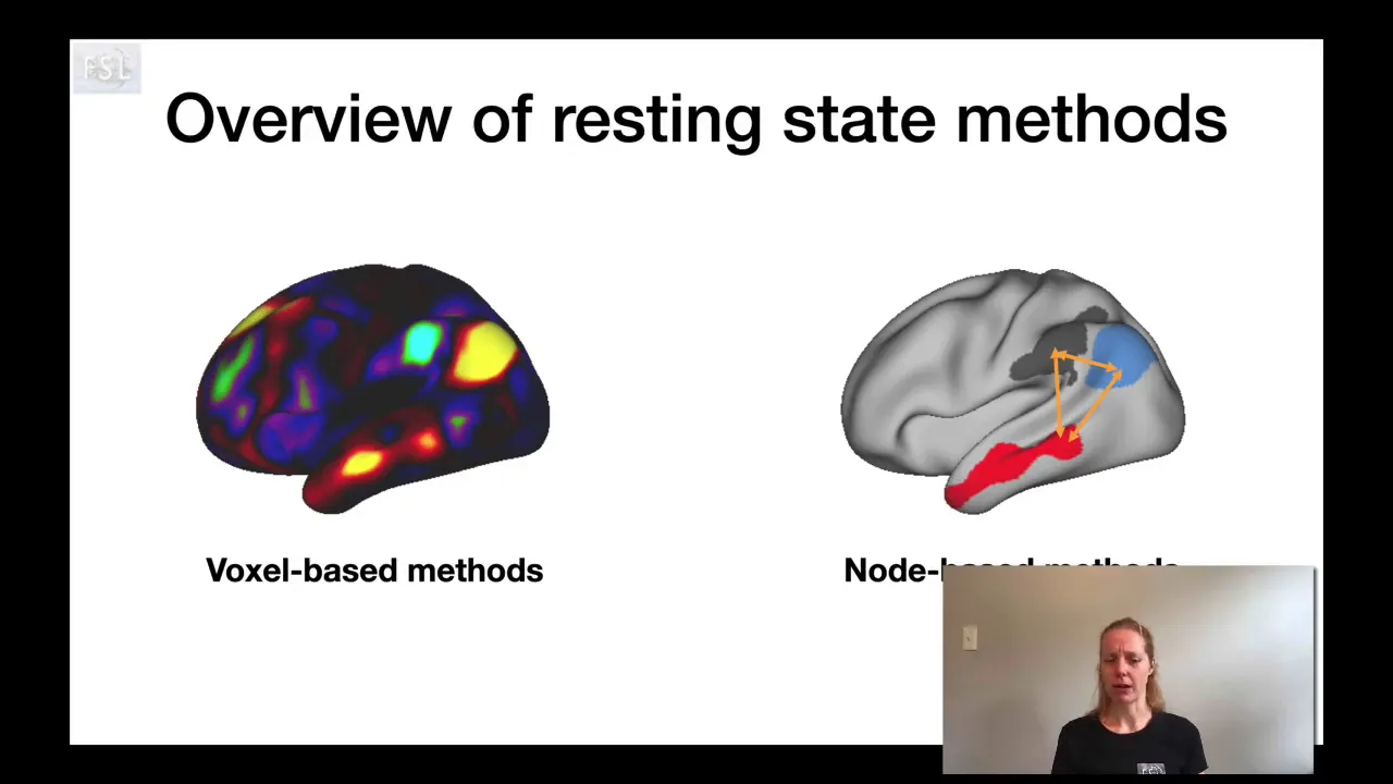
Resting-state fMRI (rs-fMRI) and the exploration of functional connectivity and default mode networks (DMN).
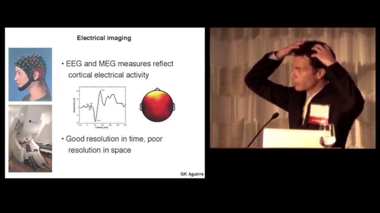
The limitations of fMRI (such as low temporal resolution) and how it is combined with high-temporal-resolution techniques like EEG or MEG.
Clinical and cognitive neuroscience applications of fMRI, such as pre-surgical mapping and studying psychiatric disorders.
3.3K views149likes4:59Original Release: 2022-04-25
this is gonna be my brain telling your brain how other brains study the brain brains are tricky things to study you can't just cut into someone's head and have a look around and even if you could that wouldn't help you to figure out how the brain actually works to control the body the brain functions by sending electrical signals but you can't see electrical signals so how the hell are you meant to investigate the brain with magnetic resonance imaging m r i an incredible machine that uses magnetic fields to image the brain you may have heard of an mri machine and you may have even had a brain scan but here's the thing all mri scans fall into one of two categories structural mri scans or functional mri scans structural mri scans usually just referred to as mri scans reveal the structure of the brain with quite beautiful images like this that reveal a detailed structure of what the brain looks like and whether there are any medical abnormalities such as a tumor then there's also functional mri that reveals crucial details about which parts of the brain are more active at certain times or during certain activities if structural mri is like a 3d camera that takes one photo of your brain functional mri is like a 3d video camera that takes multiple pictures of your brain across time to show how your brain is changing but how could this technology actually work it all starts with neurovascular coupling what the hell is neurovascular coupling well that's a great question the key cells in your brain are neurons they're responsible for transmitting electrical signals down these long pipes called axons sending these signals requires a lot of energy the more work these neurons need to do to process and send signals the more energy they require so how do neurons get more energy your body obtains energy by combining glucose a type of sugar molecule with oxygen to produce carbon dioxide water and access energy a process called respiration so to gain access to more energy neurons need more glucose and more oxygen which they get from the rich supply of blood to your brain so say you start listening to some music very best the neurons that process and interpret sound in your brain have to start working extra hard this causes the blood vessels that lie close to those neurons to increase in size delivering a larger blood supply to those neurons that is neurovascular coupling the activity of groups of neurons being coupled to the size of the blood vessels that supply that part of the brain because of neurovascular coupling increased blood flow to a certain area of the brain is a proxy for increased neuronal activity in that area altogether this means we have areas of the brain that are more active with more oxygen rich blood and areas of the brain that are less active with less oxygen rich blood go if only there was a difference in the magnetism of blood containing different quantities of oxygen oh wait there is which brings us perfectly to our fmri which can measure something called the bold signal the blood oxygen level dependent signal an mri machine is a huge magnet that can measure subtle changes in the magnetic field oxygen-rich blood called oxygenated blood is diamagnetic meaning it's slightly repelled by a magnetic field less oxygen-rich blood called deoxygenated blood is paramagnetic meaning it's slightly attracted to the magnetic field these properties allow a blood oxygen level dependent a bold signal to be measured the mri machine is therefore able to distinguish where in the brain there's more oxygen-rich blood because of neurovascular coupling we can conclude that that area of the brain is more active because an fmri scan is like a video camera that can take a whole image of the brain every two ish seconds we're able to see across time which areas of the brain get more and less oxygen as a person engages with different tasks and activities so how can this be visualized well the simple way is to get a glass brain image with these blobs in it the blobs being the areas of the brain that have increased or decreased blood flow and if you want to be fancy you can map these identified areas onto a 3d model with flashing lights and cool colors but this kind of aesthetic is mostly reserved for epic scientific conferences and advertising so if you want to know which part of the brain is more active for someone with a neurological condition or when they're happy or sad or excited or tapping their fingers stick them in an mri machine provoke that emotion or activity give them an fmri scan and see where their brain begins to light up anyway thank you so much for watching let me know if you have any questions and i'll see you in the next one
Up Next

21:01
Quantum Biology Explained: The European Robin's Quantum Compass
@upandatom
108.9K views•2020-03-13

12:53
21cm Hyperfine Transition in Neutral Hydrogen: Radio Astronomy Basics
@AaronRobertParsons
12.4K views•2011-10-13

32:32
NMR Spin Physics I: Zeeman Effect, Resonance Condition & Larmor Frequency
@nptel-indianinstituteofsci8064
2.3K views•2024-01-17

27:14
Entropy and the Second Law of Thermodynamics Explained
@veritasium
27.5M views•2023-07-01
Related Study Plans & Knowledge Roadmaps
Structured learning paths in Physics
physics
Nuclear Physics (Decay, Fission & Reactors)
01Atomic Structure and the Binding Force
02Radioactive Decay and Half-Life
03Nuclear Fission Mechanics and Chain Reactions
+ 2 more learning modules
12h5 modules
Explore Roadmap
physics
Soft Matter (Polymers, Colloids & Rheology)
01Foundations of Soft Matter & Thermal Forces
02Polymer Physics and Macromolecules
03Colloids, Surfactants, and Self-Assembly
+ 2 more learning modules
38h5 modules
Explore Roadmap
physics
Analytical Mechanics: Lagrangian & Hamiltonian
01Mathematical Foundations: Calculus of Variations
02From Newtonian Mechanics to Constraint Dynamics
03The Principle of Least Action & Lagrangian Mechanics
+ 3 more learning modules
28h6 modules
Explore Roadmap
physics
General Relativity (Spacetime & Cosmology)
01Foundations of Relativity: Special Relativity
02The Geometry of Spacetime: Equivalence Principle & Curvature
03Einstein's Field Equations: The Mathematics of Gravity
+ 2 more learning modules
55h5 modules
Explore Roadmap