This video demonstrates the cesium chloride purification method for isolating bacteriophages from bacterial cultures. The process involves creating a plate lysate by infecting bacterial plates with phage, recovering the phage from the top agar layer, infecting fresh bacterial cultures at an MOI of 1-5 until lysis occurs, precipitating phage particles using sodium chloride and polyethylene glycol, centrifuging to pellet the phage, resuspending in buffer, adding cesium chloride to create a density gradient, performing ultra-centrifugation to concentrate the phage into a visible band, and finally isolating the phage band for storage in glass vials.
Bacteriophage Purification via Cesium Chloride Gradient
Added:Deep Dive
Fundamental biology of bacteriophages, including their structure (capsid, tail, and genome) and their life cycles (lytic vs. lysogenic).

Bacteriophage; Discovery, Structure, Life Cycle (Lytic Cycle Lysogenic) Economic importance
108.1K
Bacteriophages are viruses that exclusively infect bacteria, demonstrating extreme host specificity where each phage targets only particular bacterial species or strains. Structurally, they consist of nucleic acid (double-stranded circular DNA ~150nm) surrounded by protein capsids, featuring characteristic morphological components including heads, collars, base plates, and six tail fibers visible under electron microscopy. Classified into nine families and thirty-two subfamilies based on physiological properties, bacteriophages follow a complete lytic life cycle: attachment to bacterial receptors, penetration by DNA injection, biosynthesis of viral components using hijacked cellular machinery, maturation and assembly of new phage particles, and finally lysis of the bacterial cell to release progeny phages. This cycle ensures rapid propagation and represents the fundamental mechanism by which bacteriophages propagate within bacterial populations.
Click to watch →

أحياء السادس العلمي | منهج 2026 | الفصل الثالث | 3| التكاثر في الفايروسات
74.5K
Bacteriophage is a specific type of virus that infects E. coli bacteria, with the term meaning 'bacteria eater' in Greek. Bacteriophage viruses have a distinctive structure consisting of three main parts: a head (containing genetic material), a neck (connecting the head to the tail), and a tail (with fibers for attachment). The lytic cycle of virus reproduction consists of five distinct stages: (1) Attachment - the virus attaches to the host cell surface using specialized tail fibers that bind to specific receptor sites on the bacterial cell wall, (2) Penetration - the virus injects its genetic material into the host cell by secreting enzymes that weaken the cell wall and create a hole, (3) Biosynthesis - the host cell's machinery is hijacked to produce viral components, (4) Maturation - new virus particles are assembled from the synthesized components, and (5) Release - new viruses are released from the host cell, often causing the cell to burst. A single bacteriophage virus can produce 100-200 new virus particles within approximately 25 minutes during the lytic cycle.
Click to watch →
acellular life | variety of life | biodiversity | mdcat lecture
12.3K
Bacteriophages (bacteria-eating viruses) have complex structures with heads, tails, and tail fibers. The tail contains contractile proteins that inject viral DNA into bacteria. Life cycles include: (1) Lytic cycle - virus replicates and destroys host cell, releasing new viruses; (2) Lysogenic cycle - viral DNA integrates into host genome as prophage, replicating with host DNA. Induction by UV radiation or chemicals can convert lysogenic to lytic cycle.
Click to watch →

MDCAT Biology | Acellular Life (Viruses) + HIV/AIDS | Complete UHS Lecture | Dr. Muhammad Shehzad
171
Bacteriophages (bacteria viruses) have a distinctive structure with a head containing DNA or RNA, a tail with a central core and surrounding sheath, and six tail fibers. The base plate contains enzymes like lysozyme that help the virus penetrate the host cell. Bacteriophages have two life cycles: the lytic cycle (viruses replicate and destroy the host cell) and the lysogenic cycle (viruses integrate into host DNA and replicate with the host). In the lytic cycle, the host cell bursts and releases new viruses. In the lysogenic cycle, the virus remains dormant until conditions trigger replication.
Click to watch →

Bacteriophage | Structure And Replication Of Bacteriophage In Lytic And Lysogenic Life Cycle
10.8K
Bacteriophages are viruses that infect bacteria, consisting of a protein head containing DNA and a tail with contractile sheath, base plates, and tail fibers. The tail fibers attach to specific bacterial receptors, enabling the virus to bind to the host cell. Upon attachment, the tail sheath contracts, allowing viral DNA injection into the bacterial cytoplasm. Bacteriophages have two life cycles: the lytic cycle (virulent) and the lysogenic cycle (temperate). In the lytic cycle, viral DNA hijacks host machinery, degrades host DNA, and produces approximately 200 new viruses within 20-25 minutes, followed by host cell lysis. In the lysogenic cycle, viral DNA integrates into the bacterial chromosome as a prophage, replicating passively with host DNA. This dormant state can be induced by adverse conditions (UV radiation, chemicals), causing the prophage to excise and enter the lytic cycle.
Click to watch →
Deep Dive
Principles of centrifugation, specifically the differences between differential centrifugation and density gradient centrifugation.

Biophysical Techniques | Centrifugation🧬| IIT JAM, GAT-B, CUET PG 2026 #unacademy
2.4K
Centrifugation methods classify into differential and density gradient approaches. Differential centrifugation separates particles based on size and density through sequential spinning at progressively increasing speeds: initial low-speed separates nuclei, medium-speed isolates mitochondria and lysosomes, high-speed separates microsomes, and very high-speed isolates ribosomes and viruses. Density gradient centrifugation provides superior resolution using media with varying densities. Rate zonal centrifugation layers samples atop pre-formed gradients, with particles migrating at different rates based on mass and size, forming distinct bands. Isopycnic centrifugation achieves exceptional separation by exploiting intrinsic density differences, with particles migrating until reaching their isopycnic point where density matches surroundings, forming sharp bands. This technique was pivotal in the Meselson-Stahl experiment demonstrating semi-conservative DNA replication.
Click to watch →

Analytical Techniques In Plant Sciences || Lecture- 5|| Botany Honours
398
Centrifugation techniques are divided into two types based on separation purpose. Differential centrifugation separates particles by size and density through sequential centrifugation at increasing speeds. Density gradient centrifugation separates particles based on buoyant density using a gradient medium like sucrose or cesium chloride. Particles move through the gradient until reaching equilibrium where their density equals the medium density.
Click to watch →
Centrifugation - Separation Technique | Class 11 Biotechnology Chapter 12 | 2022-23 | NCERT
18.7K
Two primary centrifugation techniques exist for separating particles: (1) Differential centrifugation separates particles with different sizes and different densities by exploiting their varying sedimentation rates at high speeds; (2) Density gradient centrifugation separates particles with similar sizes but different densities using a density gradient medium, allowing particles to migrate to positions matching their own density. These techniques enable precise separation of complex biological samples, such as separating sperm from seminal fluid or isolating specific cellular components for research and medical applications.
Click to watch →

Centrifugation
273.7K
Three primary centrifugation techniques serve different separation needs. Differential centrifugation gradually increases speed to sequentially pellet particles by mass, requiring prior knowledge of particle sizes. Velocity gradient centrifugation layers samples atop pre-formed sucrose gradients (5-30%), allowing particles to migrate until reaching equilibrium density where net velocity becomes zero. Density gradient centrifugation mixes samples uniformly with cesium chloride before centrifugation, requiring 36-48 hours for self-assembly. Cesium chloride is preferred for DNA separation because its density matches DNA, enabling precise isolation of different DNA species. Increasing rotor speed shifts band positions but does not shorten runtime in density gradient centrifugation.
Click to watch →

CENTRIFUGE-WORKING PRINCIPLE | COMPONENTS | TYPES
140
Differential centrifugation separates subcellular organelles by sequential spinning at increasing speeds: 600g for 10 minutes pellets nuclei, 15,000g for 5 minutes pellets mitochondria and other organelles, and 100,000g for 60 minutes pellets plasma membranes. Density gradient centrifugation creates a density gradient (e.g., sucrose solution) before loading samples. Components separate based on density matching the gradient position. This technique is used for DNA separation, where heavy DNA settles at higher density positions while lighter DNA floats at lower density positions.
Click to watch →
Deep Dive
The physical concept of buoyant density and how salts like Cesium Chloride (CsCl) form self-generating gradients under high centrifugal force (isopycnic separation).
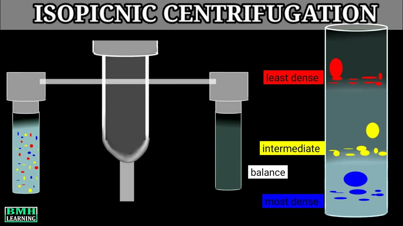
Isopicnic Centrifugation | Cscl density gradient centrifugation |
28.5K
Isopicnic centrifugation (also called buoyant density or equilibrium density gradient centrifugation) separates particles based solely on their buoyant density, which is their effective density in solution relative to the surrounding fluid medium; unlike other gradient centrifugation methods, the density gradient forms during the centrifugation process rather than being pre-established, allowing particles to migrate until they reach equilibrium positions where their buoyant density matches the local medium density, independent of centrifugation time.
Click to watch →

Molecular Basis of Inheritance | Part 5 | NCERT Line to Line | Thannambikkai Batch
110K
Cesium chloride density gradient centrifugation separates DNA by density. When centrifuged at high speed, cesium chloride forms a density gradient. Heavy DNA (N15-N15) sediments to the bottom, light DNA (N14-N14) remains at the top, and hybrid DNA (N15-N14) sediments at an intermediate position. This technique allows visualization of DNA density differences, providing evidence for the semi-conservative replication mechanism.
Click to watch →

Cesium Chloride Density Gradient Centrifugation
55.5K
Cesium chloride density gradient centrifugation is a technique that separates DNA molecules based on their density by creating a continuous density gradient in a centrifuge tube; during high-speed centrifugation, cesium ions form a concentration gradient from top to bottom (increasing density from ~1 g/mL to ~1.7 g/mL), causing DNA molecules to migrate to positions where the solution density matches their own density, allowing separation of DNA samples that differ only slightly in mass, such as those labeled with different nitrogen isotopes.
Click to watch →

Isopycnic centrifugation
77.1K
Isopycnic centrifugation is a molecular biology technique that separates biomolecules (such as nucleic acids like DNA) based on their buoyant density rather than molecular mass; the process involves creating a uniform mixture of sample and gradient-forming substrate (typically cesium chloride), which then forms a density gradient during centrifugation, causing molecules to migrate and settle at specific positions corresponding to their density, allowing for precise isolation and collection of different molecular species.
Click to watch →

Plasmid DNA Isolation Methods:Alkaline lysis, EtBr-CsCl density gradient, Isolation on basis of Size
1.6K
CsCl density gradient centrifugation separates DNA based on buoyant density. A cesium chloride gradient (1.55-1.75 g/cm³) is created in a centrifuge tube. After cell lysis, the lysate is layered on top and centrifuged. Components separate by density: proteins float at the top, RNA pellets at intermediate positions, and DNA bands at 1.70 g/cm³. For conformational separation, cells are pre-treated with ethidium bromide (EtBr), which intercalates into DNA. EtBr binds preferentially to linear and open circular DNA (reducing density by ~0.125 g/cm³) compared to supercoiled DNA (~0.085 g/cm³ reduction). This creates separate bands for plasmid and genomic DNA. Butanol removes EtBr, and dialysis eliminates CsCl salt, yielding highly pure plasmids.
Click to watch →
Deep Dive
Basic aseptic laboratory techniques and standard safety protocols for handling biological agents and hazardous chemicals like CsCl.

Infection Practical Slides
1.6K
This section covers laboratory safety and sterilization protocols. Aseptic technique for invasive procedures (IV catheterization, wound care) includes hand antisepsis (alcohol, chlorhexidine), skin antisepsis, and sterile field maintenance. Surgical aseptic technique is used for major procedures and central line insertion. Laboratory waste disposal: sharps in safety boxes, biohazardous waste in red bags (incinerated), regular waste in regular trash. PPE includes latex gloves (routine), surgical gloves (sterile procedures), and N95 masks (airborne pathogens). Sterilization methods include autoclaving (121°C, 15-20 min) for heat-stable items, chemical sterilization (ethylene oxide) for heat-sensitive items, filtration for liquids, and red heat for bacteriological loops. Biohazardous waste (red bags, safety boxes) must be incinerated. Sterilization prevents ex vivo contamination, while antimicrobial susceptibility testing guides in vivo treatment.
Click to watch →

Procedimentos de manipulação de Agentes Biológicos
164
Safe manipulation of biological agents requires following systematic protocols including: registering laboratory access, wearing appropriate PPE such as disposable lab coats and gloves; using biosafety cabinets as primary containment devices with proper pre-use preparation (cleaning with 70% alcohol, UV sterilization for 15 minutes), maintaining continuous laminar airflow during operations, and ensuring complete cabinet emptiness after each experiment; disposing of waste through autoclaving at 121°C for 30 minutes for inactivation, followed by triage for reuse and proper disposal according to ANVISA RDC 222/2018 regulations.
Click to watch →

Chapter 12 Aseptic technique and sterile compounding
468
Aseptic technique is directly associated with universal precautions. Products classified as hazardous include chemotherapeutic agents, radioactive compounds, and various hazardous chemicals such as phenol and glacial acetic acid. The most important consideration is that touching your face or glasses with sterile gloves introduces microbes—the most common way to contaminate a CSP. Medications containing microbes or unwanted debris can cause dangerous infections or death when administered to patients.
Click to watch →

BT733_Topic083
338
Biosafety Level 1 (BSL-1) is the lowest biosafety category for handling biological agents that are not consistently known to cause disease in immunocompromised adults, with minimal potential hazard to the environment, lab workers, and community. Standard microbiological practices include: hand washing before and after procedures, wearing gloves and protective clothing (lab coats, aprons), avoiding eating/drinking/smoking in the lab, using bulbs instead of mouth pipettes, disposing of sharps in proper containers, decontaminating work surfaces and equipment with 70% alcohol, and properly disposing of cultures through autoclaving. No special equipment like biosafety cabinets is required, but all workers must be fully trained in handling biological agents and institutional policies must be enforced.
Click to watch →

K3 Keselamatan Dan Kesehatan Kerja Dilaboratorium Mikrobiologi Farmasi (Safety Induction)
1.2K
Laboratory safety standards include: hand washing with soap and running water before/after procedures, prohibiting eating/drinking in labs, careful sample handling to prevent splashing, surface disinfection with 1-3 minute contact time, proper waste classification (medical vs. non-medical), clear sample labeling, equipment sterilization before use, treating all microorganisms as potentially pathogenic, and wearing gloves when handling pathogenic materials. All laboratory personnel must know safety equipment locations including first aid kits, emergency showers, and fire extinguishers. Any accidents must be reported to supervisors. Desinfection eliminates most pathogenic microorganisms from surfaces. Three levels exist: high-level (kills almost all except some spores), intermediate-level (kills vegetative bacteria, most viruses, fungi), and low-level (kills most vegetative bacteria but not resistant organisms). Methods include chemical (alcohol, chlorine, hydrogen peroxide) and physical (heat, radiation, filtration).
Click to watch →
Prerequisite Knowledge
- Concept 01Fundamental biology of bacteriophages, including their structure (capsid, tail, and genome) and their life cycles (lytic vs. lysogenic).
- Concept 02Principles of centrifugation, specifically the differences between differential centrifugation and density gradient centrifugation.
- Concept 03The physical concept of buoyant density and how salts like Cesium Chloride (CsCl) form self-generating gradients under high centrifugal force (isopycnic separation).
- Concept 04Basic aseptic laboratory techniques and standard safety protocols for handling biological agents and hazardous chemicals like CsCl.
Subsequent Learning
- Step 01Methods for downstream desalting and dialysis to safely remove toxic Cesium Chloride from the purified phage suspension.
- Step 02Techniques for characterizing the purified phages, such as Transmission Electron Microscopy (TEM) for structural analysis and plaque assays for determining titer.
- Step 03Phage genomic and proteomic analysis, including viral DNA/RNA extraction, sequencing, and annotation.
- Step 04Therapeutic applications of purified phages, including the formulation of phage cocktails and methods for endotoxin removal to meet regulatory standards.
- Step 05Alternative scalable purification methodologies, such as chromatography and Tangential Flow Filtration (TFF), for industrial and clinical-grade production.
Plate Lysis
Culture Infection
Band Purify
Plate Lysis
0:09Playing Section
- 1
Prepare plate lysate to amplify phage from bacterial stocks.
- 2
Mix phage with bacteria in top agar for confluent lysis.
Chromatography and Filtration as Alternatives to Cesium Chloride Ultracentrifugation
While cesium chloride (CsCl) gradient ultracentrifugation is a traditional gold standard for purifying bacteriophages in laboratory research, it faces significant criticism regarding scalability, toxicity, and phage viability, particularly for therapeutic applications. CsCl is highly toxic to humans and must be meticulously removed to prevent adverse reactions in patients undergoing phage therapy. Furthermore, the extreme osmotic pressure and density of the gradient can denature or inactivate sensitive phage strains. Consequently, modern bioprocessing increasingly favors scalable, non-toxic alternatives such as chromatography (monolithic, ion-exchange, or size-exclusion) and Tangential Flow Filtration (TFF). These methods are more easily adapted to Good Manufacturing Practices (GMP) and provide a gentler, more efficient route for producing clinical-grade phage preparations without the hazards associated with cesium salts.
Deep Dive
Methods for downstream desalting and dialysis to safely remove toxic Cesium Chloride from the purified phage suspension.
Plasmid DNA Isolation Methods:Alkaline lysis, EtBr-CsCl density gradient, Isolation on basis of Size
1.6K
After gradient centrifugation, the supercoiled plasmid band is collected and treated with butanol to remove bound EtBr, which partitions into the organic phase while DNA remains in the aqueous phase. The DNA-CsCl solution is then dialyzed against buffer to remove CsCl salt, leaving highly pure plasmid DNA. This EtBr-CsCl method is preferred when extremely pure plasmid preparations are required for downstream applications like cloning or sequencing.
Click to watch →
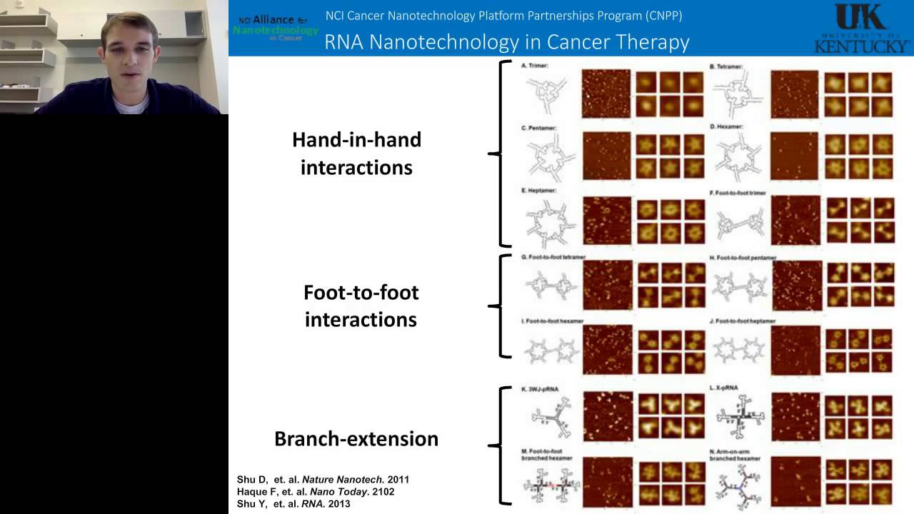
Daniel Jasinsk - Large scale purification of RNA and RNA based Nanoparticles by Preparative Ultracen
423
For large-scale RNA purification after in vitro transcription (1-2 mL scale), cesium chloride equilibrium density gradient ultracentrifugation is employed. The protocol involves mixing the sample with cesium chloride medium density solution, then loading high and low density cesium chloride solutions. After ultracentrifugation, the gradient forms and particles separate based on density. RNA, being the densest molecule in the solution (compared to proteins, salts, and unincorporated NTPs), migrates to the bottom of the gradient. Fractions are collected from top to bottom (200-300 microliters each), and RNA is identified by gel analysis. The cesium chloride must be removed afterward through dialysis into Tris-EDTA buffer or quick-drop dialysis for immediate use.
Click to watch →
The Most Dangerous Object I Have Ever Held In My Hands
1.6M
This segment covers the complete process of producing pure cesium metal. Cesium is isolated from caesium chloride through chemical processing, with the creator achieving yields of 85% and 80% across two batches, totaling approximately 146 grams. The initial purification involves transferring cesium into a still and sealing it by melting off the transfer tube. A sophisticated vacuum system is established using stainless steel tubing, KF2 flanges, and dual high-vacuum valves—one connected to a vacuum pump and another to an argon cylinder. The system undergoes multiple argon flushing cycles (at least four times) to remove moisture and create an inert atmosphere. Before distillation begins, the glassware is heated to 250°C for one hour to desorb any residual moisture from the glass surface, preparing it for handling reactive cesium.
Click to watch →

Phage isolation and concentration by cesium chloride (CsCl) gradient
5.1K
This video demonstrates a protocol for isolating and concentrating bacteriophages using cesium chloride (CsCl) density gradient ultracentrifugation, which separates phages based on their uniform density within a CsCl gradient, allowing researchers to concentrate large quantities of a single phage species into a highly concentrated, stable sample suitable for long-term storage after dialysis and proper handling procedures.
Click to watch →

Analytical aspects of Toxicology Part 4
358
Dialysis is used for separation of ions from biological matrices. About 10g of tissue is cut into small pieces and placed in a cellophane membrane bag shaped appropriately. The bag is slowly rotated in a beaker containing 100mL of distilled water using an electrical motor or mechanical device. Dialysis occurs rapidly, and after about an hour, the water in the beaker is replaced with fresh water and the bag is rotated for another half hour. The water is then taken out, mixed with previous fractions, and evaporated on a water bath to a small volume. The filtered water content contains the anions of interest and may be tested for toxic anions if necessary.
Click to watch →
Deep Dive
Techniques for characterizing the purified phages, such as Transmission Electron Microscopy (TEM) for structural analysis and plaque assays for determining titer.

VIP Webinar Series (13th)- Phage Isolation, Culture, and Characterization
1.2K
Comprehensive phage characterization includes: Efficiency of Plating (EOP)—serial dilution to find highest dilution producing confluent lysis; Routine Test Dilution—the highest dilution failing to produce confluent lysis (standard for future testing); Single Step Growth Curve—analyzing sigmoid curves to determine latent period (time to maximum growth rate) and burst size (phages released per cell); Host Range Determination—testing coverage across bacterial species within genus or family; PFGE for genome sizing; Electron microscopy for morphological identification. Different phages show varying lytic activities: clear confluent lysis, opalescent lysis, no reaction, individual plaques, or resistant mutants. Pulse Field Gel Electrophoresis (PFGE) extends DNA sizing beyond standard electrophoresis (0.2-50 kb to 200 bp-12 Mb). Procedure: mix phage suspension with low melting agarose, create plugs, release DNA by overnight shaking at 55°C, wash plugs, perform restriction digestion if needed, load plugs in agarose gel, run at appropriate voltage, stain with ethidium bromide. Transmission Electron Microscopy uses negative staining with uranyl acetate on carbon-coated grids. ~96% of examined phages are tailed phages with characteristic components: head, tail tube, sheath, base plate, spikes, and tail fibers. Different families have distinct morphologies: T4 phages have contractile tails, lambda phages have different tail structures, filamentous phages have long flexible tails. Modern sequencing technologies include Sanger (first generation, ~1000 bp reads), Illumina (next generation, ~300-400 bp fragments requiring assembly), and Nanopore (single-molecule real-time sequencing). Workflow: template preparation, fragmentation, barcoding, PCR amplification, sequencing, and bioinformatics assembly. Coverage of at least 9x ensures reliable reconstruction. Bioinformatics identifies open reading frames (ORFs) coding for structural proteins (capsid/head/tail assembly), enzymes (DNA synthesis), and other functions. Comparative genomics enables species identification and evolutionary analysis.
Click to watch →
Bacteriophage Plaque Assay for Phage Titer UPDATED
49.7K
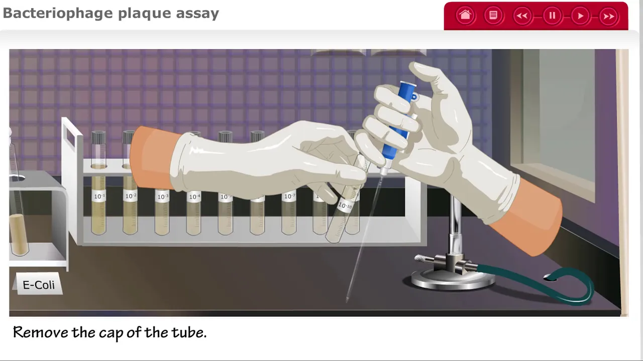
The bacteriophage plaque assay is a laboratory technique used to determine the concentration (titer) of bacteriophages in a sample by serially diluting the phage stock, mixing each dilution with a bacterial culture (E. coli) in soft agar, pouring onto petri plates, and incubating; the number of clear plaques formed indicates the original phage concentration, with more dilute samples yielding fewer plaques.
Click to watch →

Bacteriophage Purification Technique | How to purify phages | Step-by-Step Guide
658
Bacteriophage purification involves isolating individual plaques from infected bacterial cultures through repeated rounds of plaque picking, filtration, and serial dilution to obtain a pure phage lysate suitable for titer determination; the process includes dissolving isolated plaques in LB broth, filtering the suspension through a 22-micron syringe filter, preparing serial dilutions (5-7 dilutions), mixing with host bacterial suspension, and pouring onto LB agar plates for overnight incubation at 37°C to visualize clear plaques, with the procedure repeated 4-5 times to achieve high-purity phage preparation.
Click to watch →

Module 35 Determination of Phage Titre T4 by Plating Method Using E coli
145
The plaque assay is a fundamental microbiological technique used to determine the concentration of bacteriophages (such as T4) in a sample by counting clear zones (plaques) formed when phages infect and lyse bacterial cells (E. coli K12 or strain B) on agar plates; each plaque represents one or more phage particles that infected a single host cell, and the titre is calculated by dividing the number of plaques by the dilution factor and volume of sample used, expressed as plaque forming units per milliliter (PFU/mL).
Click to watch →

My Hero & Me - From waste water to phage therapy: exploiting phage-antibiotic synergies
232
Comprehensive phage characterization includes transmission electron microscopy, whole-genome sequencing, one-step growth curves determining burst size and latent period, and long-term stability testing. Receptor identification involves isolating phage-resistant mutants (typically with loss-of-function mutations in receptor genes) and sequencing to identify candidate receptor molecules. For ECC phages, researchers identified receptors including glycosyltransferase genes associated with LPS structure, UTP transferases modifying peptidoglycan and outer membrane antigens, and outer membrane proteins involved in iron uptake and virulence. Efficiency of plating (EOP) measurements reveal how well phages infect different host strains, guiding cocktail composition decisions.
Click to watch →
Deep Dive
Phage genomic and proteomic analysis, including viral DNA/RNA extraction, sequencing, and annotation.
Webinar AMR 1: Mengatasi Resistensi Bakteri: Tantangan dan Solusi melalui Bakteriofaga di Indonesia
1K
Phage genomic analysis involves extracting phage DNA and sequencing the genome. The process includes: (1) DNA extraction using methods that remove bacterial DNA contamination; (2) Sequencing using platforms like Illumina; (3) Genome assembly to reconstruct the complete phage genome. The JKT1 phage genome is 43,000 base pairs with 53.7% G+C content, containing 57 ORFs (open reading frames), of which 27 are known proteins and 30 are hypothetical proteins.
Click to watch →

Foro de Investigación Subgraduado | Departamento de Ciencias y Tecnología - Inter Investiga
111
Bacteriophage isolation involves collecting environmental samples (such as soil from Isabela, Puerto Rico), mixing with liquid solution, incubating in a shaker, and centrifuging. The supernatant is filtered through a 0.22-micrometer filter to isolate bacteriophages from other particles. Two methods exist: direct isolation (immediate plaque assay) and enriched isolation (incubation with host bacteria to increase concentration). Multiple rounds of purification yield clonal populations (genetically identical phages) confirmed by consistent plaque morphology. The purified phage is then named (e.g., 'Cyro') and prepared for genomic analysis. Genomic annotation involves identifying and assigning functions to genes within a phage genome using bioinformatics tools. The process transforms raw DNA sequences into meaningful biological information by identifying open reading frames (ORFs), start codons, and stop codons. Tools like Glimmer generate maps of potential coding regions, while HMMER identifies protein similarities to predict function.
Click to watch →

Phage Research Workflow Explained | Norgen’s Phage DNA Isolation Kit
251
Phage research follows a comprehensive workflow from discovery to application: phages are isolated from diverse sources like soil, water, and sewage; characterized by host range, morphology, and stability; analyzed genomically using high-quality DNA extraction and sequencing technologies (Illumina, PacBio, Oxford Nanopore); studied for their interaction mechanisms (lytic vs. lysogenic cycles) and applications in combating antimicrobial resistance through phage cocktails, engineered phages, and combination therapies with antibiotics; applied across human infection therapy, agriculture, animal farming, wastewater management, and phage display technology for protein development.
Click to watch →

Rob Edwards - Advances and Challenges in Understanding the Virosphere | S03 MVIF22
207
Cross-assembly combines metagenomic samples using sequence assembly, then plots coverage depth across samples to identify viral genomes. Genome assembly generates assembly graphs containing valuable information often ignored in standard analysis. FABLES analyzes these graphs to identify individual phage genomes by calculating read coverage, linearizing fragments with different coverage patterns, and deconvoluting complex cases into separate genomes. This reveals complete circular genomes, repetitive sequences used by lysogenic phages for integration, and complex cases with multiple components. Despite good annotation tools, phage genomes still contain many genes with unknown functions. Researchers have developed approaches by linearizing genomes and orienting them so integrase is always at one end, revealing strong conservation in gene order. Long Short-Term Memory (LSTM) models can predict functions based on gene order rather than sequence similarity. AlphaFold predictions can validate these annotations by generating protein structures and comparing them against known structures in databases.
Click to watch →
Phage Genome Annotation with Deborah Jacobs-Sera | IBRC & Phage Directory
2.5K
Phage genome annotation requires manual curation of computational predictions (using tools like Glimmer, GeneMark, tRNA Scan SE, and HHpred) because phage genomes are highly diverse with many unique genes, and researchers must answer three fundamental questions for each gene: whether it is a gene, its start position, and its function, using comparative genomics and multiple lines of evidence to assign functions to the approximately 30% of phage genes that have known functions.
Click to watch →
Deep Dive
Therapeutic applications of purified phages, including the formulation of phage cocktails and methods for endotoxin removal to meet regulatory standards.

Removing endotoxin from phage with octanol - Sep 2022 - Jessica Sacher
182
This video demonstrates a laboratory technique for removing endotoxins from concentrated phage lysate by adding octanol, shaking the mixture for one hour, chilling, centrifuging, and collecting the aqueous phase containing the phage; the method can be repeated if initial endotoxin removal is insufficient.
Click to watch →
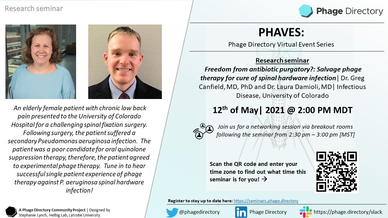
Salvage phage therapy for cure of spinal hardware infection—Dr. Laura Damioli & Dr. Greg Canfield
303
Phage manufacturing follows standardized protocols: bacteria are grown to OD600 0.1-0.6, infected at MOI 0.01, allowed to amplify for 6 hours, then filtered through 0.22 μm filters. Lysates are concentrated using 100 kDa cutoff centrifuge concentrators. Titer is determined via double agar layer method. Endotoxin concentration is measured using Limulus amebocyte lysate (LAL) assay with acceptable limits of ≤5 EU/mL per gig. Sterility testing follows USP 71 guidelines, and exotoxin A levels are tested via Western blot (sensitive to <1 pg/mL). Phages are diluted in PBS with magnesium sulfate to 1×10^10 PFU per dose.
Click to watch →
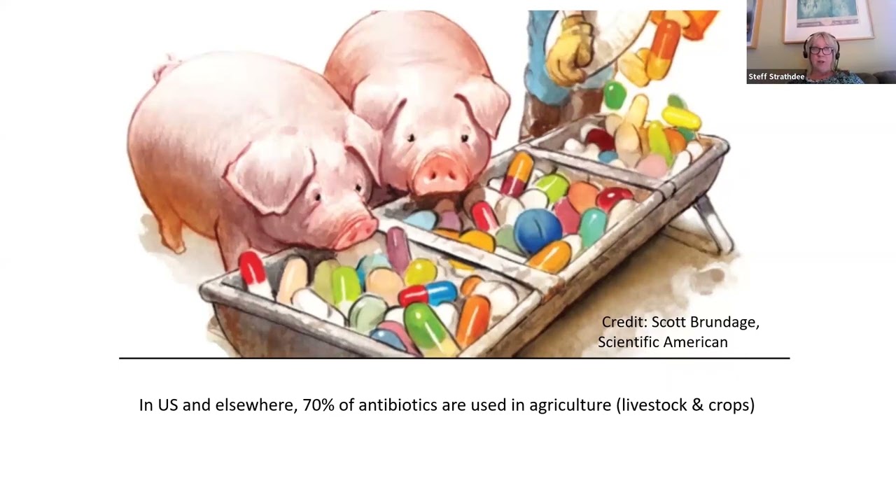
From Bog to Bedside: The Story Behind the First Dedicated Phage Therapy Center in North America
596
Finding phages that match a patient's specific bacterial isolate is challenging because phages must match the exact strain, not just species. Researchers searched sewage, barnyard waste, and ship bilges worldwide. Multiple phages were needed in cocktails to prevent resistance. Endotoxin purification was critical—requiring specialized weekend work to meet FDA safety thresholds before treatment could proceed.
Click to watch →

Bacteriophage | Wikipedia audio article
161
Phage therapy has continued since the Cold War in Georgia and Central/Eastern Europe. The first regulated randomized double-blind clinical trial was reported in June 2009 in the Journal of Wound Care, evaluating a bacteriophage cocktail for infected venous ulcers. The FDA approved it as a Phase 1 trial, demonstrating safety but not efficacy, with authors noting that standard wound care chemicals (lactoferrin, silver) may have interfered with phage viability. Another trial in August 2009 in Clinical Otolaryngology showed bacteriophage preparations were safe and effective for chronic ear infections caused by Pseudomonas aeruginosa. In 2015, a psychology professor's wife (Stephanie Stradling) used FDA-approved emergency phage treatment for her husband Tom Patterson, who had a resistant Acinetobacter baumannii infection while traveling in Egypt. After three days of treatment at UC San Diego Health, he woke from his coma. Since 2006, the FDA and USDA have approved several phage products: LMP 102 IntraLytx for ready-to-eat poultry and meat, and Listex (developed by Mike Rios) for killing Listeria monocytogenes on cheese, granted GRAS status and approved for all food products in July 2007. In 2011, the FDA cleared the first phage-based diagnostic product: KeyPath MRSA, a blood culture test using phage cocktails to detect Staphylococcus aureus and determine methicillin resistance in 5 hours versus 2-3 days for standard methods. Government agencies in the West have explored phages for counteracting bioweapons and toxins like anthrax and botulism. Other applications include spray application in horticulture for protecting plants from bacterial disease, bio-sites for environmental surfaces in hospitals, and preventative treatments for catheters and medical devices.
Click to watch →

Actualicémonos en Bacteriófagos: una estrategia exitosa para el control de bacterias
545
Strategies to overcome bacterial resistance include phage cocktails, combinations with antibiotics, genetic engineering, and combining with disinfectants. Phage therapy began in 1917 but declined with antibiotic introduction. Current studies use various administration routes: topical, intravenous, transnasal, intrarectal, transurethral, and oral. A 2022 study compiled 63 successful cases treating infections by Pseudomonas, Klebsiella, Enterococcus. Most cases used phage cocktails at high concentrations (10^6-10^9 phages/mL). Outcomes included clinical cure, reinfection, and rare adverse events. Phage therapy is particularly effective for chronic periprosthetic infections with biofilms. For drug-resistant infections, phages treat ESBL-producing Enterobacteriaceae, carbapenem-resistant Klebsiella pneumoniae, and Pseudomonas aeruginosa. Regulatory bodies recommend phage use for life-threatening infections with no alternatives. Quality control requires verifying phage activity, ensuring absence of resistance/virulence/integration genes, sterility, and endotoxin removal.
Click to watch →
Deep Dive
Alternative scalable purification methodologies, such as chromatography and Tangential Flow Filtration (TFF), for industrial and clinical-grade production.

Bioprocessing Part 3: Purification
362.1K
Industrial bioprocessing purification employs two primary techniques—chromatography and filtration—to isolate target proteins from complex biological mixtures. Chromatography utilizes column-based separation where resin beads interact with molecules based on physical properties: size-exclusion separates by molecular dimensions, ion-exchange separates by charge, and hydrophobic-interaction chromatography separates by hydrophobicity using high-salt conditions to expose protein patches. Filtration, specifically Tangential Flow Filtration (TFF), separates molecules by size using ultrafiltration membranes, enabling concentration (removing water) and diafiltration (buffer exchange). The purification workflow typically involves pre-filtration, multiple chromatography steps, TFF processing, and final filtration, all managed through batch records with operator verification to ensure product purity and consistency.
Click to watch →

TECNIC's Tangential Flow Filtration (TFF) solutions for biomolecule purification and separation
906
Tangential Flow Filtration (TFF) is an advanced membrane-based separation technique that differs from traditional dead-end filtration by having fluid pass across the membrane surface rather than through it, which increases filtration efficiency and extends membrane lifespan; TFF systems are highly scalable, ranging from 0.1 to 65 square meters of filtration surface area, and can be configured for either single-use or multi-use applications with automated process control, making them versatile for both defiltration and concentration processes in biomolecule purification.
Click to watch →
Tangential Flow Filtration: Principles, Membranes, and Techniques
88K
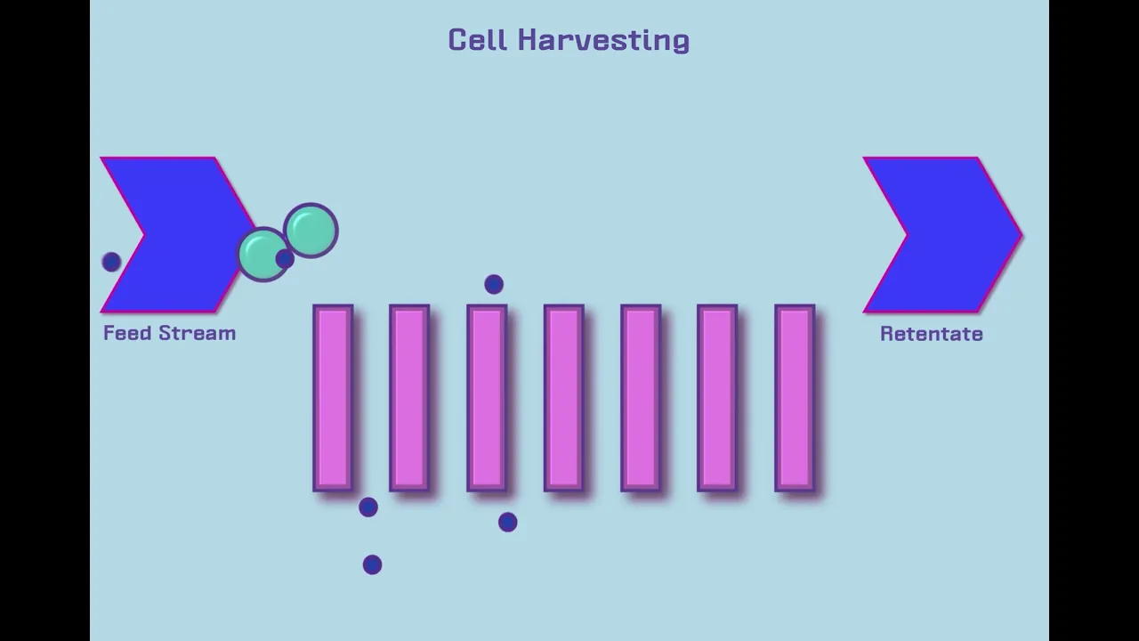
Tangential flow filtration (TFF) is a membrane separation technique where fluid flows parallel to the membrane surface rather than through it, allowing biomolecules to be separated based on size; the system includes a feed reservoir, pump, filter, retentate and permeate lines, and pressure gauges, with two main membrane types—hollow fiber and cassette—offering pore sizes ranging from microfiltration (0.1 micron+) for separating cells and debris to ultrafiltration (<0.1 micron) for concentrating and purifying proteins in the 3-1000 kDa range, commonly used in cell harvesting, lysate clarification, protein concentration, and diafiltration processes.
Click to watch →
Development of an Efficient Manufacturing Process for Adenovirus
1.3K
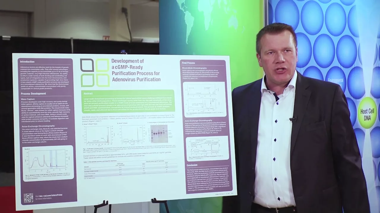
A two-column purification process using mixed-mode chromatography (Nuvia cPrime) followed by anion exchange chromatography (Nuvia Q) effectively purifies recombinant adenoviruses for clinical applications, achieving high yield, low host-cell protein and DNA contamination, and scalable manufacturing suitable for producing clinical-grade viral vectors.
Click to watch →
Scalable Strategies for pDNA, mRNA, and LNP Purification Supported by Rapid At-Line Analytics
150
Monolith chromatography provides scalable purification solutions for lipid nanoparticles across all manufacturing scales. The O column can capture particles directly after encapsulation with inline dilution, removing free nucleic acids and enriching encapsulation efficiency. Elution in formulation buffer concentrates particles with buffer exchange and purification from free nucleic acids. This shearless process imparts minimal stress on particles, preserving particle integrity. Dynamic binding capacity of 3.7 mg encapsulated RNA per milliliter of column volume enables scaling from analytical (0.1 ml) to production (40 liter) scales while maintaining consistent process parameters.
Click to watch →
Plate Lysis
Culture Infection
Band Purify
Plate Lysis
0:09- 1
Prepare plate lysate to amplify phage from bacterial stocks.
- 2
Mix phage with bacteria in top agar for confluent lysis.
Chromatography and Filtration as Alternatives to Cesium Chloride Ultracentrifugation
While cesium chloride (CsCl) gradient ultracentrifugation is a traditional gold standard for purifying bacteriophages in laboratory research, it faces significant criticism regarding scalability, toxicity, and phage viability, particularly for therapeutic applications. CsCl is highly toxic to humans and must be meticulously removed to prevent adverse reactions in patients undergoing phage therapy. Furthermore, the extreme osmotic pressure and density of the gradient can denature or inactivate sensitive phage strains. Consequently, modern bioprocessing increasingly favors scalable, non-toxic alternatives such as chromatography (monolithic, ion-exchange, or size-exclusion) and Tangential Flow Filtration (TFF). These methods are more easily adapted to Good Manufacturing Practices (GMP) and provide a gentler, more efficient route for producing clinical-grade phage preparations without the hazards associated with cesium salts.
Fundamental biology of bacteriophages, including their structure (capsid, tail, and genome) and their life cycles (lytic vs. lysogenic).
Principles of centrifugation, specifically the differences between differential centrifugation and density gradient centrifugation.
The physical concept of buoyant density and how salts like Cesium Chloride (CsCl) form self-generating gradients under high centrifugal force (isopycnic separation).
Basic aseptic laboratory techniques and standard safety protocols for handling biological agents and hazardous chemicals like CsCl.
Methods for downstream desalting and dialysis to safely remove toxic Cesium Chloride from the purified phage suspension.
Techniques for characterizing the purified phages, such as Transmission Electron Microscopy (TEM) for structural analysis and plaque assays for determining titer.
Phage genomic and proteomic analysis, including viral DNA/RNA extraction, sequencing, and annotation.
Therapeutic applications of purified phages, including the formulation of phage cocktails and methods for endotoxin removal to meet regulatory standards.
Alternative scalable purification methodologies, such as chromatography and Tangential Flow Filtration (TFF), for industrial and clinical-grade production.
14.2K views323likes5:49@BCMTAILORLabsOriginal Release: 2021-02-23
phage purification can be a very difficult process the idea is to grow a lot of it isolate it from the bacterial products and concentrate it so that you can have it in whatever buffer that you choose one of the very first phages that i purified was hp3 this was a phage that was originally isolated from a goose duck mixture at hermann park it's been used multiple times to treat different e coli infections in patients our technician geraldo sanchez has purified 70 plus phages which will be used to treat multiple infections in different people today i'm going to take you through how to purify phage starting from the beginning starting with a staph aureus phage which will be used to treat a staph aureus infection in a patient so my first step is to make a plate lysate i add some already made phage lysate that i had or purified phage to my top auger in a previous video i showed you how to collect a plaque and make a plate lysate and to that i had 100 microliters of the bacteria that i want to purify on i mix it with the top auger and i add it to my bottom auger the next day your plate should be completely clear like you didn't even add bacteria emmeline heckman is an amazing technician who will be showing us how to recover the phage from the plate emeline is using a sterile cell scraper to scrape off the top agar layer from the plate this top agar layer should have all the phage that you need to make a plate lysate i usually use about three plates in order to make a lysate to infect 50 to 100 ml culture next we grow our bacteria to an od 600 of 0.1 to that we're going to add our phage at an moi of one to five and grow for four to five hours or until clear now it's time to take a break i suggest some stretching maybe some exercise and some yoga breathing it's going to be a long process so now your culture should look clear probably more clear than when you first started them you could add chloroform and clear them up even more then we do it again we grow our cultures to an od 600 of 0.1 and we infected at moi of one to five now your culture is clear you could add chloroform and clear it up even more in all i made four liters of lysate to that cooled lysate i'm going to add 30 grams per liter of sodium chloride so 120 grams to dissolve next i'm going to add 7.5 percent polyethylene glycol or 300 grams to dissolve stir overnight in the cold room the next day you're going to pellet your precipitated phage which means you have to centrifuge all four leaders your now pelleted fade will look like streaks on the side of the bottles resuspend and phage buffer then add chloroform mix and centrifuge after centrifugation collect the top portion above the pellet next i add cesium chloride at.75 grams per mil with gentle heated mixing your refractometer reading should be at 1.381 now you can prepare for ultra centrifugation now you should have a very bright band that should contain your phage very talented technician kyle wiesner will show us how he isolates a phage band first he pipettes up everything above the band then he pipettes up the phage band leaving the rest of the cesium chloride buffer behind so as not to dilute the phage you can then store the phage in a glass vial you
Up Next
55:41
Phage-Antibiotic Synergies in Biofilm Infections | Research Webinar
@BSVoM_official
232 views•2023-02-17
14:12
Quantitative Real-Time PCR (qPCR): Principle, Method & Data Analysis
@animatedbiologywitharpan
129.2K views•2023-09-06
54:52
Bioinformatic Analysis of Therapeutic Phages for Phage Therapy
@BCMTAILORLabs
3.5K views•2021-05-03

16:04
CRISPR and Genetic Engineering: How Gene Editing Works and Why It Matters
@kurzgesagt
30.5M views•2016-08-10
Related Study Plans & Knowledge Roadmaps
Structured learning paths in Biotechnology
biotechnology
AAV Production: Transfection, Prep & qPCR
01Introduction to Gene Therapy and AAV Biology
02Mammalian Cell Culture: Suspension HEK293 Cells
03Upstream Bioprocessing: Triple-Transfection Optimization
+ 2 more learning modules
18h5 modules
Explore Roadmap
biotechnology
Building Gut-on-a-Chip: PDMS, Cells & TEER
01Foundations of Microfluidics and Gut Anatomy
02Cleanroom Fabrication & PDMS Soft Lithography
03Epithelial Cell Culture and On-Chip Seeding
+ 2 more learning modules
26h5 modules
Explore Roadmap
biotechnology
Optimizing CFPS: E. coli Extract & Yield
01Fundamentals of Molecular Biology and Translation
02Introduction to Cell-Free Protein Synthesis (CFPS)
03Preparing E. coli Crude Lysate (The Extract)
+ 2 more learning modules
19h5 modules
Explore Roadmap
biotechnology
Haematococcus PBR: Design, Stress & Astaxanthin
01Biology and Life Cycle of Haematococcus pluvialis
02Bench-Scale Photobioreactor (PBR) Design and Engineering
03Culture Media Formulation and Inoculation
+ 2 more learning modules
15h5 modules
Explore Roadmap