PTSD involves dysregulation of the brain's threat response system, characterized by decreased activity in the medial prefrontal cortex (which normally inhibits the amygdala) and increased amygdala activity, leading to disproportionate fear responses to trauma-related stimuli; however, some patients also exhibit emotional suppression due to opposing mechanisms where heightened prefrontal cortex activity dampens emotional expression regions, demonstrating the complex and variable neurocircuitry underlying this disorder.
PTSD Neurocircuitry Explained: A 2-Minute Neuroscience Overview
Added:Deep Dive
Basic neuroanatomy of the limbic system, specifically the structure and primary functions of the amygdala and the prefrontal cortex.

14. Limbic System
1.9M
The limbic system contains several major structures: (1) Amygdala - a cluster of neurons involved in fear, anxiety, and aggression; (2) Hippocampus - critical for learning and memory; (3) Septum - a midline structure involved in inhibiting aggression; (4) Mammillary bodies - involved in memory consolidation and maternal behavior; (5) Thalamus - relays sensory information to the cortex; (6) Ventral tegmental area and nucleus accumbens - part of the dopamine reward system; (7) Prefrontal cortex - the cortical component of the limbic system. Dutch neuroanatomist VY NAA proposed in the 1950s that the prefrontal cortex should be classified as part of the limbic system based on anatomical connectivity, a view later validated. The prefrontal cortex is proportionately largest in humans among all species and is the most recently evolved brain region. Research across approximately 150 primate species shows that prefrontal cortex size correlates strongly with social group size - species living in larger social groups have proportionately larger prefrontal cortices. This suggests the prefrontal cortex evolved primarily for social intelligence, gossip, tracking social relationships, and appropriate social behavior rather than purely cognitive functions.
Click to watch →

Amygdaloid Complex Anatomy: Regions, Nuclei, and Neural Pathways
570
The amygdala is a limbic system structure located in the frontal medial temporal lobe, consisting of six main nuclei (lateral, basolateral, paralamellar, basal medial, central, and ventral cortical) that collectively contain 12-15 million neurons, with the basolateral complex being the largest at approximately 3.24 million neurons; it receives blood supply from the anterior choroidal artery and drains via the lower Croydon main or choroid plexus vein, and is primarily responsible for processing fear, aggression, and emotional responses through its efferent pathways including the amygdalo-fugal pathway and stria terminalis, while receiving afferent input from olfactory structures and temporal lobe regions.
Click to watch →

How to Fight the Demon of Anxiety
28.7K
The amygdala (limbic system) is the brain's fight-or-flight center that constantly scans for danger and worst-case scenarios. The prefrontal cortex is the brain's center for love, care, intelligence, and rational decision-making. The amygdala responds faster than the prefrontal cortex, which is why people often react emotionally before thinking logically. This biological difference explains why emotional reactions can override rational judgment.
Click to watch →

【LIVE】 정목스님의 조계사 청년회 실시간 법문
104.8K
The speaker explains three key brain structures involved in decision-making and self-control: (1) The amygdala (편도체) - shaped like an almond, responsible for fear, danger detection, and emotional responses; (2) The limbic system (변연계) - responsible for emotions, attachment, and basic survival instincts; (3) The prefrontal cortex (전전두엽) - the newest part of the brain, responsible for rational decision-making, impulse control, and self-regulation. The speaker explains that the amygdala and limbic system trigger impulsive desires and emotions, while the prefrontal cortex helps us control these impulses and make rational choices.
Click to watch →
![PSICOLOGÍA - Bases biológicas de la conducta [CICLO FREE]](https://i.ytimg.com/vi/l5Bxh7DHiRo/maxresdefault.jpg)
PSICOLOGÍA - Bases biológicas de la conducta [CICLO FREE]
39.6K
The limbic system (hippocampus, amygdala, hypothalamus) handles emotional responses and memory formation, evolving beyond basic survival functions. The thalamus serves as a sensory relay station directing information to appropriate cortical areas: visual to occipital lobe, auditory to temporal lobe, sensory/motor to parietal/frontal lobes. The prefrontal cortex, located in the frontal lobe, distinguishes humans from other animals—it enables thinking, reasoning, planning, language, moral judgment, and personality formation. The famous Phineas Gage case demonstrated that prefrontal damage dramatically alters personality while leaving basic intelligence intact. The brain exhibits lateralization: left hemisphere specializes in analytical, logical, mathematical, and verbal processing, while the right hemisphere excels in creative, artistic, intuitive, and spatial thinking. Broca's area (left frontal lobe) controls speech articulation, while Wernicke's area (between parietal and temporal lobes) governs language comprehension.
Click to watch →
Deep Dive
The physiological mechanisms of the body's 'fight-or-flight' survival response, including the activation of the sympathetic nervous system.

Ресурсное состояние
40.5K
When the nervous system perceives danger, the sympathetic nervous system activates the fight-or-flight response. This mobilizes energy for action, shifting focus from the face and heart to the limbs. Physiological changes include increased heart rate, elevated blood pressure, rapid breathing, sweating, stress hormone release, widened eyes, and muscle tension. Speech becomes loud and fast, and complex emotions become difficult to experience. Facial expressions reflect fear or anger. This system prepares the body for immediate action but is limited in social interaction and can become chronically activated, leading to chronic anxiety and physical health problems.
Click to watch →

MSN, PEDIA, PHARMA | NORCET Series #852 | NORCET & All Nursing Exams Special Class | Raju Sir
13.1K
The sympathetic nervous system is responsible for the 'fight-or-flight' response, which prepares the body for emergency situations. When activated, it increases heart rate, dilates airways, redirects blood flow to muscles, and releases energy stores. This system works in opposition to the parasympathetic nervous system, which promotes 'rest and digest' functions. The hypothalamus serves as the regulatory center for the autonomic nervous system, coordinating these responses to maintain homeostasis.
Click to watch →

Real Doctors play OPERATION game with Dr Ali Abdaal
185.1K
The sympathetic nervous system controls the body's fight-or-flight response. When activated, it causes the heart to beat quicker, breathing to increase, and blood to be diverted from digestive organs to muscles. This physiological response creates the sensation of 'butterflies in the stomach' when people feel nervous or are about to engage in physical exertion.
Click to watch →
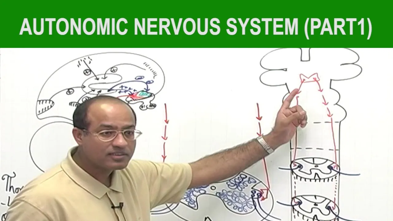
Autonomic Nervous System | Neurology | Neuroanatomy | Part 1🧠
578.9K
The sympathetic nervous system is a thoracolumbar outflow division of the autonomic nervous system that mediates the fight-or-flight response during stress. When the central nervous system perceives a threat, the hypothalamus activates descending polysynaptic pathways that stimulate preganglionic sympathetic fibers from T1-L2/L3 spinal cord segments. These preganglionic fibers synapse at sympathetic chain ganglia and prevertebral ganglia, releasing norepinephrine from postganglionic nerve endings. The adrenal medulla simultaneously releases epinephrine into circulation. Tissues are categorized by their adrenergic receptor types: alpha-1 and beta-1 receptors stimulate tissues helpful during stress (skeletal muscles, heart), while beta-2 receptors inhibit tissues not helpful (GIT, bladder, blood vessels to skin and viscera). This coordinated response increases cardiac output, redirects blood flow to muscles, dilates airways, and prepares the body for immediate action against perceived threats.
Click to watch →
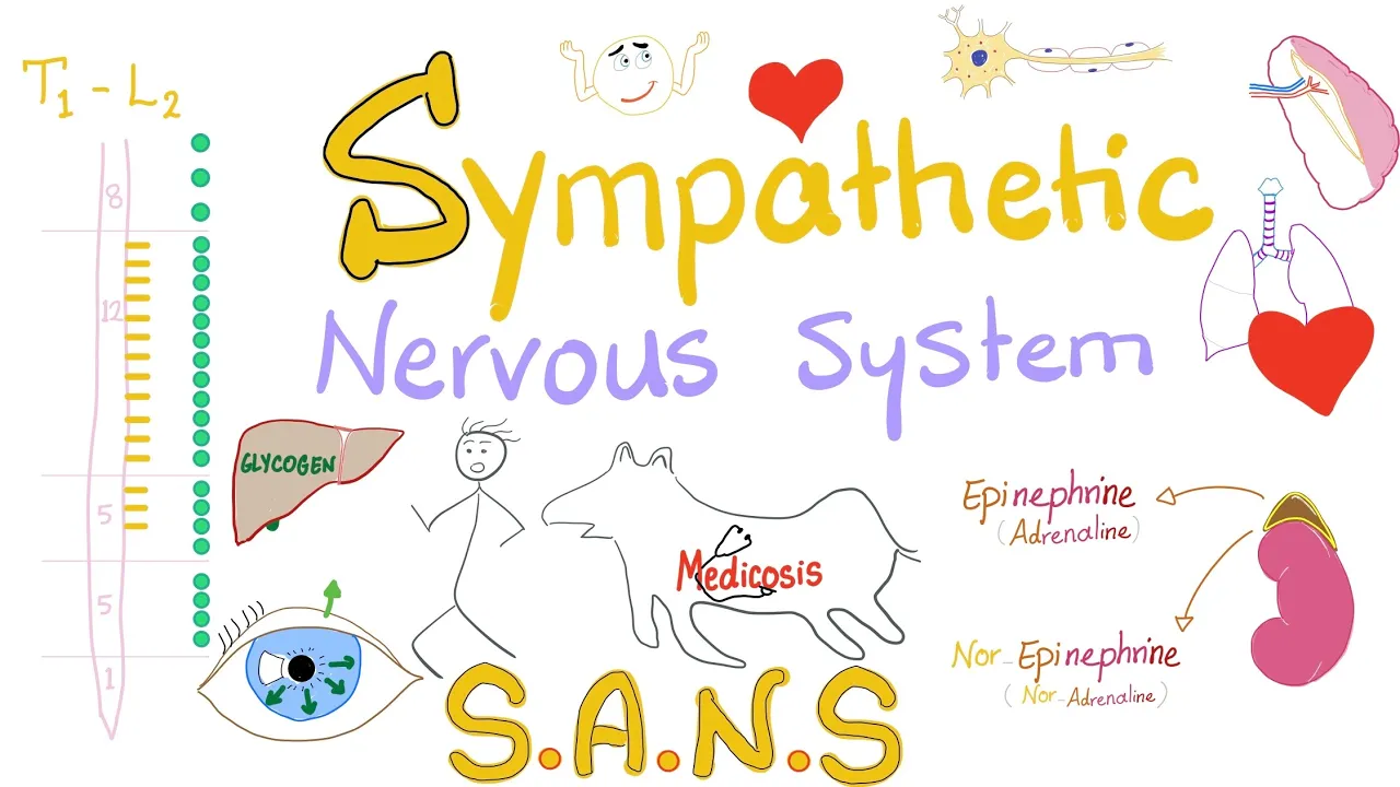
Sympathetic Nervous System | Fight and Flight | Origin, Relay, and Response | Physiology.
62.9K
The sympathetic nervous system represents the body's rapid-response mechanism for survival situations. Anatomically, it originates from thoracolumbar spinal cord segments (T1-L2/3), with preganglionic fibers secreting acetylcholine and postganglionic fibers secreting norepinephrine/epinephrine. Neural organization follows the reflex arc model: sensory stimulus → receptor → afferent fiber → integration center → efferent fiber → effector organ. The system utilizes paravertebral ganglia alongside the vertebral column and prevertebral ganglia (celiac, superior mesenteric, inferior mesenteric) positioned along major blood vessels to minimize tissue piercing. Physiologically, sympathetic activation produces catabolic responses including glycogenolysis and lipolysis for energy mobilization, while cardiovascular effects include increased heart rate, cardiac contractility, and vasoconstriction of systemic vessels with selective coronary and skeletal muscle vasodilation.
Click to watch →
Deep Dive
A fundamental clinical definition of Post-Traumatic Stress Disorder (PTSD) and its primary diagnostic symptoms.

פוסט טראומה "בגובה העיניים" ברקע ה-7 באוקטובר
20.9K
PTSD (Post-Traumatic Stress Disorder) is a psychiatric disorder resulting from exposure to traumatic events. The diagnosis requires Criterion A: direct exposure to actual or threatened death, serious injury, or sexual violence. Four symptom clusters must be present: (1) Intrusion symptoms including flashbacks and nightmares, (2) Avoidance behaviors that shrink the person's world, (3) Negative changes in thinking and mood, and (4) Hyperarousal including being easily startled and difficulty sleeping. These symptoms vary in severity between individuals but share common characteristics.
Click to watch →
TRASTORNO POR ESTRÉS POSTRAUMATICO | TEPT | Diagnóstico, síntomas y tratamiento | DSM 5 TR (Resumen)
16.9K
PTSD is defined as a syndrome characterized by clinically significant alterations in cognition, emotional regulation, or behavior affecting social, occupational, and academic functioning. It results from psychosocial, experiential, neuronal, or hereditary factors. The disorder develops after exposure to traumatic events involving death, serious injury, or violence through direct experience, witnessing, or learning about close others' trauma. Core symptoms include intrusion (recurrent memories, nightmares, flashbacks), avoidance, negative alterations in cognition/mood, and alterations in arousal/reactivity. Two age-based classifications exist: one for children under six and another for those six and older.
Click to watch →

The Trauma of Work - a painted video essay
133K
PTSD (Post-Traumatic Stress Disorder) is defined as a condition of persistent mental or emotional stress occurring as a result of injury or severe psychological shock. In other words, PTSD is when your brain and body have perpetual severe reactions to a dramatic event, even long after the event has passed. The primary PTSD symptoms include hypervigilance (extreme alertness to surroundings, almost like paranoia about the environment, constantly assessing for potential threats and danger), dissociation (a lack of alertness, referring to a strong disconnection between your senses, environment, people around you, resulting in extreme detachment from day-to-day life), and intrusive thoughts (hurtful painful memories that keep popping into your head unwanted).
Click to watch →
Analisis Funcional de la Conducta de un caso de Estrés Postraumático
194
Post-Traumatic Stress Disorder (PTSD) is a specific psychological disorder characterized by particular symptoms experienced by individuals exposed to traumatic events. The American Psychological Association defines trauma as an emotional response to a terrible event such as war, natural disasters, assaults, robberies, torture, or kidnappings. According to DSM-5, PTSD diagnosis requires four criteria: (1) Exposure to a traumatic event involving death or threats to physical integrity, reacting with intense fear; (2) Re-experiencing trauma through intrusive memories, flashbacks, and physiological responses; (3) Avoidance and emotional numbing, including inability to recall important aspects of the event and pessimistic worldview; (4) Increased arousal with insomnia, irritability, hypervigilance, and exaggerated startle responses. Symptoms must persist for more than one month and cause significant clinical distress or impairment.
Click to watch →

Stress: O que é, sintomas e tratamentos
38.7K
PTSD is a chronic psychiatric condition requiring exposure to life-threatening events followed by specific symptom clusters. The diagnostic framework includes intrusion symptoms (intrusive thoughts, nightmares, flashbacks), avoidance behaviors (avoiding reminders, social withdrawal), negative alterations in cognition and mood (guilt, hopelessness, emotional numbness), and arousal/reactivity symptoms (hypervigilance, startle responses, insomnia). Diagnosis requires symptoms persisting beyond 30 days post-trauma, distinguishing normal stress reactions from clinical disorder.
Click to watch →
Deep Dive
The concept of neurocircuitry, particularly how inhibitory and excitatory signals regulate communication between different regions of the brain.
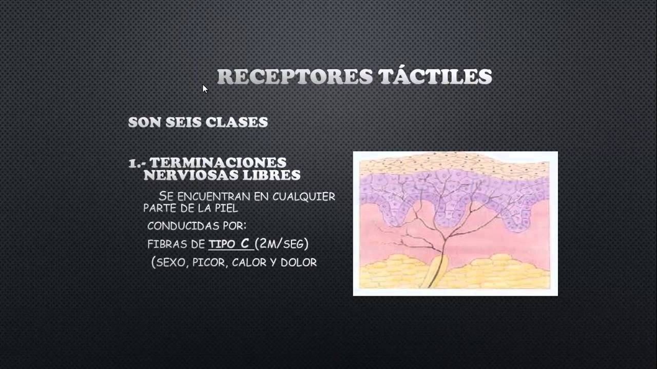
capitulo 46 de fisiologia receptores sensoriales
54.6K
Neuronal circuits have both excitatory and inhibitory output signals. An input signal to a neuronal group causes excitatory signals to follow one direction and inhibitory signals to go to another location. The reciprocal inhibition circuit involves excitatory synapses that excite one pathway and inhibitory synapses that inhibit antagonistic pathways (such as muscles).
Click to watch →

Intro to Neuroscience
908.7K
Neuroscience is the interdisciplinary study of the brain using tools from anatomy, biochemistry, physiology, and molecular biology to understand normal and pathological nervous system function; neurons communicate through electrical signaling (resting potential ~-60mV, action potentials via all-or-none principle) and chemical synaptic transmission (excitatory postsynaptic potentials depolarize, inhibitory postsynaptic potentials hyperpolarize), with common neural circuit motifs including feed-forward excitation/inhibition, convergence, divergence, lateral inhibition, and recurrent inhibition that enable complex behaviors like locomotion and memory formation.
Click to watch →

Cómo Funciona un Cerebro INFELIZ según la Neurociencia con Nazareth Castellanos
33.6K
Neurotransmitters serve as the biochemical basis of thought, transmitting information between neurons. They can be excitatory (increasing electrical charge to promote firing) or inhibitory (decreasing charge to prevent firing). Inhibition is nearly as important as excitation because it regulates neuronal firing rates and prevents uncontrolled signal propagation. Without proper inhibition, electrical discharges could spread uncontrollably throughout the brain, as seen in epilepsy. The brain requires both excitation and inhibition to function properly.
Click to watch →
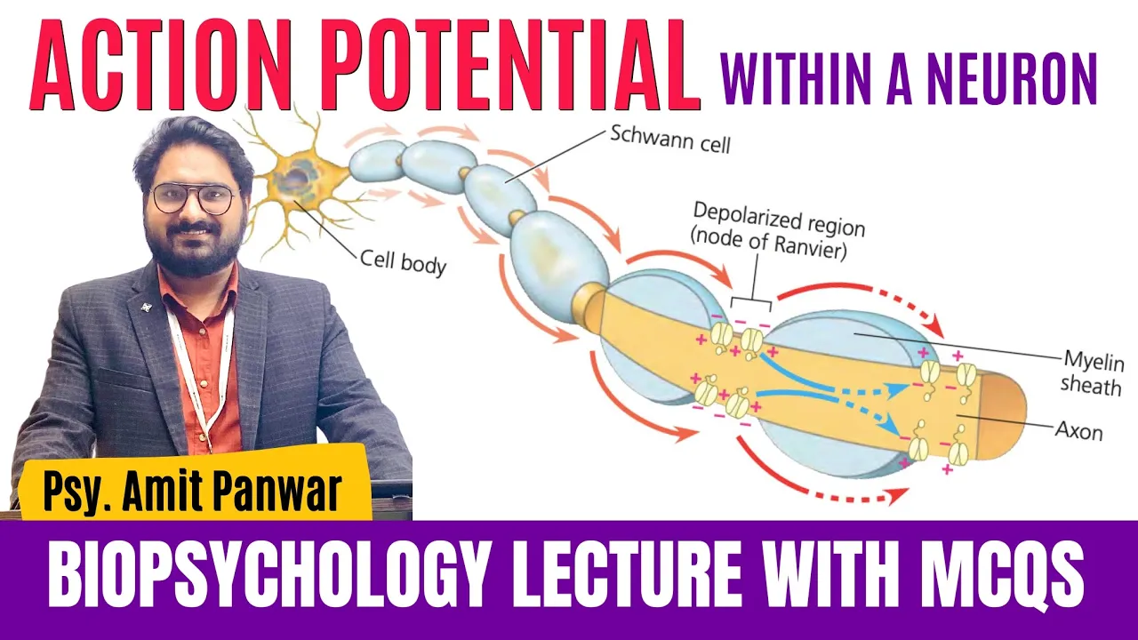
Action Potential - Communication Within a Neuron - Complete Lecture by Psychologist Amit Panwar
12.5K
Neurons transmit two types of signals: (1) Excitatory signals - promote signal transmission and cause the receiving neuron to fire, (2) Inhibitory signals - block or reduce signal transmission. For example, touching a hot surface sends excitatory signals to withdraw the hand, while the brain may send inhibitory signals to prevent dropping a hot object.
Click to watch →
![[Intro to Computational Neuroscience W1D3] Neuroscience Basics](https://i.ytimg.com/vi/BJyRA1iGQ3Y/maxresdefault.jpg)
[Intro to Computational Neuroscience W1D3] Neuroscience Basics
2.1K
Dendrites receive and integrate information from tens of thousands of synapses, with basal dendrites near the soma and apical tufts extending away. Signals attenuate as they travel from distal to proximal regions, modeled by cable theory treating dendrites as cylindrical structures with capacitance and resistance. In CA1 hippocampal neurons, entorhinal cortex input to distal dendrites (sensory information) does not reliably propagate to the soma, while Schaffer collateral input to proximal dendrites (memory information) does. Cortical circuits contain excitatory neurons with recurrent connections and inhibitory interneurons (10-20% of neurons) that balance excitation. Inhibitory synapses are categorized as axo-axonic, axo-somatic, or axo-dendritic. Common inhibitory types include somatostatin-expressing and parvalbumin-expressing interneurons, with VIP-expressing interneurons targeting other interneuron subtypes. Excitatory and inhibitory neurons combine into various circuit configurations: feed forward excitation for information relay, feed forward inhibition for limiting downstream excitation, convergence for integrating multiple inputs, divergence for broadcasting signals, lateral inhibition for sensory edge enhancement, recurrent inhibition for negative feedback loops, and feedback excitation for self-sustaining switch-like behavior.
Click to watch →
Prerequisite Knowledge
- Concept 01Basic neuroanatomy of the limbic system, specifically the structure and primary functions of the amygdala and the prefrontal cortex.
- Concept 02The physiological mechanisms of the body's 'fight-or-flight' survival response, including the activation of the sympathetic nervous system.
- Concept 03A fundamental clinical definition of Post-Traumatic Stress Disorder (PTSD) and its primary diagnostic symptoms.
- Concept 04The concept of neurocircuitry, particularly how inhibitory and excitatory signals regulate communication between different regions of the brain.
Subsequent Learning
- Step 01The neurobiology of trauma recovery, focusing on how evidence-based therapies like CBT, Prolonged Exposure, and EMDR restore prefrontal regulation over the amygdala.
- Step 02The involvement of other critical brain regions in PTSD, such as the hippocampus's role in memory consolidation and context-processing deficits.
- Step 03Pharmacological interventions for PTSD and how selective serotonin reuptake inhibitors (SSRIs) affect neurochemistry and neuroplasticity.
- Step 04The relationship between chronic trauma, hypothalamic-pituitary-adrenal (HPA) axis dysregulation, and systemic cortisol levels.
PTSD Basics
PTSD Symptoms
Brain Hypothesis
Regulation Failure
Emotional Numbness
Complex Circuits
PTSD Basics
0:04Playing Section
- 1
PTSD develops after a traumatic event.
- 2
Intrusive symptoms like nightmares and memories occur.
- 3
Avoidance of trauma reminders is a key symptom.
The Neuroreductionism Critique and Socio-Cultural Framework of Trauma
While neurocircuitry models (focusing on the amygdala and prefrontal cortex) provide valuable biological insights, critics argue that this "neurocentric" view is overly reductionist. This perspective cautions against treating PTSD purely as a brain-based wiring defect, which risks pathologizing adaptive survival responses and ignoring the critical role of subjective meaning, personal history, and socio-cultural context. Proponents of socio-cultural and bio-psycho-social frameworks emphasize that trauma is a complex, lived experience, not just a biological state. Factors such as systemic oppression, interpersonal support systems, and the individual's narrative interpretation of the event are vital to understanding and healing trauma. Focusing exclusively on brain circuitry can lead to over-medicalized treatments while neglecting somatic therapies, community healing, and the structural interventions needed to address the root causes of trauma.
Deep Dive
The neurobiology of trauma recovery, focusing on how evidence-based therapies like CBT, Prolonged Exposure, and EMDR restore prefrontal regulation over the amygdala.

Trauma - Wie Traumata das Gehirn verändern und ihre moderne Therapie
315.8K
The medial prefrontal cortex acts as an antagonist to the amygdala, capable of inhibiting it effectively within 100-300 milliseconds. For this inhibition to occur, the prefrontal cortex must be informed that it is doing so—this happens implicitly or explicitly during virtually all forms of trauma therapy. Without this inhibition, only weak amygdala suppression occurs, resulting in strong anxiety responses.
Click to watch →

Se Sanó sin Medicinas, Solo con Su Mente con Juan Lucas Martín
89.3K
Trauma creates lasting changes in the brain's stress response system. When traumatic memories are triggered, the amygdala (the brain's alarm center) activates, releasing cortisol and adrenaline. These hormones prepare the body for fight-or-flight but cause damage when released chronically. The prefrontal cortex, which helps evaluate the present moment, can be activated through techniques like EFT to calm the amygdala. This neurobiological understanding explains why trauma can lead to physical illness and why processing trauma is essential for health.
Click to watch →
La ANSIEDAD te eligió sin permiso… pero SANAR es tu decisión
15.4K
During overwhelming experiences, the amygdala takes control and records danger information while the hippocampus disconnects (explaining fragmented traumatic memories). The prefrontal cortex (rational part) decreases in function, making the brain more emotional than logical. Trauma is remembered through sensations, not words - the amygdala activates milliseconds before the cortex processes information. Healing involves recovering prefrontal function through intentionality and voluntariness. The body remembers trauma even when the mind forgets - this is why trauma is an involuntary limbic learning that persists.
Click to watch →

#62 Dr. Karen Lamb on Trauma and Highly Sensitive People
369
Three evidence-based therapies serve different functions in trauma recovery: Cognitive Behavioral Therapy (CBT) and Dialectical Behavioral Therapy (DBT) provide practical crisis management tools for regulating the nervous system during panic attacks or overwhelming situations. EMDR (Eye Movement Desensitization and Reprocessing) is highly effective for processing significant life events, with research showing it completely resets the brain from PTSD patterns whereas talk therapy produces only minor shifts. The therapeutic alliance—the relationship between provider and client—is critically important across all approaches and can itself be healing. Homeopathy works synergistically with these therapies by addressing physiological responses.
Click to watch →
How Trauma and PTSD Change the Brain
1.4M
Several therapeutic approaches help the brain and body rewire and restore inner sense of safety and clarity. Writing exercises turn on the prefrontal cortex, clarify present safety, help the hippocampus process memories, and send messages to the amygdala that one is actually safe. Other treatments include CBT (Cognitive Behavioral Therapy), EMDR (Eye Movement Desensitization and Reprocessing), and Somatic Experience. Research using MRI scans shows that mindfulness practice correlates with growth in the hippocampus and shrinking of the amygdala, essentially reversing the effects of trauma.
Click to watch →
Deep Dive
The involvement of other critical brain regions in PTSD, such as the hippocampus's role in memory consolidation and context-processing deficits.

Нейробиология ПТСР
1.9K
The amygdala and hippocampus are critical limbic system structures in PTSD. The amygdala detects threats and triggers fear responses, becoming hyperactive in PTSD while the prefrontal cortex fails to regulate it. The hippocampus consolidates memories and provides contextual information. In PTSD, high stress hormones disrupt memory consolidation, causing fragmented memories that trigger flashbacks. Chronic stress causes hippocampal atrophy, reducing its ability to contextualize memories and organize them chronologically.
Click to watch →
Post Traumatic Stress Disorder, The Brain and The Memory | Israel Liberzon
7K
The most promising model proposes that PTSD results from dysfunction in the hippocampal-prefrontal contextual processing system. This system modulates fear responses based on environmental context—allowing the brain to distinguish between threatening and safe situations. Experimental studies demonstrate that PTSD patients fail to use contextual information appropriately, responding with fear in both conditioned and unconditioned contexts. This unified framework explains diverse PTSD symptoms including inappropriate fear expression, emotional numbing, intrusive memories, and nightmares. The model integrates hippocampal-prefrontal circuit abnormalities with HPA axis dysfunction and genetic/epigenetic vulnerabilities, providing a comprehensive explanation for PTSD pathophysiology.
Click to watch →
Neurobiology & Treatment PTSD in Refugees and Other Populations | Mental Health Professional Webinar
232
The amygdala detects threats and triggers fight-or-flight responses, while the hippocampus processes context and helps form fear memories. Together with the prefrontal cortex, these structures determine whether a threat is real or safe. In PTSD, amygdala activation increases to trauma-related stimuli, while hippocampal volume and activation decrease, particularly in context processing. The insula shows increased activity correlating with symptom severity. Critically, these brain changes are plastic and can improve with effective treatment, countering the misconception that mental illness is permanent.
Click to watch →

Stress Depression Anxiety PTSD Part 2
5.7K
Other symptoms of PTSD include memory loss such as amnesia for some aspects of the traumatic event, flashbacks, and short-term memory loss. There are also physical effects on the brain. Memory deficits in PTSD suggest the hippocampus is involved, and researchers have found that those with PTSD tend to have a smaller right hippocampus. Additionally, activation of the hippocampus in response to fearful stimuli like fearful faces is different for PTSD sufferers, who show lower activation in the amygdala. However, twin studies suggest that the smaller hippocampal volume may have existed prior to the traumatic experience and the development of PTSD.
Click to watch →

Presentation Trailer Documentary "PTSD. Invisible Scars. Children"
217
The hippocampus is a key brain region identified in both adults with combat-related PTSD and children with developmental trauma. It plays an important role in memory consolidation and retrieval, particularly bringing contextual memories into recall. It also plays a crucial role in emotional regulation and is one of the most stress-susceptible structures in the body. High cortisol levels can alter the branching and morphology of neurons within the hippocampus.
Click to watch →
Deep Dive
Pharmacological interventions for PTSD and how selective serotonin reuptake inhibitors (SSRIs) affect neurochemistry and neuroplasticity.

Антон Волосовець
192
Pharmacological treatment includes SSRIs (first-line), anxiolytics, zinc, vitamin D, and sleep aids. Benzodiazepines are risky due to addiction potential. Antidepressants should be used when psychotherapy is not possible, the patient is unstable, or results are insufficient. Escitalopram (Cipralex) is a selective serotonin reuptake inhibitor that naturally stimulates existing serotonin, improving mood, reducing anxiety, and helping with somatic obsessive states. It has no sedative effect, making it suitable for many situations. It should always be paired with psychotherapy, which reprograms the brain from PTSD state to normal reaction to stimuli.
Click to watch →

Посттравматичний стресовий розлад (ПТСР).
2.3K
Pharmacological treatment for PTSD includes: (1) Selective serotonin reuptake inhibitors (SSRIs) such as fluoxetine, sertraline, and paroxetine; (2) Serotonin-norepinephrine reuptake inhibitors (SNRIs) such as venlafaxine; (3) Tricyclic antidepressants such as amitriptyline; (4) Other medications for specific symptoms. SSRIs and SNRIs are first-line treatments for PTSD. Benzodiazepines are sometimes used for acute anxiety symptoms but are not recommended for long-term treatment due to risks of dependence, cognitive impairment, and potential worsening of PTSD symptoms. Antipsychotics are sometimes used for PTSD, particularly for patients with prominent psychotic symptoms or severe agitation, but evidence for their effectiveness is limited. Anticonvulsants such as topiramate and gabapentin are sometimes used for PTSD symptoms, particularly for sleep disturbances and anxiety, but evidence for their effectiveness is limited. Effective PTSD treatment typically involves a combined approach that includes both pharmacological and psychotherapeutic interventions. The choice of treatment depends on individual factors, including symptom severity, comorbid conditions, and patient preferences. Treatment should be individualized and may require adjustments over time based on response. PTSD frequently occurs with other mental health conditions, including: major depressive disorder, anxiety disorders, substance use disorders, and personality disorders. Comorbidity is common and may complicate diagnosis and treatment. The presence of comorbid conditions may influence the course of PTSD and require comprehensive treatment approaches.
Click to watch →
Episode 204-USMLE And The Military (Very HY for the USMLEs)
31.4K
PTSD treatment is divided into pharmacological and psychotherapeutic approaches. SSRIs (sertraline, fluoxetine, paroxetine) are first-line medications. SNRIs (venlafaxine) have also been shown to help, particularly in military populations. These medications address the underlying neurochemical imbalances associated with PTSD.
Click to watch →

Complex Psychiatric Cases: Diagnosis and Treatment
77.3K
Serotonin reuptake inhibitors (SSRIs) are a mainstay pharmacological treatment for PTSD, working partly by increasing levels of brain-derived neurotrophic factor (BDNF). Research is investigating whether combining behavioral interventions with pharmacological approaches targeting different pathways might improve outcomes. This represents a future direction in personalized treatment strategies for PTSD.
Click to watch →

Post Traumatic Stress Disorder (PTSD) Explained
210.6K
Medications are useful adjuncts to psychotherapy for PTSD treatment. Selective serotonin reuptake inhibitors (SSRIs) like paroxetine and sertraline, and serotonin-noradrenaline reuptake inhibitors (SNRIs) like venlafaxine are first-line pharmacological options. Clonidine, an alpha agonist, helps reduce trauma-related nightmares. Trazodone or promethazine may be used for insomnia. Some studies suggest adding antipsychotics like risperidone to antidepressant therapy may improve outcomes in certain patients.
Click to watch →
Deep Dive
The relationship between chronic trauma, hypothalamic-pituitary-adrenal (HPA) axis dysregulation, and systemic cortisol levels.

Powerful Strategies to Undo PTSD: Rewiring the Traumatized Brain
407.2K
Trauma fundamentally alters brain function through the hypothalamus-pituitary-adrenal (HPA) axis, which controls the stress response. When trauma remains unprocessed, the HPA axis stays chronically activated rather than functioning normally. This persistent activation leads to excitotoxicity, where excessive stress hormones (cortisol, glutamate, norepinephrine) damage neurons and cause hippocampal shrinkage. The amygdala-prefrontal cortex connection strengthens, creating hyper-vigilance. Simultaneously, the vagus nerve weakens, reducing the body's ability to activate relaxation responses. Systemically, chronic HPA activation causes cortisol to lose its anti-inflammatory properties, triggering widespread inflammation. The gut microbiome shifts toward microbes producing fight-or-flight neurotransmitters. Hormonal imbalances affect thyroid function and gonadal hormones, altering neurotransmitter availability. Sleep disruption compounds these effects by activating neural inflammation and suppressing neurogenesis. Psychologically, emotional dysregulation emerges as the HPA axis reduces sensitivity to prevent constant activation, yet this same reduction impairs positive emotional experience. When triggered, survivors experience dramatic neurochemical surges shifting from flat affect to intense anxiety or anger. Cognitive impairments include pessimism, disempowerment, inflexible thinking, and difficulties with memory and learning. Separation anxiety develops as survivors fear loved ones will be victimized. Trust issues emerge, particularly with people resembling perpetrators. Seemingly benign environmental stimuli can trigger traumatic memories and re-traumatization, perpetuating chronic stress states. Effective recovery requires strengthening vagal tone through respiratory vagus nerve stimulation (deep breathing), anti-inflammatory nutrition following Mediterranean principles, aerobic exercise promoting neurogenesis, quality sleep, optimal hormone balance, and engaging in hobbies and social activities releasing oxytocin as a neuroprotectant.
Click to watch →

Neurobiology of PTSD: Genetics, Epigenetics, and Emerging Treatments
99K
The hypothalamic-pituitary-adrenal (HPA) axis is fundamentally dysregulated in PTSD. Normally, CRH triggers cortisol release through a negative feedback loop. In PTSD, individuals have elevated CRH levels despite lower cortisol—a pre-existing vulnerability rather than trauma consequence. Lower cortisol levels predict who will develop PTSD after trauma exposure. Vietnam veterans decades later still show elevated T3/T4 levels, contributing to chronic anxiety and sympathetic symptoms. This HPA dysregulation represents a core biological marker distinguishing PTSD from other stress responses.
Click to watch →
Discover How Trauma Rewires the Brain
353.8K
This comprehensive section covers the complete neurobiological cascade of trauma response. The HPA axis serves as our threat response system, controlling stress reactions and regulating digestion, immune function, mood, and reproductive systems. Individual responses vary based on personal resources, resilience, proximity to trauma, similarity to victims, and prior trauma history. Children require specific support due to limited life experience and egocentric thinking. Persistent HPA activation causes cortisol release for fight-or-flight energy while suppressing immune, digestive, and reproductive systems. Chronic activation leads to digestive problems, reduced immunity, mood disturbances from low serotonin and GABA, and blood sugar imbalances. When activation continues excessively, the body reduces cortisol production as a protective mechanism, creating hypocortisolism seen in chronic fatigue, burnout, and PTSD. This paradoxical state causes either flat/depressed or hyperaroused responses. Abnormally activated HPA axis creates simultaneous cortisol presence and systemic inflammation, contributing to autoimmune issues, allergic reactions, irritable bowel syndrome, and reduced stress tolerance. Core neurochemical features include abnormal regulation of catecholamines (norepinephrine, dopamine from tyrosine), serotonin (multiple receptor types), and GABA systems. Norepinephrine accounts for classic PTSD symptoms including hyperarousal, startle responses, and fear memory encoding. Poor serotonin transmission causes impulsivity, hostility, depression, and suicidality. GABA is reduced in PTSD patients, explaining emotional dysregulation. Endogenous opioids may cause numbing and dissociation, explaining why some PTSD patients self-medicate with opioids.
Click to watch →
Do You Have Adrenal Fatigue? Top 3 Causes
50.5K
Prolonged elevated cortisol triggers two critical dysregulation mechanisms: corticotropin-releasing factor receptor downregulation and HPA axis sensitization. Cells become desensitized to cortisol signals, while the brain becomes hyper-responsive, creating dangerous cortisol spikes and crashes. Three primary factors drive this dysfunction: emotional stress perception (negative trauma causes worse responses than positive excitement), sleep deprivation (impairs CRF regulation and neuroplasticity recovery), and sugar consumption (inflammation disrupts brain-adrenal communication). Effective management requires addressing these root causes through controlled emotional responses, consistent quality sleep, reduced sugar intake, and maintaining proper mineral balance (potassium, sodium, magnesium) to support aldosterone, norepinephrine, and adrenaline production.
Click to watch →

🚨Como BAJAR el CORTISOL en 5 PASOS SENCILLOS: resultados INMEDIATOS | Mr Doctor
23.8K
Cortisol release is not autonomous but results from the hypothalamic-pituitary-adrenal (HPA) axis. When the brain perceives a threat, the hypothalamus releases CRH, which stimulates the pituitary to release ACTH, which then commands the adrenal glands to produce cortisol. This system includes negative feedback mechanisms that automatically shut down production when cortisol levels reach a threshold, functioning like a thermostat. When stressors persist without resolution (demanding bosses, economic uncertainty, social isolation), the HPA axis becomes constantly stimulated and overworked. Over time, brain receptors lose sensitivity, the feedback mechanism breaks down, and the body continues producing cortisol at elevated levels. This condition is called allostatic load or adrenal fatigue. Chronic high cortisol levels can damage the hippocampus over time, potentially affecting memory function.
Click to watch →
PTSD Basics
PTSD Symptoms
Brain Hypothesis
Regulation Failure
Emotional Numbness
Complex Circuits
PTSD Basics
0:04- 1
PTSD develops after a traumatic event.
- 2
Intrusive symptoms like nightmares and memories occur.
- 3
Avoidance of trauma reminders is a key symptom.
The Neuroreductionism Critique and Socio-Cultural Framework of Trauma
While neurocircuitry models (focusing on the amygdala and prefrontal cortex) provide valuable biological insights, critics argue that this "neurocentric" view is overly reductionist. This perspective cautions against treating PTSD purely as a brain-based wiring defect, which risks pathologizing adaptive survival responses and ignoring the critical role of subjective meaning, personal history, and socio-cultural context. Proponents of socio-cultural and bio-psycho-social frameworks emphasize that trauma is a complex, lived experience, not just a biological state. Factors such as systemic oppression, interpersonal support systems, and the individual's narrative interpretation of the event are vital to understanding and healing trauma. Focusing exclusively on brain circuitry can lead to over-medicalized treatments while neglecting somatic therapies, community healing, and the structural interventions needed to address the root causes of trauma.
Basic neuroanatomy of the limbic system, specifically the structure and primary functions of the amygdala and the prefrontal cortex.
The physiological mechanisms of the body's 'fight-or-flight' survival response, including the activation of the sympathetic nervous system.
A fundamental clinical definition of Post-Traumatic Stress Disorder (PTSD) and its primary diagnostic symptoms.
The concept of neurocircuitry, particularly how inhibitory and excitatory signals regulate communication between different regions of the brain.
The neurobiology of trauma recovery, focusing on how evidence-based therapies like CBT, Prolonged Exposure, and EMDR restore prefrontal regulation over the amygdala.
The involvement of other critical brain regions in PTSD, such as the hippocampus's role in memory consolidation and context-processing deficits.
Pharmacological interventions for PTSD and how selective serotonin reuptake inhibitors (SSRIs) affect neurochemistry and neuroplasticity.
The relationship between chronic trauma, hypothalamic-pituitary-adrenal (HPA) axis dysregulation, and systemic cortisol levels.
190.2K views3.2Klikes1:59@neurochallengedOriginal Release: 2022-08-16
Post-traumatic stress disorder, or PTSD, is a condition that develops after someone experiences a traumatic event.
It involves the occurrence of intrusive symptoms like nightmares or distressing memories that are linked to the trauma and may cause the person to feel like they are reliving aspects of the traumatic event.
These symptoms also lead to the avoidance of things that remind a person of the trauma.
PTSD may cause various other issues such as difficulty sleeping, negative emotions like fear, guilt, or sadness, trouble concentrating, and irritability.
Although the neurocircuitry underlying PTSD is still not completely clear, one supported hypothesis suggests that PTSD involves decreased activity in the medial prefrontal cortex and increased activity in subnuclei of the amygdala that are involved in the identification of threats.
According to this hypothesis, the medial prefrontal cortex normally acts to regulate amygdala function, inhibiting it when there is not an immediate threat to devote attention to.
In an individual with PTSD, however, the amygdala might be hyperactive and provoke a fearful reaction in response to trauma-related stimuli.
The medial prefrontal cortex fails to inhibit this unnecessary amygdala activation, causing patients to experience responses that are disproportionate to the threat that trauma-related stimuli currently pose.
Some patients with PTSD, however, also experience the suppression of emotions, which causes symptoms like social detachment and emotional numbness.
This might be caused by an opposing mechanism where increased activity in the medial prefrontal cortex dampens activity in regions such as the amygdala and other areas involved in emotional expression.
Thus, the neuroscience of the disorder is complex and the neurocircuitry involved likely depends on the symptoms a particular patient displays.
Additionally, more recent research has suggested a role for other networks that span larger areas of the brain in bringing about the symptoms of PTSD.
Up Next

102:32
Neuroscience of Anxiety, Dopamine & Personal Growth | Huberman
@JordanBPeterson
5.1M views•2022-10-13

21:55
Deciphering Neural Circuits Controlling Anorexia in Mice | Richard Palmiter
@AllenInstitute
2.3K views•2011-10-10
1:59
How Smell Works: Olfaction Explained in 2 Minutes | Neuroscience
@neurochallenged
303.7K views•2018-01-31

13:03
How Exercise Benefits Your Brain: Science Explained
@TED
11.4M views•2018-03-21
Related Study Plans & Knowledge Roadmaps
Structured learning paths in Neuroscience
neuroscience
Appetite Regulation: Brain Circuits & Hormones
01Foundations of Endocrinology and Brain Anatomy
02Introduction to Energy Homeostasis
03Peripheral Signals: Leptin, Ghrelin, and Insulin
+ 2 more learning modules
14h5 modules
Explore Roadmap
neuroscience
Neuroeconomics: Value, Risk & Brain Mechanisms
01Foundations of Neuroscience and Brain Anatomy
02Introduction to Neuroeconomics and Subjective Value
03Dopaminergic Prediction Errors & Reinforcement Learning
+ 3 more learning modules
18h6 modules
Explore Roadmap
neuroscience
Spatial Navigation: Cells, Grids & Brain Maps
01Foundations of Brain Anatomy and Memory
02Place Cells and Head-Direction Cells
03Grid Cells: The Brain's Coordinate System
+ 2 more learning modules
12h5 modules
Explore Roadmap
neuroscience
BCIs: Neural Decoding & Prosthetic Control
01Neuroscience Foundations: How the Brain Signals Intent
02Signal Acquisition: Recording the Brain's Activity
03Neural Decoding: Translating Brain Waves into Data
+ 2 more learning modules
25h5 modules
Explore Roadmap