












The vestibulocochlear nerve (CN VIII) is a purely sensory cranial nerve consisting of two parts: the vestibular division (maintaining equilibrium/balance) and the cochlear division (facilitating hearing). The vestibular system detects motion and spatial orientation through the semicircular canals (detecting rotational movements via endolymph movement in ampullae) and otolith organs (utricle for horizontal, saccule for vertical movements via otoconia). The cochlear system processes sound through the cochlea, where vibrations from the oval window create waves that move the basilar membrane, stimulating hair cells in the organ of Corti. The vestibular nerve projects to the vestibular nuclear complex (superior, medial, lateral, and inferior nuclei), giving rise to four pathways: vestibulo-ocular reflex (stabilizing eye gaze via MLF to extraocular muscles), vestibulospinal reflex (maintaining posture via lateral and medial tracts), vestibulocerebellar pathway (to flocculonodular lobe), and vestibulo-cerebral pathway (to thalamus and cortical regions including frontal eye fields, somatosensory cortex, parieto-insular vestibular cortex, posterior parietal cortex, and hippocampus). The cochlear nerve projects to the ventral and dorsal cochlear nuclei, crosses to form the lateral lemniscus, passes through the superior olivary complex for sound localization, reaches the inferior colliculus, and finally projects to the medial geniculate body and primary auditory cortex in the superior temporal gyrus.
Vestibulocochlear Nerve (CN VIII): Anatomy, Pathway & Functions
Added:What’s up, Taim Talks Med here. Let’s continue our Cranial nerve series.
Cranial nerves are twelve pairs of nerves that exit the brain and the brainstem, and in this segment, we’ll talk detailed about the eighth cranial nerve, the vestibulocochlear nerve.
And we’ll do that by first making a quick scheme of the vestibulocochlear nerve pathway.
Then we’ll break down the basic principles of the inner ear because you need to have an understanding of how the sensation of equilibrium and sound is perceived in order to understand these nerves better.
Then we’ll look into the component and the pathway of the vestibulocochlear nerve, and then talk detailed about the vestibular system and the auditory system.
Alright, we’ll break this down as easy as we can, so let’s start making a little scheme.
Now, the the vestibulocochlear nerve is purely a sensory nerve. It has no motor function. And it consists of two parts. A vestibular part, which is responsible for maintaining a sense of equilibrium and balance. And it has a cochlear part, which facilitates hearing.
We’ll start this scheme off with a little structure here called the inner ear, this is the membranous labyrinth of the inner ear. We’ll talk about this in detail in a couple of minutes, but the inner ear consists of a coclear part and a vestibular part. From the coclear part, there will be dendrites of nerves located within the organ of corti, in the cochlea. These dendrites take signals towards the spiral ganglion, from where axons then run as the coclear root.
From the vestibular part of the inner ear, you’ll find nerve endings coming from the saccule, or macula of the saccule. Saccule is just a sac of fluid. Macula of the saccule is an accumulation of hair cells that detect vertical linear acceleration. So nerves comes from the macula of the saccule as the saccular nerve. There’s one coming from the posterior ampullary crest, as the posterior ampullary nerve. They two form an inferior division. Then we got a nerve coming from the macula of the utricle as the utricle nerve, then we got the lateral and the anterior ampullary nerves, from the lateral and the anterior ampullary crests. They form the superior division.
The superior and the inferior divisions will take the signals towards the vestibular ganglion located in the lateral end of the internal acoustic meatus.
Axons from the ganglion leave as the vestibular root. The vestibular root and the cochlear root now travel together as the vestibulocochlear nerve. As the 8th cranial nerve. And they’re going to leave through the Internal acoustic meatus.
Alright. Now what. CN VIII will run past the angle between the cerebellum and Pons and enters the brainstem at the pontomedullary junction. Meaning the junction between the pons and medulla oblongata. Then they will separate as the cochlear nerve and the vestibular nerve. The cochlear nerve will run off and synapse with the ventral and the dorsal cochlear nuclei. Or the anterior and the posterior cochlear nuclei. In proper theory there are three divisions of the cochlear nuclei: anteroventral (AVCN), dorsal (DCN), and posteroventral coclear nuclei but we’ll keep it simple and detailed enough to understand the concept of this nerve.
The Vestibular nerve will reach something called the vestibular nuclear complex. The vestibular nuclear complex, there are four parts of it, there’s the superior, medial, lateral, and the inferior part. And they lie in the lower part of pons, as well as the upper medulla and they’re closely related to the floor of the fourth ventricle.
Now, now that we know that. What happens? Let’s do the vestibular part first.
Here's what I want you to remember to basically understand what we’re going to go through later.
It’s really easy to remember it like this. From the vestibular nucleus. Information can go up, it can go down, it can go back, and it can go all the way up to the cerebral cortex. Now.
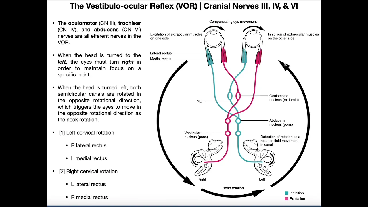
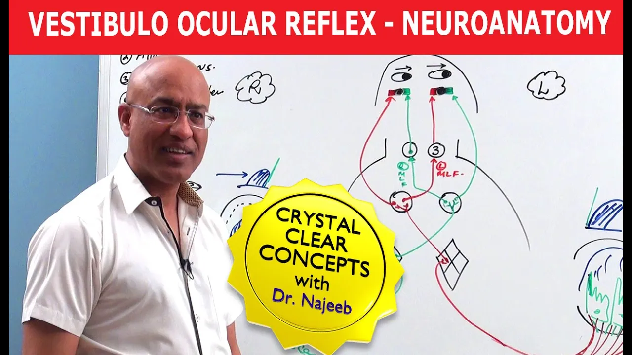
The medial nucleus sends off ascending fibers to the motor nuclei of the extraocular muscles ipsilaterally and contralateral via the medial longitudinal fasciculus (MLF), which helps facilitate the vestibulo-ocular reflex. Some fibres come from the superior nuclei as well but for the majority of it, we associate the MLF with the medial vestibular nucleus. Alright?
Now, that’s for the fibers that go up. The medial nucleus also helps mediate the vestibulospinal reflex by controlling head and neck movements, though the medial vestibulospinal tract.
The lateral nucleus also sends fibers down as the lateral vestibulospinal tract, mediating the vestibulospinal reflex and maintain posture and balance. So those are the fibers that go down.
The lateral vestibulospinal tract from the lateral nucleus, going down ipsilaterally for the body and limb, and the medial vestibulospinal tract from the medial nucleus, which is a part of the MLF going down both ipsilaterally and contralaterally to control the head and neck movement. Alright.
Fibers that go back go from the inferior vestibular nucleus primarily, through the inferior cerebellar peduncle and then to the cerebellum, to involve it into the vestibulocerebellar system.
We’re gonna talk about this detailed later but it goes to the flocculonodular lobe.
Fibers also can come directly from the vestibular nerve before it enters the brainstem. These are direct cerebellar fibers. This is a two way connection, so fibers will also come from the cerebellum and then into the vestibular nucleus again, to help influence the vestibulospinal tracts. Because the cerebellum is more involved in things like posture, muscle tone and balance. Right? So the cerebellum is a key component here.
Then we got fibers that go all the way up to the Ventral Posterior group of thalamus.
These are fibers that primarily come from the superior vestibular nuclei. They then project to the internal capsule and further radiate into different areas of the cerebral cortex.
It could be to the primary somatosensory cortex to map the body location and movement signals, it could be the frontal eye fields to control the eye movement and receive vestibular motion information. Could be the hippocampus and the parahippocampal area for spatial orientation and navigation. Parieto-Insular cortex which responds to the body and head motion information, and the posterior parietal cortex aswell. We’ll mention these briefly later.
For the Cochlear part information is going to cross and ascend as the lateral lemniscus. But they can also go ipsilaterally and ascend. They may synapse with the superior olivary complex, which plays a role in localizing the direction of sound. Cochlear fibres may synapse with cells that lie within the lemniscus itself, which form the nucleus of the lateral lemniscus. As fibers from both sides cross, they form a mass of fibres called the trapezoid body, containing cell bodies with nuclei which the cochlear nerve may also synapse with. The lateral lemniscus will go up and synapse with the inferior colliculus. From here fibers may go down as the tectospinal tract, which plays a role in the auditory reflexes. Fibers may also go up through the brachium of the inferior colliculus, towards the medial geniculate body next to the thalamus. Fibers may also come directly from the lateral lemniscus towards the medial geniculate body. Fibers will then radiate towards the superior temporal gyrus which is believed to be the primary auditory cortex.
So. That was the scheme. It’s a relatively large scheme and a lot of information to squeeze in, so I tried to separate these two nerves in order to make it easier to study and understand, so keep in mind that this is just schematic without structures actually being anatomically correctly placed. Now. Let’s start over here. Let’s briefly understand the ear anatomy a bit.
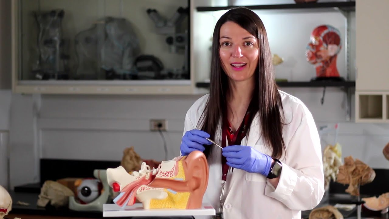
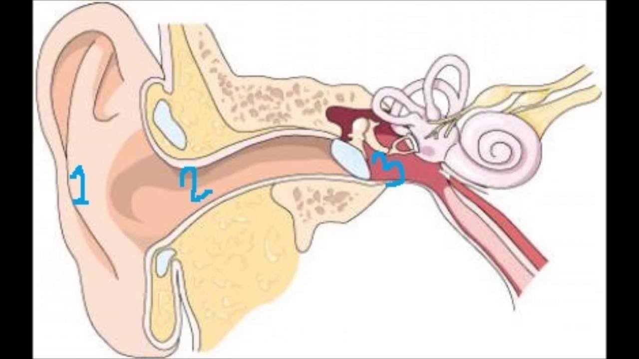
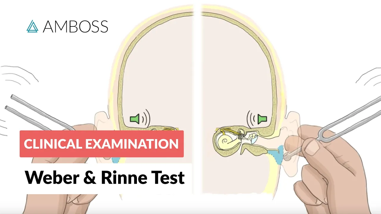
--------------------------------------------------------------------------------- To understand the vestibulocochlear nerve, we need to have a sense of how balance and hearing are perceived, how the inner ear actually works. And I’m not gonna go in too much detail into this we’ll do that in a separate video. But here we see the auricle, the external acoustic meatus and the tympanic membrane. These three are what we refer to as the external ear.
Behind the tympanic membrane, we see the three ossicles called Malleus, Incus and Stapes. We call this area the middle ear. The stapes is going to vibrate and mechanically push on a structure called the oval window, of the inner ear. Otherwise known as the vestibulocochlear organ.
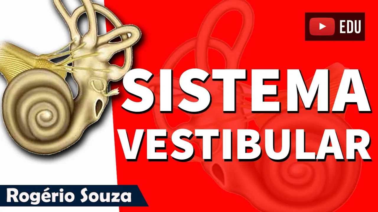
From the vestibulocochlear organ, you’ll find the vestibular nerve and the cochlear nerve going through the internal acoustic meatus. And so that is what we call the inner ear. Let’s focus on the anatomy of the inner ear for a few minutes. Alright. Now. We grossly divide the inner ear into the vestibular component, Semi-circular component, and the Cochlear component.
The vestibular and the semicircular canals are primarily responsible for maintaining equilibrium, and cochlea processes hearing. This blue structure you see here is a hard shell, a bony component we call the bony labyrinth. If we now fade the bony labyrinth, you’ll see the wall of the bony labyrinth here, so grossly, we divide the bony part into vestibule, semicircular canals and the cochlea. Notice that within the bony labyrinth, you have another structure, another membrane. This is what we call membranous labyrinth. And the membranous labyrinth is also divided into parts according to their function.
Within the vestibule, we’ll find the utricle, the saccule, and the ampulla. Within the semicircular canals, you’ll find the semicircular ducts. The anterior, posterior and the lateral parts of it.
Within the cochlea, you’ll find the cochlear duct. And then up here we have the endolymphatic duct that end as the endolymphatic sac, lying deep to the dura mater in the posterior cranium.
One thing to keep in mind is that the membranous labyrinth is a closed system, it's a poutch. And the fluid within it is removed by reabsorption via the epithelium of the endolymphatic sac.
So that’s generally how we divide the bony and the membranous labyrinth.
Now, between the bony and the membranous labyrinth, there’s a cavity. And this cavity, you see here is filled with a fluid known as the perilymph, which is similar in ionic composition to that of the extracellular fluid. So it’s low in potassium, and high in sodium. The lumen of the membranous labyrinth contains another fluid known as endolymph, which has an ionic composition similar to cytosol, so it’s high in potassium and low in sodium. And these two substances do not communicate with each other. Alright. Now, we divide the inner ear into two systems. The vestibular system and the auditory system. The vestibular system is a sensory system responsible for providing our brain with information about motion, head position and spatial orientation. It’s able to do all that because of the vestibular labryrinth and the vestibular nerve. The vestibular labrynith again consist of three semicircular canals. Each of the canals can detect movements such as nodding up and down, shaking side to side or tilting left and right. The semicircular canals remember they’re filled with a fluid called endolymph.
When the head is rotated it causes the movement of endolymph through the canal that corresponds to the plane of movement. The endolymph flows into an expansion of the canal called the ampulla, within which there are hair cells, the sensory receptors of the vestibular system. At the top of each hair cells are a collection of small hairs called stereocilia. The movement of the endolymph causes movement of these stereocilia which leads to the release of neurotransmitters to send information about the plane of movement to the brain. The vestibular system uses two other organs, known as otolith organs, to detect forward and backward movement and gravitational forces.
There’re two otolith organs in the vestibular labyrinth. One located in the utricle, which detects movements in the horizontal plane, and the saccule, which detects movements in the vertical plane. Whitin the utricle and the saccule, hair cells detect movements when crystals of calcium carbonate called otoconia shift in response to it, leading to movement of the hair cells below the otoconia to stimulation of the vestibular root of the vestibulocochlear nerve.
Now the cochlear system is a little special. When soundwaves travel through the canal of the ear, they hit the tympanic membrane and causes it to vibrate. This vibration prompts the vibration of the ossicles that transmits the vibration into the oval window, which sits in the wall of the cochlea. If we take a small chunck, a section of the coclea, you’ll see that it consists of three fluid filled canals that run parallel to one another. The scala vestibuli, the scala media and the scala tympani. Scala vestibuli and scala tympani lie under the bony labyrinth and contain perilymph. And Scala media lie within the membranous labyrinth and contain endolymph.
When oval window is depressed by the ossicles it creates waves that travel through the fluid of the cochlea. These waves causes a structure called the basilar membrane to move as well.
In this way the basilar membrane accurately translates the frequency of sounds picked up by the ear into representative neural activity and sends it to the brain through the cochlear nerve.
The translation of sound frequency to neural impulse happens in the organ of corti. It sits on top of the basilar membrane and contain receptor cells called hair cells.
Alright, so that is the general idea of how the inner ear works. We will look into this in detail in a separate video but I wanted you guys to have the general idea before you start studying the vestibulocochlear nerve. Alright. So. Here we see the bony labyrinth.
Inside of it we see the membranous labyrinth. The vestibular root will innervate each part of the vestibular system containing receptor cells, so the ampullae, utricle and the saccule. Then the fibers will travel to the vestibular ganglion, located within the internal acoustic meatus of the temporal bone. This ganglion is separated into the superior and an inferior division. The superior division receives input from the utricle and the superior and lateral semicircular ducts.
The inferior division receives input from the saccule and the posterior semicircular duct.
Within the cochlea, you’ll find the spiral ganglion that sends out fibers innervating the spiral organ of corti, so there are multiple of them, there’s hundreds of these guys that come together and form a root, called the cochlear root of the vestibulocochlear nerve. So, we said that the ganglion for the cochlear root is located within the cochlea itself, but the ganglion for the vestibular root, the vestibular ganglion, is located within the temporal bone. So let’s add the temporal bone. Now. Here we got the cochlear root of the vestibulocochlear nerve. Here we got the vestibular root of the vestibulocochlear nerve, as well as the vestibular ganglion.
After the vestibular ganglion. These two roots are going to unite and form the vestibulocochlear nerve. From here, they’re going to go out from the temporal bone towards the brainstem through the internal acoustic meatus. And keep in mind that the seventh cranial nerve also goes through here, the facial nerve. They’re then going to go into the brainstem through the pontomedullary junction, and synapse with their respective nulei. Alright now. Let’s first do the vestibular pathway.
The vestibulocochlear nerve is going to separate and synapse with the vestibular nuclear complex.
There’s different components to that vestibular nuclear complex. There’s the superior part of it, lateral, medial and the inferior. There are four major pathways that come from this nuclear complex. There’s the vestibulo-ocular reflex pathway that stabilizes eye gaze, the Vestibulospinal pathway that stabilizes posture, Vestibulo-cerebellar pathway, and the vestibulo-cerebral pathway.
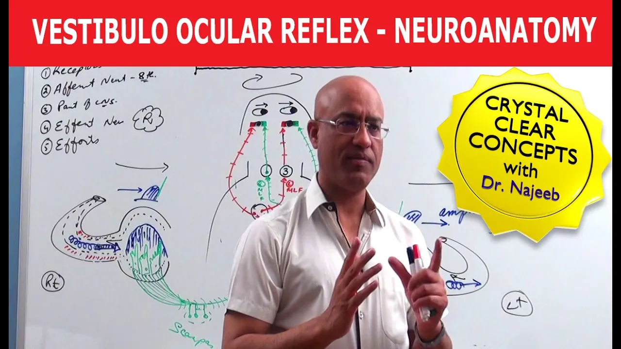
The vestibulo-ocular reflex is an interesting reflex. Let me show you an example, just turning on the camera here. Now, I’m looking at the camera, I’m looking at you guys. As I’m moving my head, I am able to fixate my gaze at the camera while moving my head in a horizontal direction.
This is what the vestibulo-ocular reflex is all about. You don’t really think about it but it’s quite interesting. Let’s see how this works. Let’s add the eyes along with the extraocular muscles.
Within the brainstem, you’ll find nuclei of the 6th cranial nerve and the 3rd cranial nerve.
From the medial vestibular nucleus, fibers are gonna synapse with the contralateral 6th nerve nuclei. And if you’ve followed my videos, you already know that the 6th cranial nerve is the abducent nerve innervating the lateral rectus muscle. The 6th cranial nerve is now going to cross over and give stimulation to the 3rd cranial nerve, the oculomotor nerve, and it’s going to innervate the medial rectus muscle. While this is happening, the contralateral parts of it is going to send inhibitory neurons to the other side. So that was how my vestibular system and my eyes were able to work together to fixate my eyes while moving my head.
So here’s the thing, when we’re doing horizontal movements, that is mainly gonna be regulated by the 6th cranial nerve and the 3rd cranial nerve. But when you’re jumping up and down or turning your head up and down, that is controlled by the 4th nerve nucleus and the 3rd nerve nucleus. All of those nuclei contribute to the vestibulocochlear reflex, and those fibers are all named medial longitudinal fasciculus. So that’s the vestibuloocular reflex.
Then we have the vestibulospinal reflex. The vestibulospinal reflex is a reflex that helps you maintain your posture and resist gravity. And there are two fibers. Form the lateral vestibular nucleus, there’s the lateral vetibulospinal tract. Fibres descend ipsilaterally though the anterior funiculus of the same side of the spinal cord, synapsing on the extensor antigravity motor neurons. Then from mainly the medial vestibular nuclei, we got the medial vestibulospinal tract that descend bilaterally. And it’s gonna innervate muscles of the cervical and upper thoracic spine. What’s special with the medial vestibulospinal tract is that it’s going to descend in the medial longitudinal fasciculus.
Alright, let’s move on to the vestibulocerebellar reflex.
The cerebellum you know is in control of our posture, our muscle tone and our balance. So what happens is, fibers from the inferior vestibular nuclei are gonna travel through the inferior cerebellar peduncle. So here’s the cerebellum, and here’s the fiber travelling through the inferior cerebellar peduncle. There are fibers that come directly into the vestibulocerebellum aswell, but those fibers are going to go to the flocculonodular lobe and the vermis of the cerebellum to give the cerebellum he vestibular sensation. This is important this is a two way connection, meaning fibers also come from nucleus fastigius from the cerebellum into the vestibular nuclear complex to involve the cerebellum to influence the vestibulospinal tract.
Alright then lastly we got the vestibulocerebral pathway, or the vesitbulothalamic pathway, which sends information to cortical regions of the brain known to be involved with vestibular processing.
So fibers will go up to the thalamus, specifically the ventral posterior medial part of the thalamus, as you see here. And then from here they can go to the internal capsule which sends fibers further to different cortical regions. It is believed that it’ll go to the frontal eye fields, which controls the eye movements through the paramedian pontine reticular formation and receive vestibular motion information. It can go to the primary somatosensory cortex helping with basically mapping the bodys location and movement. It can go to the Parieto-Insular Vestibular Cortex, which helps with basically responds to body and head motion information, it can go to the posterior parietal cortex, which is the motion perception and responds to both visual and vestibular motion cues, and it can go to the hippocampus and parahippocampul regions, which helps with spatial orientation and navigation functions.
Alright so that is briefly the vestibular part of the vestibulocochlear nerve.
Let’s do the cochlear part, or the hearing pathway. Here again we got the brainstem, and the vestibulospinal tract coming in through the pontomedullary junction. The cochlear nerve will then diverge and synapse with the dorsal and ventral cochlear nuclei within the upper part of the medulla. We’ll keep this as simple as possible and give you the brief idea of how this works.
So imagine these nuclei receive fibers from different parts of the spiral organ in a definite sequence according to the frequency of the sound waves. From here, most of the fibers cross to the opposite side and form the lateral lemniscus, but some remain uncrossed.
The crossing of fibers of the two sides form a mas of fibers called the trapezoid body.
Some fibers from the cochlear nucleus terminate in the superior olivary complex, which plays a role in localizing the direction of sound by calculating the time difference in arrival of inputs from right and left cochlea. Some cochlear fibers that do not synapse in the superior olivary nuclear complex join the lateral lemniscus after relaying in scattered groups of cells lying within the trapezoid body. In addition to that, some cochlear fibers sends information to cells tat lie within the lemniscus itself, which form the nucleus of the lateral lemniscus.
But now, what happens from here? What happens is, some fibers fo the lateral lemniscus ascend to the midbrain and terminate in the inferior colliculus. Fibres arising in the colliculus enter the inferior brachium to reach the medial geniculate body, like this.
Keep in mind that some fibres of the lateral lemniscus reach the medial geniculate body without going through the inferior colliculus. Now why is the inferior colliculus important? It’s important because it also sends out fibers down called the tectospinal tract. You know whenever someone says something, or yells something, and you turn your head that way and focus your head on wherever the voice came from? That reflex or movement when you move your head and neck so you can fixate your gaze somewhere from where that auditory stimulus came from. That’s called the auditory reflex.
And this is helped through something called the tectospinal tract. Now. From the medial geniculate body there are radiations that will be sent out from this medial geniculate body called auditory radiations. And they’ll be sent out towards the primary auditory cortex, or the superior temporal gyrus. So it makes you aware of the actual sounds, helps you perceive the sound around you.
There are other areas around it called auditory association areas giving you the ability to determine the meaning of the sound. But there’re other structures here aswell. You know in order to comprehend the sound, there’s a structure here posterior to it called Wenicke’s area.
This area plays a role in comprehension of speech. So if someone is talking to you, you’re hearing them, you become aware of the sound, that’s the primary job of the primary auditory cortex, right? And you use association areas sending fibers back and forth to give the sound meaning.
Then the wernicke’s area are gonna receive some of those fibers to help us comprehend this speech, then what do you wanna do? You don’t wanna just sit there and not respond to the speech.
The wernicke’s area talk to another area of the brain called the brocas area, primarily located on the left hemisphere. So right here in the frontal lobe there’s another nucleus called brochas area. He’s the one that controls the muscles of speech. So what happens is, you get the sound stimulus, goes to the primary auditory cortex to perceive the sound stimulus.
You also have the auditory association areas to help us associate that sound stimulus to previous memory and help us to understand the importans of it. Then it can go to wernickes area to help us comprehend that speech. Now from the wernicke’s area, through the arcuate fasciulus, it connects to the brocas area. And the brocas area is then going to tell the primary motor cortex alogn with other parts of the cortex, to go to muscles that control speech, so that you’re able to talk back to respond to the person. Alright. So that was a brief overview of the vestibulocochlear nerve pathway. Here again is the scheme of this nerve. It’s just schematic, not exactly anatomically correct but I hope it’ll help you gain a good understanding of this nerve.
So, we now covered the vestibulocochlear nerve. The next video is going to be about the ninth cranial nerve, the glossopharyngeal nerve. Thank you so much for watching another one of my videos. If you enjoyed, learned something from it, please remember to like, comment your favourite moment, subscribe. Turn on those notifications. If you are looking for other ways to support, go ahead and check out the link in the description box. Have fun ya’ll. Peace.
Up Next

Fiber Pathways of the Cerebrum: Association, Projection, Commissural Tracts
@AANSNeurosurgery
39.5K views•2016-09-29

Deciphering Neural Circuits Controlling Anorexia in Mice | Richard Palmiter
@AllenInstitute
2.3K views•2011-10-10

Vagus Nerve (CN X): Anatomy, Nuclei & Functions Explained
@Alilamedicalmedia
305.2K views•2022-10-31

How Exercise Benefits Your Brain: Science Explained
@TED
11.4M views•2018-03-21
Related Study Plans & Knowledge Roadmaps
Structured learning paths in Neuroscience