


















Mechanical ventilation is categorized into two fundamental types: fully supported modes (such as Assist/Control and PRVC, which set respiratory rate, tidal volume, FiO2, and PEEP) and weaning modes (including CPAP, Pressure Support, and T-tube trials, which reduce ventilator support to assess patient readiness for extubation); successful weaning requires daily spontaneous breathing trials coordinated with nursing and respiratory therapy, with success criteria including patient comfort, stable vital signs, and acceptable arterial blood gas results showing no hypoxemia, hypercapnia, or acidosis.
Mechanical Ventilation Basics for ICU House Officers
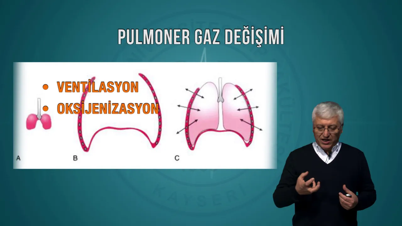
Added:Basic respiratory physiology, including lung compliance, airway resistance, and the mechanics of spontaneous breathing.
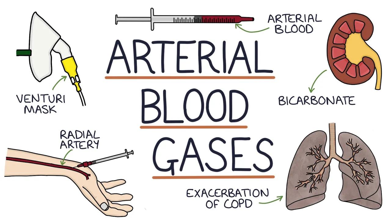
Interpretation of arterial blood gas (ABG) results and the classification of hypoxemic versus hypercapnic respiratory failure.
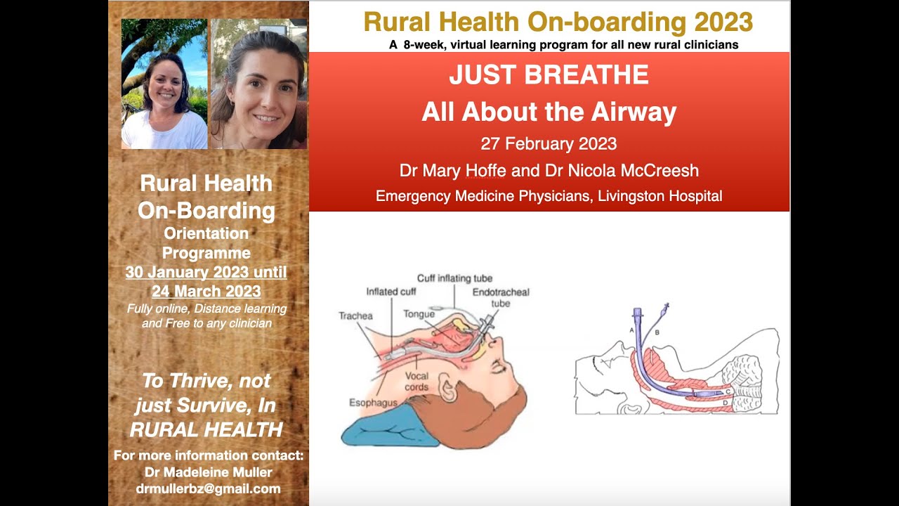
Clinical indications for endotracheal intubation and the fundamental anatomy of the upper and lower airways.
The physiological effects of positive pressure ventilation on hemodynamic stability, specifically venous return and cardiac output.
Advanced mechanical ventilation strategies, including lung-protective ventilation for ARDS (low tidal volume) and obstructive lung strategies for COPD/Asthma.
Identification and troubleshooting of patient-ventilator dyssynchrony (e.g., double-triggering, autotriggering, and flow starvation).
A systematic clinical approach to troubleshooting ventilator alarms (such as high peak airway pressures) using the DOPE mnemonic.
The protocolized steps for extubation, managing post-extubation stridor, and transitioning patients to non-invasive ventilation (NIV) or high-flow nasal cannula (HFNC).
842.8K views6.9Klikes18:17Original Release: 2015-05-16
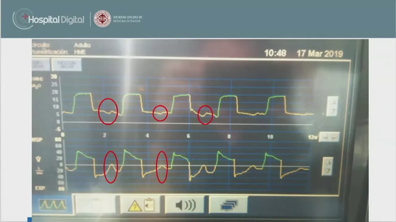
this is our educational video focusing in on introduction to the patient who requires mechanical ventilation this is aimed at a house officer with their first experience being in an intensive care unit next to me here is the ventilator and later on in the video we're going to be focusing in on some of the specifics of how you can make adjustments to the ventilator when you're here in the intensive care unit in most units you'll be working in and specifically this particular intensive care unit you actually shouldn't be making specific changes to the ventilator you should be coordinating with the nursing staff and the respiratory therapists as well as the supervising intensivist and so that everybody's on the same page it can often be quite confusing if numerous members of the multidisciplinary team are making adjustments in terms of specifics and how we want you to think about patients who are on mechanical ventilation we try to break things down into two fundamental types of mechanical ventilation fully supported modes and then weaning modes fully supported modes the classic mode is assist control and there are certain variables that are set when a patient is on assist control you said a respiratory rate a tidal volume and fio2 and a peep level and we'll go over some of that more when we talk about the ventilator but for you as a trainee as a house officer it's important to understand some basic settings when a patient first comes into the intensive care unit perhaps from the emergency department or from the operative theatre you would want that patient to be on your units version of assist control so that you have set them on usually starting at a hundred percent oxygen and working with the respiratory therapist to wean the oxygen down you'll set them on some sort of reasonable rate twelve fourteen sixteen breaths per minute you'll set a tidal volume usually in the 300 to 400 milliliter range sometimes higher but as you'll see for patients with a RDS where you're aiming at six MLS per kilo predicted body weight and their tidal volumes will be much lower than that and almost all patients are set on a peep level physiologic of starting out at five centimeters of water and for the majority of patients specifically here in a surgical intensive care unit you can leave them on these low levels of peep and they won't require further adjustments patients who develop acute respiratory distress syndrome often require higher levels of mechanical ventilation in this particular intensive care unit our most commonly used ventilator mode is what's known as prvc pressure regulated volume control and you can get through the whole rotation and think of it as a form of assist control but it's actually a complex mode and it's what's called a dual mode of mechanical ventilation in which you are setting it as though it is a volume targeted mode but it's really a pressure targeted mode where the computer in the ventilator is titrating the pressure target up and down to deliver the tidal volume that is set by the clinician and although that can be a bit of a mouthful the idea is there are real benefits from a patient standpoint of using a pressure targeted mode of mechanical ventilation first of all it gives them what's called demand flow as they breathe in more they get the flow they need since you're focusing in on a particular pressure and you get most of your flow upfront in addition as the patient's a pulmonary compliance changes the amount of tidal volume they'll get can change from breath to breath if you're using a mode like prvc there are some times where you will see the intensivist and the rest para pused work with each other and disengage the patient off of prvc and formally switch them to either a volume control mode or a pressure targeted mode like pressure control and it really is like taking a car and switching them from an automatic transmission to more of a stick shift where we want to specifically either dial in a pressure or dial in a volume and have a volume delivered rather than have the ventilator dial up and down on a pressure to have it try and get that tidal volume that we dialed in what we'd like to do here is give you a tour of the maquette servo live ventilator this is the ventilator that we happen to use in all of our adult intensive care units here at the Medical Center this is a standard setting we have an artificial lung here and this is as you can see the patient is set on prvc pressure regulated volume controlled the patient is set with an fio2 of 60% there's a peep of five set and you would change these by dialing the little knobs here up and down there's a set rate of 15 and the tidal volume is set at approximately at exactly 450 milliliters if you press this button here this is the standard mode that you'll see on this particular ventilator and this is telling you what the patient is actually getting so for example you can see that the tidal volume is going in our 454 MLS and the tidal volumes coming back are 458 MLS these numbers should be very close to each other and if they're not something needs to be looked into either the patient has a problem with their endotracheal tube balloon or the patient has some sort of Bronco pleural fistula of some kind where air is leaving the patient not coming back through the lumen of the endotracheal tube this is documenting that the patient is still on 60% oxygen that they are actually getting the oxygen you want them to be getting and there will be discrepancies here sometimes for example when you first make changes to the amount of oxygen that you're requesting as the oxygen blender dials up and down here is important documenting the ite ratio and this is normal the 1 2 3 ratio in terms of inspiration and exhalation and this is important I'm always asking this on rounds what are we set at and what are we breathing at because this is very important depending on what the arterial blood gas shows are we over ventilating the patient are we under ventilating the patient are we over sedating the patient next we're going to demonstrate how you do an inspiratory pause to show a plateau pressure as we've talked about on rounds the differential diagnosis of patients that have a high peak airway pressure includes those patients that have a high peak airway pressure with a high plateau pressure and those that have a high peak airway pressure with a normal plateau pressure so my assistant here during the next breath is going to do an inspiratory pause and you can see that the plateau pressure is 16 and the peak airway pressure was 19 and so the overall is that that is particularly its remaining elevated but the point is is that neither the peak nor the plateau were abnormal in this particular case if somebody has a high peak airway pressure and a high plateau pressure it means the problem is intrinsic either to the lung or to the chest wall and if it's a high peak airway pressure with a normal plateau pressure you've taken away flow and therefore it's problems with airway resistance or somebody's biting on the endotracheal tube we've now set it up so that the ventilator with my colleague here is in pressure support mode in that mode the patient is designating and triggering all of the breaths and you can tell that yourself as the clinician by seeing this little purple uptick which is the patient triggering event we'd like to take a few moments here and describe to you what each of these waveforms refers to this is called the pressure scaler this is called the flow scaler and this is called the volume scaler as you can see starting from the bottom with each breath the volume scaler goes up and as the patient exhales it goes back down to zero the green here showing that with each breath during inspiration it's positive and during exhalation it's negative and it's important that you see that it goes back down to zero indicating that we don't feel there's any obvious auto-peep that's an important issue and this is the pressure scaler showing that the pressure goes up and although it does go back down quickly during exhalation because the peep is set at five it doesn't go back to zero this is an example a ventilator to synchrony this is an important thing for you to be aware of in the intensive care unit and there isn't one particular answer the patient needs to be assessed to make sure that the tube has not been dislodged or has does not have mucus plugging or that the patient isn't in pain or having breath stacking for other reasons it's very important that the patient be quickly assessed if you see this phenomenon here we have a patient on pressure support ventilation during a spontaneous breathing trial we're watching closely for any evidence of either tachypnea or brittany a' to see if there's problems where the patient may be failing as you can see there are fairly prolonged periods between breaths this is a sign that the patient may be over sedated or still have residual narcotics on board or sedatives and you can see that the tidal volumes generated are quite small this may show a sign this may be a sign of neuromuscular weakness or again that the patient has not fully recovered from anesthesia it's very important to be able to recognize some of these abnormalities on the ventilator in this particular ventilator if we wish to do a formal CPAP trial as you can see here you set the pressure support to zero and you keep the people what you would like the patient can take breaths at whatever rate they wish the demand valve is open and as you can see the ventilator is doing its best to keep the pressure at the mouth opening at five centimeters of water you can see that there is flow going into and out of the patient and tidal volumes are being generated but they're not being particularly generated at a particular tidal volume nor is a particular pressure set above the peep level this is an example of what the servo eye looks like if you set the patient on pressure control again as I mentioned before you don't set a tidal volume you set a pressure control level above a certain peep and here you for example we're on pressure control 12 over a peep of five with a total pressure target of 17 the respiratory rate is set and the fio2 is set and you set an inspiratory time which is equivalent to setting an i to e ratio which you can see here is one two three but again this is another fully supported mode and you would do this for example in a patient who had severe acute lung injury where you wanted to not allow the peak and plateau pressures to become very elevated this is an example of what is known on many ventilators as aprv airway pressure release ventilation it is somewhat confusing at first because for those of us who care for patients who are critically ill day-in day-out we're used to setting a respiratory rate a tidal volume and an fio2 and a peep and although you do set an fio2 in this mode almost everything else is different and the basic way when we teach about this this particular mode is high levels of CPAP with release breaths and there are four variables that you use when you're setting up a patient on this particular mode appiy high at E high a pillow and at E low and these are some of the basic settings that you would choose a pea high of 15 or 20 centimeters of water a peep of zero and the idea is that you never actually get down to that level of peep before your next breath begins at E high which is set at five or six seconds and a time for release breaths which is usually less than a second there are more details that you can read about in terms of this mode how you exactly set the time for the release breaths depending on whether or not the patient has a chronic obstructive pulmonary disease or acute lung injury in terms of the rapidity with which the air leaves the patient as you can see here on the pressure scaler it is high levels of continuous positive airway pressure with with release breaths and the concept is that the patient is exchanging carbon dioxide and oxygen throughout the cycle and that's the theory behind aprv or by vent is that you are constantly doing what you can to open up small alveoli and small Airways without injuring the overall lung parenchyma one of the important things to mention about by vent or aprv is that it is another one of these modes that is considered very patient friendly and the patient as you can see here can take their own breaths throughout the respiratory cycle and there are some ventilators in this mode where you can actually add a pressure support level to each one of these breaths the next thing I'd like to speak about which is very important is the concept of a daily spontaneous breathing trial as we've talked about in some of the other videos this is coupled with a daily sedation vacation but in this particular video I'd like to focus in on the spontaneous breathing trial component it's very important when you're on this rotation early in the morning that you coordinate closely with the nursing staff and the respiratory therapist and coordinate explicitly I'd like to coordinate a spontaneous breathing trial on this patient there are multiple right answers you can either put the patient on low levels of pressure support and we'll talk about pressure support in a little bit you can put the patient on formal CPAP continuous positive airway pressure or for certain patients such as subgroup of patients who have a low ejection fraction you might want to put them on a formal t-tube trial and I'll talk about three different types currently the first is a formal t-tube trial this is the oldest way of doing a spontaneous breathing trial in which you coordinate with the respiratory therapist and politely ask them if they could put the patient disconnect them from the ventilator and they put corrugated tubing and they patient breathes on their own one might say well what about the work of breathing of the endotracheal tube itself and and there have been studies looking at this and the basic idea is if the patient can't tolerate a tea tube trial they may have difficulty being extubated so the advantages of a tea tube trial is that you have taken all of the support away from the patient you are really doing your best to see if this patient can do well off of the ventilator as I mentioned before the reason you would do this in a patient who had evidence of congestive heart failure or decreased systolic function is that low levels of positive pressure ventilation can often be enough to have a look like they're doing well and then when you extubate the patient they can develop flash pulmonary edema and require immediate or urgent reintubation so that's a t-tube trial usually we'll watch a patient for 30 40 minutes or so and if they're comfortable and they don't appear to have either subjective or objective criteria of failing including becoming too kipnuk tachycardic anxious agitated hypoxic the idea here is if the patient looks good after 30 or 40 minutes and you check an arterial blood gas and they don't show evidence of either hypoxemia or hypercapnia or acidosis then the patient should be extubated this is the concept of a spontaneous breathing trouble we're not slowly weaning patients from the ventilator as was done 15 or 20 years ago continuous positive airway pressure can be set on the ventilator and the idea is as I've been taught this is sticking your head out of a car window going 50 miles an hour the machine isn't trying to determine when you're breathing in and out it's just there to keep a certain amount of pressure at the mouth this is another reasonable way to do it the most commonly used approach at least in this intensive care unit is low levels of pressure support so let me talk for a couple of minutes now about pressure support ventilation and the way I like to describe this is if I'm the ventilator and you're the patient pressure support mode the variables that are said are different from assist control in pressure support you still set an fio2 and you set a peep but you don't set a respiratory rate and you don't set a tidal volume you set a pressure support level over a certain people 'evil and what the Machine does is sense when you're taking a breath by the change in flow in the circuit and then it quickly delivers flow until it gets to that pressure target that you've asked for and then it starts to decelerate that flow when the flow rate gets to 25% of the peak inspiratory flow rate the ventilator shuts off and you exhale that's what pressure support ventilation is it's a very patient comfortable mode it's a very patient friendly mode some of the downsides are you're not dialing in a particular tidal volume however that having been said most modern ventilate have a minute ventilation alarm that if your minute ventilation goes too low it will alarm and click into a fully supported mode so from our perspective there are two kinds of ventilator modes fully supported modes such as assist control or prvc and weaning modes and weaning modes that we've described are three major modes that can be used during spontaneous breathing trials including pressure support ventilation formal CPAP trials and t-tube trials
Up Next
55:06
SEVA VentRounds: Reviewing Ventilator Settings for Effective Mechanical Ventilation
@clevelandclinic
1.4K views•2021-04-02

48:47
Graphic Medicine: Comics for Collaborative Healthcare Communication
@nationalpatientadvocate
189 views•2023-12-04

10:28
Neuroanatomy: Central and Peripheral Nervous System Divisions Explained
@AKLECTURES
136.2K views•2014-09-20

3:05
Stages of Labor and Vaginal Birth | Childbirth Animation
@nucleusmedicalmedia
52.1M views•2017-08-18
Related Study Plans & Knowledge Roadmaps
Structured learning paths in Medicine
medicine
Abnormal LFTs: Diagnostic Workup & Pathology
01Liver Anatomy and Basic Physiology
02Introduction to Liver Function Tests (LFTs)
03Hepatocellular vs. Cholestatic Patterns
+ 3 more learning modules
12h6 modules
Explore Roadmap
medicine
Analyzing Blood Smears: RBC, WBC & Platelets
01Introduction to Blood Components & Smear Preparation
02Normal Morphology of Blood Cells
03Erythrocyte Pathologies & Morphological Variants
+ 2 more learning modules
12h5 modules
Explore Roadmap
medicine
Mechanical Ventilation: Waveforms & Alarms
01Anatomy and Physiology of Breathing
02Introduction to Mechanical Ventilation & Core Modes
03Ventilator Settings and Waveform Interpretation
+ 2 more learning modules
12h5 modules
Explore Roadmap
medicine
Neuro Exam: Localization & Stroke Syndromes
01Foundations of Neuroanatomy and Vascular Supply
02The Neurological Examination: Step-by-Step
03Principles of Neurological Localization
+ 3 more learning modules
18h6 modules
Explore Roadmap