



![USMLE Step 1 - Renal Physiology [High Yield BRS Concepts]](https://i.ytimg.com/vi_webp/Ob_g35rqOmA/maxresdefault.webp)


![RENINA-ANGIOTENSINA ALDOSTERONA [Renal 02]](https://i.ytimg.com/vi_webp/bBHDNldjONU/maxresdefault.webp)

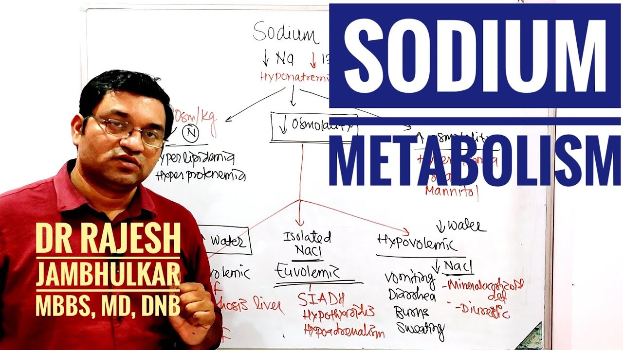


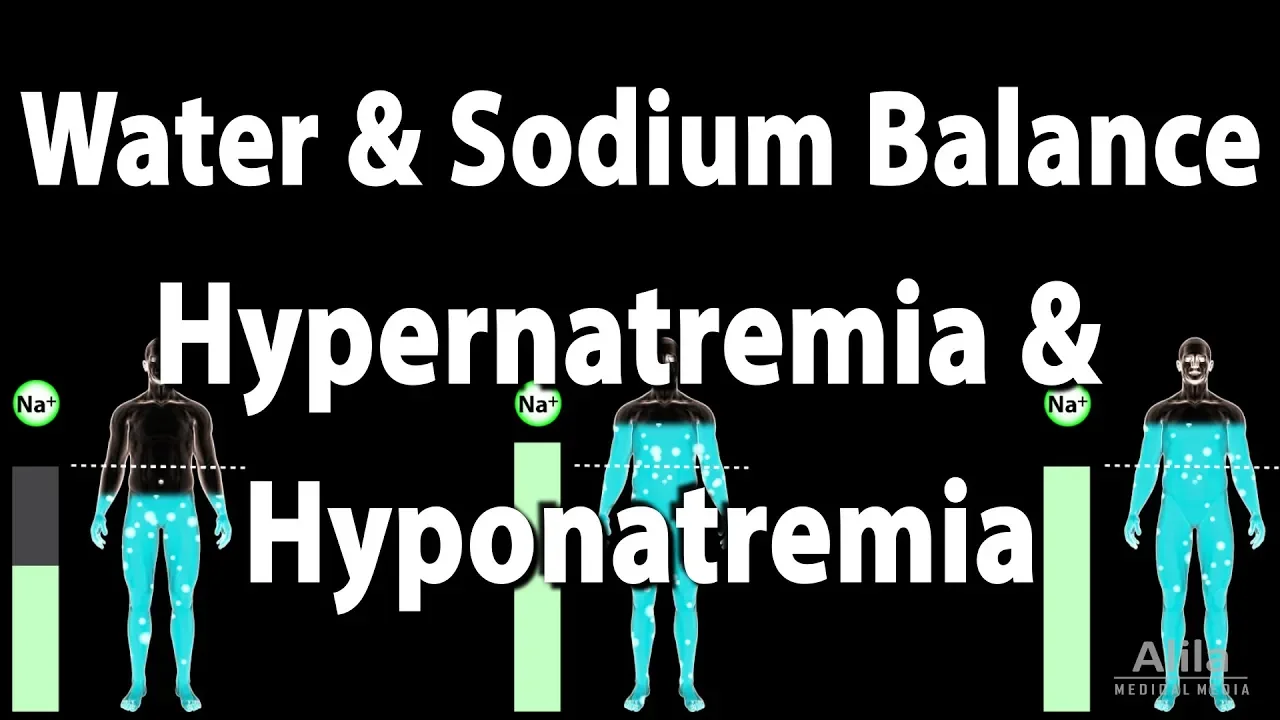







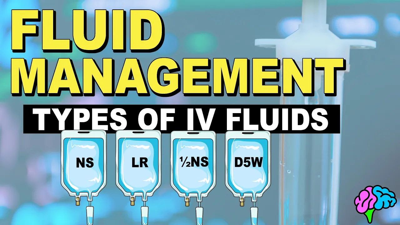
Hyponatremia (serum sodium <135 mEq/L) is primarily caused by increased circulating ADH or renal sensitivity to ADH combined with free water intake, leading to dilutional hyponatremia. It is classified into three types based on plasma osmolality: hypertonic hyponatremia (>290 mOsm/kg) from effective osmoles, isotonic hyponatremia (275-290 mOsm/kg) as a laboratory artifact from high lipids/proteins, and hypotonic hyponatremia (<275 mOsm/kg) as true physiological hyponatremia. Further classification by volume status includes hypovolemic hyponatremia (extra-renal causes like vomiting/diarrhea, renal causes like thiazides/mineralocorticoid deficiency), hypervolemic hyponatremia (heart failure/cirrhosis/nephrotic syndrome), and euvolemic hyponatremia (SIADH being the most common cause). Clinical features include neurological symptoms from cerebral edema, with acute symptomatic hyponatremia being a medical emergency. Treatment varies by type: IV saline for hypovolemic, diuretics for hypervolemic, and underlying cause treatment for SIADH. Critical to avoid rapid correction (>8-10 mEq/L/24h) in chronic cases to prevent osmotic demyelination syndrome.
Hyponatremia: Causes, Diagnosis & Treatment | Clinical Guide
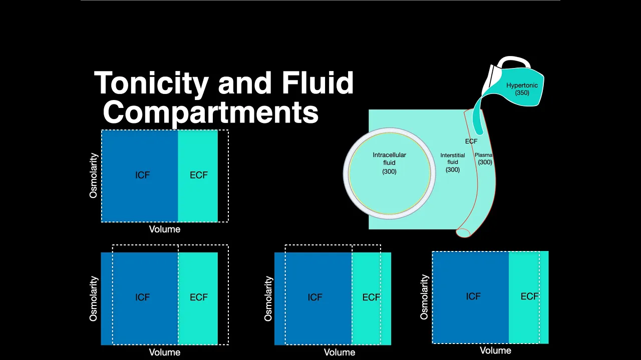
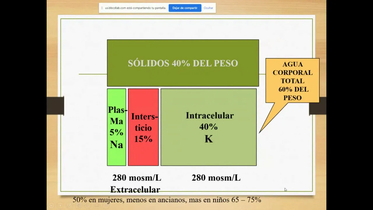
Added:[Music] hi friends today we are going to discuss a very important topic in fluid and electrolytes that is hyponatremia first of all we should knowe that disorders of serum sodium concentration are caused by abnormalities in water homostasis this lead to changes in the relative ratio of sodium to that of body water so the change in this ratio that is to that of serum sodium to that of body water is responsible for the sodium disorders namely hyponatremia or hyponatremia so the two key factors in the defense of serum osmolarity are first one is water intake and second is circulating AVP levels so the defect in any one or both of these leads to hyponatremia or hyperia so let's begin our disc discuss of hypona in this discussion we will first have a look on intro then we will have in detail discussion of hypo volic hypona hyper volic hypona UIC hypona low solute intake hyponatremia which is a very rare cause clinical features of hyponatremia then we will move on to diagnostic evaluation in which we will see the history examination Radiology blood investigations and urinary investigations finally we will come to treatment in which we will first see what is the treatment according to the type how water deprivation helps potassium correction increase in dietary solute intake what is the treatment of siadh acute symptomatic hyponatremia and the treatment of chronic hyponatremia first of all let's have a look on some terminology sodium determines ECF volume this is a very important point and this ECF volume comprises of our intravascular volume as well as extra vascular or interstitial volume so hypo volia means volume is depleted and the total body sodium will be deficit since the sodium is determining the ECF volume on the other hand hyper volumia means volume overloaded State and there is excess total body sodium on the other hand u volumia means volume is normal and there is normal total sodium so this does not mean that these patients cannot have hyponatremia since the sodium levels will be measured in only intravascular volume that is serum sodium this do not correlate with volume of the patient volume status of the patient since the volume status is of extracellular fluid volume which comprises both intravascular as well as interstitial also known as extravascular fluid volume so this concept is clear now hydration impacts the cell volume if a person is overhydrated then the water intoxication will happen this will lead to cell swelling on the other hand dehydrated patient will have water depletion leading to cell shrinkage so starting with the introduction hyponatremia is defined as the plasma sodium concentration less than 135 m per liter it is a very common disorder which is occurring in around 22% of hospitalized patients and this is a very large figure when we talk about the cause it is mainly due to increased circulating AVP that is ADH or increased renal sensitivity to AVP along with increased intake of free water so what will this AP do this will retain the free water so so the free water will retain and will lead to dilution leading to hyponatremia rarely hyponatremia can occur due to low salt intake in the diet so what is the most common reason leading to hyponatremia in this cases this is due to nonosmotic release of ADH since in the hypo Alia lecture we discussed that ADH can release due to osmotic stimuli and non-osmotic stimuli mainly due to hypo volia so here the hypo vol IA also known as nonosmotic stimuli will lead to release of ADH now coming on to classification broadly it is classified into hypertonic hyponatremia that is along with HP hyponatremia the total tonicity is increased second is isotonic hypona that is despite hyponatremia the tonicity is normal and third one is hypon hypotonic hyponatremia and this is known as true hypon which we will be discussing in this lecture so let's briefly discuss all three entities that is hypertonic hypona which is said when the osmolality is more than 290 mosmol per kg of water isotonic hypona which is when the osmolality is normal that is 275 to 290 and hypotonic or true hyponatremia when the plasma osity is less than 275 mosmol per kg so this height hypertonic hypon nmia is due to the presence of another effective osmo that causes free water to move from the ICF to the ECF leading to hyponatremia so this free osmol can be a osmotic diuretic like manitol glycine or marked hypoglycemia so what these conditions will do this these will cause free water to move from the ICF to ECF leading to D dilutional hypon netrium so the treatment will be correction of the underlying condition or to remove the offending agent now coming on to the isotonic hypon Neta this is also known as pseudo hyponatremia because it is a laboratory artifact this is due to marked increase in serum lipid or proteins which results in reduction of fraction of serum that is water and is showing artif ially low serum sodium so since the plasma is reduced in the sample and lipid and proteins are increased the relative sodium concentration will be decreased in this the plasma water sodium concentration osity are unchanged that is normal but the measured sodium concentration in the total plasma volume will be reduced because the given sample contains less plasma water and will also contain lipids and proteins so it is very important point that only Flame photometry or indirect photo potentiometry can lead to this pseudo hyponatremia or isotonic hyponatremia so it can be corrected when we use direct potentiometry and ion specific electrodes these will avoid this misdiagnosis now coming on to hypotonic hyponatremia this occurs when there is plasma ality is less than 275 and it is the true hyponatremia true physiological hyponatremia that will result from excess water either due to ADH stimulation or impaired water excretion so in this hypertonic hyponatremia what happens is there is a presence of effective osmo in this case this is glucose so this will lead to increased in osmolality or tonicity so the plasma osity will be increased this leads to water flow from from ICF to ECF ICF to ECF due to presence of this etic osmo and this water will lead to dilution of the plasma leading to dilutional hyponatremia on the other hand in isotonic hyponatremia there will be increase solid content that is increase fat and protein content due to use of flame photometry or indirect potentiometry so the water content containing the sodium will be decreased it will be decreased in this case in this case and the sodium measurement will be reduced as in comparison to the total flute on the other hand in hypotonic hyponatremia there is no presence of effective osmo and it is a true hyponatremia and there will be water flow from ECF to ICF due to low plasma osmolality low plasma osmolarity now our whole lecture will be towards the hypotonic hyponatremia so this hypotonic hyponatremia on the basis of volume status is further divided into hypo volic hypon Neta UIC hypona or hyper volumic hypon Neta which we'll be discussing one by one now coming on to discussion of hypo volume Hy hypona this is due to True volume depletion that is low ECF volume so when there is loss of fluid volume including the sodium from the ECF volume particularly intravascular volume this will stimulate the nonosmotic release of ADH nonosmotic release of ADH now this ADH will help preserving the BP of the patient so this BP will be preserved with the help of receptors V1 a and V2 so V1 a is vascular and Baro receptor and V2 is renal receptor for ADH now due to this V2 receptor the activation of this receptor can lead to hyponatremia and the setting of increased free water intake so what is happening is due to any cause of hypo volia there will be initially decrease in total body water and as well as decrease in total body sodium due to True volume depletion equal loss will be there but this hypo volia will lead to nonosmotic release of ADH nonosmotic release of ADH and this ADH will correct the hypo volumia to a certain level so that the total body water will be somewhat increased but the total body sodium will be still decreased so we get this equation the total body water is decreased but the total body sodium is much more decreased relative to that of water this will lead to hyponatremia in the setting of hypoa so according to the urine sodium we can divide the causes if the urin sodium is less we will have extra renal causes if the urin sodium is more we will have renal causes so when the urine sodium is less than 20 M Mo this will Direct to extrenal causes extrenal causes of hypo volum hyponatremia why because due to extrenal law of fluid and sodium and the kidneys are normally functional the body will try to preserve the sodium therefore less extration of sodium in the urine so this include GI causes like vomiting diarrhea there can be insensible losses in the form of burns sweating this will lead to insensible water loss there can be third spacing of fluids example in peronal cavity other causes include pancreatitis trauma which will lead to True volume depletion and further stimulation of ADH now the important part about this extral losses is the salign treatment when we give the normal salign to these patients there will be inhibition of ADH inhibition of ADH which was released due to non-osmotic stimulation and there will be free water diuresis free water diuresis and correction of hyponatremia so it is a very important point that salign treatment will lead to free water diuresis in these patients and will lead to correction of hyponatremia indeed when there is a urine sodium concentration less than 20 m in the absence of a cause of hyper volia hyper volia this is a case of hypo hypona so whenever we get a patient with urine sodium less than 20 M and on examination he is not hyper volic there is no edema then consider the diagnosis of hypo volumic hyponatremia with a extra renal cause now when the urine sodium will be more than 20 M these will be the renal causes of hypo volumic hypona so the renal causes lead to an inappropriate loss of sodium chloride in urine leading to volume depletion and an increase in circulating AVP so what can be causes there can be diuretic exis so which diuretics can lead to hyponatremia these are thide diuretics so let's discuss why only thide diuretics leads to hyponatremia first of all use of these diuretics will lead to polydipsia and volume depletion now this volume depletion will stimulate the AVP so this increased AVP in the setting of polydipsia will lead to hyponatremia now these do not affect the countercurrent mechanism do not affect the countercurrent mechanism so that AVP can function in a normal way in water reabsorption through the collecting duct through its V2 receptor on the other hand the Loop Diuretics inhibit the sodium potassium to Chloride channel in the thick ascending limb of loop of hel and this will alter the counter current concentrating mechanism in the medula part medula part therefore Loop Diuretics do not lead to hyponatremia usually because the AP cannot act through the collecting duct because there is loss of countercurrent mechanism and renal concentration of medula so after diuretic mineralocorticoid deficiency that is elderone deficiency can lead to hyponatremia so this occurs due to primary adrenal insufficiency so the eldone will be deficient and the sodium will not be absorbed and will be lost in the urine also the associated finding will be hyper calmia that is increase serum potassium because it will not be removed from the body in exchange to that of sodium it is important to note that primary adrenal efficien deficiency leads to eldone deficiency leading to hypo volic hyponatremia on the other hand secondary adrenal insufficiency will lead to glucocorticoid deficiency and it will be a cause of UIC hypona so other cause include salt losing nephropathies like reflux nephropathy intial nephropathies post obstructive uropathy medular cystic disease and Recovery phase of these will also lead to hypo volic hyponatremia other renal cause of hypo volic hyponatremia include osmotic diuresis so any cause of osmotic diuresis be it glycosuria seen in setting of diabetes malius or ketona seen in Starvation keto acidosis diabetic keto acidosis or alcoholic keto acidosis or Bic carbonia with renal tubular acidosis and metab olic alosis so how this bicarbonate excretion will lead to hypona this will take sodium along with the negatively charged bicarbonate ions leading to loss of sodium through the kidney so all these are the causes of hypo volic hyponatremia now very important cause is cerebral salt wasting syndrome cerebral salt wasting syndrome it is a very rare cause of hypo volumic hyponatremia in this any intracranial disease can lead to clinical hypo volia and inappropriate netri orasis so till now we have discussed that hyponatremia is occurring due to stimulation of ADH along with free water intake but this is the exception in the form that it will lead to netri uresis that is loss of sodium so what intracranial diseases can lead to cerebral salt wasting syndrome these can be subarid hemorrage traumatic brain injury craniotomy entis or menitis so distinction from siadh is important s cerebral salt wasting syndrome should be distinct with SI ADH because cerebral salt wasting syndrome will respond to sodium chloride repletion when we give sodium chloride this will be corrected on the other hand SI ADH will not be corrected now coming on to hyper volumic hyponatremia in this condition there will be total body volume exis that is the ECF volume will be increased but the intravascular volume depletion will be there despite of the increased ECF volume there will be intravascular volume depletion which is seen in edima States like curosis cardiac failure rotic syndrome so this intravascular volume depletion will stimulate the ADH this stimulates the ADH and retains free water so since there is hyper volumia and we have already discussed that sodium concentration is equivalent to volume so in hyper volia there will be increased sodium concentration in the whole body so there will be increased total body sodium chloride but it will be accompanied by proportionally greater increase in total body water so there will be decreased plasma sodium means the total body water will be much more proportionally increased in comparison to the total body sodium so what will happen is if this is our total body water and this is total body sodium so in this condition the total body water is increased and total body sodium is also increased but when we compare the intravascular water it is decreased so this will stimulate the ADH this will increase the intravascular volume but the total body water will increase to a greater strength on the other hand ADH will not affect the total body sodium and it will be only single increase so there will be two Arrow increase in total body water and single Arrow increase in total body sodium so the causes can again be differentiated based on urinary sodium if it is more than 20 mm due to dysfunctional kidney and the kidneys are not able to reabsorb the sodium the causes will be acute of chronic renal failure and if the urine sodium is less than 20 mm that is urine kidneys are able to retain the sodium by reabsorption the causes will be nephrotic syndrome sosis and cardiac failure and it is important to note that in these cases the urine sodium is typically very low less than 10 m even after hydration with the normal saline but diuretic therapy obscures the diagnosis if we give diuretic in these cases the urine sodium will be falsely increased and this will obscure the diagonosis of these sosis cardiac failure and nephrotic syndrome so diuretic therapy should be withdrawn if there is any confusion between the diagnosis so that actual urine sodium can be measured now coming on to U volumic hypon netria why the name U volumic because there is normal eccf volume but strictly speaking there will be primary water gain in these conditions so these patients will be U volumic on exam that is these will not have any edema but the total body water will be increased due to activity of ADH and the total body sodium will be normal so this will lead to decrease serum sodium decrease serum sodium now all these conditions are associated with increased urine sodium more than 20 mm and the causes are as follows that is glucocorticoid deficiency due to secondary adrenal insufficiency we discussed that in primary adrenal insufficiency there is hypo volic hypo nmia on the other hand in secondary adrenal insufficiency due to glucocorticoid deficiency there is UIC hyponatremia so the secondary adrenal insufficiency occurs due to any pitutary disease and why this leads to hyponatremia normally these glucocorticoids what these do are these will have a negative feedback on ADH release so due to this glucocorticoid deficiency since these will deficient there will be increase in ADH concentration leading to water retention and hyponatremia and when we give hydrocortisone in these patients the hyponatremia will be corrected other conditions which can lead to UIC honia are hypothyroidism which will correct on achieving the U thyroid State stress drugs and Si ADH so this syndrome of inappropriate anti-diuretic hormone secretion is the most common cause of U volumic hyponatremia and we will discuss this in detail now SI ADH is the most common cause of U volumic hypona the generation of hypona in si ADH requires intake of free water now four distinct patterns of AP secretion are seen in si ADH first one is unregulated and ertic AVP secretion which is seen in 1/3 patients in this there is no obvious correlation between the serum osmolarity and the circulating AP levels second pattern is failure to suppress AVP at lower serum osmolarities third there is reset osmate seen in some patients with lower threshold osmolarity for release of AVP and left shifted osmotic response curve finally in some patients there is no detectable circulating ABP this means that either there is gain of function mutation in renal water reabsorption that is these are reabsorbed without ADH or AP and or there is some circulating anti-diuretic substance that is distinct from from the AVP now strictly speaking SI ADH patients are not UIC but these are subclinically volume expanded now when we talk about serum uric acid this is typically less than 4 mg per de in these patients because there will be increased sodium delivery to the distal tubules distal tubules and by negative feedback that is glom tubular feedback this will inhibit the proximal tubular absorption of sodium and uric acid this will lead to decrease uric acid levels on the other hand when we see the hypo volic hyponatremia hypo volumic hyponatremia there will be decreased sodium delivery to the distal tubules and stimulation of reabsorption of uric acid as well as sodium in the proximal tuule this will lead to increased uric acid levels in hypo volic hypon netri Pria which are typically more than 4 mg per dcil now this is a very big table which include all the causes of Si ADH we will discuss some important causes the most important and most common cause of Si ad are pulmonary disorders so these will include infections like bacterial pneumonia viral pneumonia pulmonary absis tuberculosis and plural infusion so these all pulmonary disorders are a very common cause of siadh some Central Nervous disorders typically meningitis sub aroid hemorrage brain tumors can also lead to siadh now when we consider the malignant diseases the most common tumor associated with siadh is small cell carcinoma of lung this is the most common cause in malignant diseases which is responsible for 75% of malignancy related siadh on the other hand out of all the cases of small cell carcinoma 10% cases are associated with siadh now the most common drugs which are responsible for SI ADH are SSRI SSRI these are selective serotonin reuptake inhibitor other causes include hereditary gain of function mutation in V2 receptor endurance general anesthesia nausea Etc now till now we have discussed all the causes of hyponatremia that is hypo volic UIC and Hyper volic which were associated with AVP activity and increased water intake now low solute intake hyponatremia is not associated with AVP and this is solely due to low intake of sodium so it is due to very low intake of dietary solutes typically seen in Alcoholics whose sole nutrition is beer so this is therefore known as beer poom Mania now this beer is very low in protein and salt content with salt content only 1 to 2 m of sodium other salt restricted diets include tea and toast diet and extreme vegetarian diets so this is associated with very low urine osmolality which is typically less than 100 to 200 mil per kg m per kg and very low serum very low urine sodium concentration Which is less than 10 to 20 M now AVP levels are not been reported to increase in these conditions but if at all increased these will suppress with saline hydration and resumption of normal di so the treatment of these patient is just resumption of normal diet and the serum sodium will correct very rapidly now coming on to clinical features now the clinical features are due to generalized cellular swelling which is due to movement of water from the hypotonic ECF to the ICF the symptoms are primarily neurological due to development of cerebral edema in the rigid skull early symptoms include nausea headache vomiting and severe symptoms include seizures brain stem herniation even coma death can occur respiratory failure in this setting is typically normocapnic or hypercapnic which is due to non-cardiogenic neurogenic pulmonary edema with normal pulmonary capillary Ved pressure now accute symptomatic hyponatremia is a medical emergency which can occur in a number of settings this can occur most importantly in iatrogenic settings which can be seen in post-operative conditions and more common in premenopausal women it can also occur when hypotonic fluts are given with a cause of increase in vasopressin so in a patient who has already increased veso pressence and hypotonic fluids are given in such patient it will lead to acute symptomatic hypon netria sudden increase in serum sodium also glycine Irrigation in turp or uterine surgery and colonoscopy preparation can lead to hyponatremia recent institution of thyoides can lead to sodium loss polydipsia can lead to hyponatremia drugs like easy and moly which have active ingredient MDMA can lead to hyponatremia because there is rapid and potent induction of both thirst and AD ad release so both thirst and ADH will be stimulated leading to hyponatremia now exercise induced hyponatremia occurs because there is non-osmotic release of ADH as well as increased free water intake now this is a very important slide showing the effects of hyponatremia on brain so how does the brain adapts to hyponatremia first of all there is increased in IAL pressure which will shunt the ECF and solutes from the interstitial space into the CSF which is later going into systemic circulation so the first mechanism is this that the interstitial space will lead the ECF and solutes to the CSF which will be later reabsorbed by the systemic circulation the second step will be there will be e flux of major intracellular ions like sodium potassium chloride from the brain cells to the ECF these will reflux to the ECF and there will be inhibition of cerebral edema due to water gain since the ECF is hypotonic in hyponatremia this will lead to water gain so the brain protects Itself by rapid adaptation that is influx of these sodium potassium chloride to the ECF now if this step one and step two fails and it is overwhelmed by rapid decrease in tonicity it can lead to acute hypon netrium acute hypon now the third step is after the 48 hours that is slow adaptation in this there is loss of organic osmolites which are of low osmolarity from the intracellular compartment to the ECF this is the third step and improper therapy that is rapid correction of sodium during this step can lead to osmotic demyelination and if there is proper therapy that is slow correction this will lead to normal brain with normal osmolality now if the hyponatremia is lasting for at least 48 hours this is known as persistent chronic hyponatremia in this case the brain will adapt by elux of organic osmolites as we have discussed these organic osmolites include creatinin betin glutamate myoinositol toin so these will reflux out of the intracellular compartment to the ECF to avoid the cerebral edema now this influx of these osmolites does not mean that the patient will be free of symptoms the patient can have vomiting confusion Cesar Etc but if the patient is asymptomatic this will be known as chronic asymptomatic hyponatremia so do we need to treat these patients since these are asymptomatic or not yes we have to treat these patients because there is increased risk of fall and bony fractures in these patients because these patients have neurological dysfunction and hyponatremia Associated reduction in the bone density so sodium correction should be done even in such patients who are asymptomatic due to Chronic hyponatremia now very important point is that rapid correction which is more than 8 to 10 m in 24 hour or 18 m per in 48 hour is dangerous and can lead to osmotic demyelination syndrome therefore slow correction should be done in the patients of chronic hyponatremia so this osmotic demalation syndrome occurs due to Rapid correction which leads to degenerative loss of oligodendrocytes so oligodendrocytes are lost in osmotic demalation syndrome the most common site is pawns therefore if it is involved it is known as Central pontine milonis when we talk of clinical features these occur one or more days after overcorrection of hyponatremia and include paraparesis quadri es dysphasia dysarthria diplopia even logged in syndrome and loss of consciousness can be seen now these all are the clinical features of central pontin myolysis now according to the other sides involve the clinical features will vary so what are these other sides these include cellum lateral genid body thalmus bamin seal cortex so the clinical features will vary according to the involvement of extra point sites and these can also involve Axia mutism parkinsonism distonia and katonia so what are the risk factors of developing osmotic demalation syndrome these are alcoholism malnutrition hypokalemia and patients of liver transplantation and what is the treatment the treatment is Rel lowering of the plasma sodium after overly rapid correction so this is the treatment that we have to reler the plasma sodium it if it is overly corrected now coming onto the diagnostic evaluation of such patients first of all detailed drug history is very crucial as we have discussed that drugs are a very important cause of sidh and other reasons of hyponatremia clinical assessment of volume status is important for classical diagnostic approach of hyponatremia as is hypo volic UIC and Hyper volumic radiological Imaging should be done to rule out pulmonary or CNS cause of hyponatremia as we have seen there can be pulmonary or CNS cause of siadh these are very important typically for pulmonary causes CT scan is better than chest x-ray because chest x-ray can miss cancers like small s carcinoma of lung now coming onto blood investigation serum osmolality has to be measured because it can be either increased normal or decreased and we have seen that decreased Serum osmolality is only associated with true hyperia on the other hand increased and normal is not true hypona now blood Uria nitrogen serum creating is important because if increased it can suggest renal dysfunction as potential cause of hyponatremia serum potassium if found elevated is seen in primary adrenal insufficiency measurement of serum glucose is very important because serum sodium Falls by 1.6 to 2.4 M with every 100 MGR per dcil rise in serum glucose serum uric acid importance is very important to differentiate between SI ADH and hypo volic hyponatremia because in si ADH it will be less than 4 MGR per decil and in hypo volic it will be more than 4 Mig per dcil thyroid adrenal and pitutary function is very important because hypothyroidism is associated with u hyponatremia secondary adrenal failure due to pitutary insufficiency also leads to UIC hypon poopin test is used to assess primary adrenal insufficiency now coming on to importance of urine electrolytes and osity urine sodium if less than 20 to 30 m is suggestive of hypo volic hyponatremia if there is absence of hyper volia like edema SI ADH typically has urine sodium more than 30 mm thide Associated hyponatremia also has increased urine sodium more than 20 M and it also has other findings which are same as siadh so one should defer making diagnosis of Siad in such patients who are taking thide until 1 to 2 weeks after discontinuing thide diuretic now beer potomania is associated with very low urine sodium which is typically less than 20 M and very low urine osity Which is less than 100 MOS per kg now it is important to differentiate poly dipsia with siadh as a cause of hypona when we consider the urine osmolality in polysia it will be less than 100 in si ADH it will be more than 400 on the other hand between 100 to 400 it will have multiple pathophysiology which can involve both SI ADH as well as polyps and combination now let's begin a very important part that is treatment of hyponatremia first of all there are some major considerations in treatment like there the presence and severity of symptoms determine the urgency and goals of the therapy for example if there is acute hyponatremia there will be symptoms which are severe like headache NOA vomiting seizure obtundation and Central herniation on the other hand chronic hyponatremia which is more than 48 hours is less likely to have severe symptoms so acute has to be treated urgently and chronic is treated slowly other consideration is chronic hyponatremia has risk of developing osmotic demalation syndrome if it is corrected at a rate more than 8 M at first 24 hour or more than 80 m in the first 48 hour so it is corrected slowly third consideration is response to treatment is highly predictable sorry highly unpredictable response to treatment of hyponatremia is highly unpredictable and frequent serum sodium monitoring during treatment is very important now treatment according to type in U volumic hyponatremia which include causes like SI ADH hypothyroidism secondary adrenal failure we have to treat the underlying cause treat the underlying cause in hypo volic hyponatremia IV hydration with normal saline is very important because it will rapidly reduce the circulating AVP level and also will cause brisk diasis therefore correct the hypon netrum now hyper volumic hyponatremia for example we have patient with cardiomyopathy we have to intensify the a inhibitor therapy in such patient so that it is not leading to intravascular hypo volia now low salt intake hyponatremia for example beer poonia is corrected by giving IV saline therapy and resumption of normal saline intake typically these patients have very high risk of osmotic demalation syndrome the patients of V poo have high risk of osmotic demalation syndrome because of associated risk factors like hypokalemia malnutrition alcoholism Etc now other treatment strategies include water deprivation water deprivation has long been used as a corner store therapy of hyponatremia but it is very difficult in conditions like SI ad because thirst is also inappropriately stimulated in such conditions now urine plasma urine to plasma electrolyte ratio that is urinary sodium plus potassium upon plasma sodium is exploited as quick indicator of electrolyte free water exion so if it is coming more than one then we have to restrict the water to less than 500 ml per day if it it is coming around one we have to restrict around 500 to 700 ml per day and if it is less than 1 then we have to restrict to less than 1 L per day so it is inversely proportional to the intake if it is more then we have to restrict more that is take less water intake now potassium replacement in hypoc calmia will serve to increase the plasma sodium concentration so potassium replacement is also important strategy for treatment of hyponatremia if it is is associated with hypokalemia increase dietary solute intake which will increase the ability to excrete free water this can be done by taking oral salt tablets or taking palatable oral UA tablets now in the patients in whom therapy with fluid restriction potassium replacement and increased solute intake fails May Merit phological therapy to increase their serum sodium so these are the following we can use fosite oral Sal tabletes decycling Ura as we have discussed and AVP antagonist so let's discuss them one by one now many patients with siadh responds to combined oral furosemide and oral Sal tablets now furosemide is typically given in dose of 20 MGR BD what this will do is it will inhibit the counter current mechanism and blunts the urinary concentrating ability as we have already discussed now oral salt tabletes are given along with fosite to contract the diuretic Associated nuris since furos fosite will lead to sodium loss we have to give oral salt tabletes along with this now demlo cycling is a potent inhibitor of principal cells this is a potent inhibitor of principal cells and it can be used in patients whose sodium levels do not increase even in response resp to the fosite but it should be kept in mind that it can lead to excessive netri orasis and direct renal toxicity leading to reduction in GFR therefore it is avoided in patients who are serotic and have increased risk of nephrotoxicity due to drug accumulation oral Ura will increase free water excretion now coming on to very important drugs that are AVP antagonist AVP antagonist these are known as WEP tense these are importantly used in patients of siadh and Hyper volumic hyponatremia due to CHF therosis Etc hyper volumic hypona this is important to note that these are not used in conditions of hypmic hyponatremia and acute hyponatremia these are not useful in such conditions so in si ad and hyper volumic hyponatremia these have aquaretic effects aquaretic effects which will increase the free water clearance lead to free water clearance so these are typically V2 receptor antagonist which are which are present on uh principal Cells Two drugs which are important are tall wepon and conpon tall vpan is a oral V2 receptor antagonist V2 receptor antagonist which is a approved by us FDA for use and the con webam is a only IV webam only IV webam which is a mixed v1a as well as V2 receptor antagonist now important side effect of ptin is hypotension which occurs due to its effect on V1 a antagonism now during weapen therapy we have to liberalize the food restriction that is the patients who are on webon therapy should take at least more and 2 L per day of fluid and the close monitoring of plasma sodium concentration is done in such patients now if this liberalization of fluid intake is not done and patients are on taking less fluid along with ptin this can lead to hypernia in such patients now the use of web 10 is restricted to 1 to 2 months 1 to two months continuously we should give only for 1 to two months because abnormalities in LF have been reported with chronic tall weapon treatment now let's come to the treatment of acute symptomatic hyponatremia first of all ABP antagonists have no role in such questions AP antagonist have no role we can give hypertonic 3% saline with osmolarity of 53 M mes and to increase the plasma sodium the rate of Correction is 1 to 2 m Mo per hour till a maximum of 2 to 6 M so for treatment of acute symptomatic hyponatremia we have to do total correction of 4 to 6 Millo with rate of 1 to 2 m per hour this will alleviate the severe symptoms after which slow correction can be followed so for this we have to calculate the sodium deficit which is 6 into body weight multiply by Target sodium minus started starting sodium and the required rate of Correction is calculated according to this formula now the increase in plasma sodium can be highly unpredictable during treatment with hypertonic saline due to rapid changes in the underlying physiology therefore plasma sodium concentration should be monitored every 2 to 4 hourly now supplementary oxygen and ment support is also important in acute symptomatic hyponatremia particularly in acute pulmonary edema or hyper CAPIC respiratory failure now IV Loop diuretics are also important for acute symptomatic hyponatremia because first of all these will treat the acute pulmonary edema acute pulmonary edema also these will increase the free water clearance free water clearance due to inhibition of countercurrent mechanism now after we have discussed treatment of acute symptomatic hyp hyponatremia the treatment of chronic hyponatremia that the rate of Correction is typically less than 8 to 10 m per 24 hour and less than 18 m per 48 hour this will avoid the risk of osmotic dilation syndrome and further lower targets are set for patients who have risk of osmotic demalation syndrome like alcoholics or hypo calics now sometimes there is despite all precautions there is overcorrection overcorrection so what to do if there is over correction first of all over correction can occur when we treat The Chronic hypo volic hyponatremia with normal saline or it can also occur when we have treated secondary adinal failure with gluc cordico replacement or web tens can also sometimes lead to over correction it is seen with approximately 10% of patients who are taking web tens and it is seen when the water intake is not liberalized so the treatment of over corruption is we have to give Agonist AVP Agonist this AVP Agonist is desmopressin acetate desmopressin DD AVP this will lead to correction of hyp hypernia also we can give free water Administration in the form of IV D 5% to correct hypernia the goal is to prevent or reverse the development of osmotic de mation syndrome
Up Next

Arterial Blood Gas (ABG) Sampling: Procedure & Guide
@SurgicalTeaching
273.4K views•2020-03-17

Graphic Medicine: Comics for Collaborative Healthcare Communication
@nationalpatientadvocate
189 views•2023-12-04

Neuroanatomy: Central and Peripheral Nervous System Divisions Explained
@AKLECTURES
136.2K views•2014-09-20

Stages of Labor and Vaginal Birth | Childbirth Animation
@nucleusmedicalmedia
52.1M views•2017-08-18
Related Study Plans & Knowledge Roadmaps
Structured learning paths in Medicine