A recent study by University College London neuroscientist Cathy Price reveals that the human brain has a unique adaptation for managing multiple languages through a specific region—the left caudate—which activates when bilingual speakers switch between languages, explaining why individuals with damaged left caudates may involuntarily switch languages while speaking.
Bilingual Brain Switch: Left Caudate in Language Control
Added:Deep Dive
Basic Neuroanatomy of the Basal Ganglia: Understanding the location and general motor/cognitive functions of the basal ganglia, specifically the caudate nucleus.

👨🏽🏫NEUROANATOMÍA-GANGLIOS BASALES🧠
167
The basal ganglia are subcortical nuclei within the cerebral hemispheres, adjacent to the internal capsule, comprising the caudate nucleus, putamen, and globus pallidus. The caudate nucleus and putamen form the striatum (entry point receiving cortical and thalamic inputs), while the globus pallidus has medial and lateral portions separated by the medial medullary lamina. The medial globus pallidus inhibits the thalamus, while the lateral portion inhibits the subthalamic nucleus. The direct pathway (medial globus pallidus) promotes voluntary movements by disinhibiting the thalamus, while the indirect pathway (lateral globus pallidus) suppresses inappropriate movements through subthalamic nucleus activation.
Click to watch →

62 Basal ganglia Neuroanatomy 62 cxt
16.7K
The basal ganglia are deep brain structures embedded in the white matter of the cerebrum, consisting of three main components: the caudate nucleus (C-shaped structure), the lentiform nucleus (divided into lateral putamen and medial globus pallidus), and the substantia nigra; these structures are interconnected by white matter tracts including the internal capsule (between caudate and lentiform nucleus), external capsule, and claustrum, forming a critical motor control system.
Click to watch →

Updated Anatomy Course: Neuroanatomy of the Basal Ganglia #anatomy #neuroanatomy #neuroscience
170
The basal ganglia are subcortical structures consisting of cell bodies of neurons that receive input from the cerebral cortex, particularly motor and somatosensory areas. They function as a 'fine tuner' for movements, modulating and refining cortical activity before movement execution. The main components include the caudate nucleus (comma-shaped), lentiform nucleus (triangular, consisting of putamen and globus pallidus), and substantia nigra (midbrain structure). On MRI, these appear as gray matter structures lateral to the lateral ventricles. The caudate nucleus consists of head, body, and tail, connecting with the putamen to form the neostriatum. The lentiform nucleus consists of putamen (laterally) and globus pallidus (medially), which can be subdivided into external and internal segments. The substantia nigra is dark due to melanin pigment and contains dopamine-producing neurons essential for motor function.
Click to watch →

NEUROANATOMIA - Núcleos da Base no Cérebro REAL
2.8K
The basal ganglia are a group of subcortical nuclei involved in motor control and cognitive functions. The main structures include: (1) Caudate nucleus - with a bulbous head and thin tail extending to the mesial temporal lobe; (2) Thalamus - a central structure with interthalamic connections, separated from basal ganglia by the internal capsule; (3) Lentiform nucleus - composed of the medial globus pallidus (lighter) and lateral putamen (darker); (4) Claustrum - a thin ribbon lateral to the putamen; (5) Internal capsule - white matter barrier with anterior limb, genu, and posterior limb; (6) External and extreme capsules - white matter bands separating basal ganglia from insular cortex. The amygdala and nucleus accumbens are associated structures at the caudate-putamen junction.
Click to watch →

Núcleos da Base e Suas Alterações │ Neurologia │ GUYTON - Capítulo 57 │ PORTH - Capítulo 15/19
32.5K
The basal ganglia are critical brain structures responsible for motor control, cognitive functions, and emotional regulation. They regulate movement initiation, interruption, and intensity while preventing unwanted movements. The basal ganglia consist of the caudate nucleus (named for its tail-like extension), putamen, globus pallidus (divided into internal and external portions), substantia nigra (in the midbrain, divided into reticulata and compacta), and subthalamic nucleus. These structures communicate through excitatory and inhibitory synapses, with the substantia nigra sending dopaminergic fibers and the subthalamic nucleus sending glutamatergic fibers. The internal capsule separates these structures, and the corpus striatum refers to the caudate and putamen together.
Click to watch →
Deep Dive
Principles of functional Magnetic Resonance Imaging (fMRI): Knowing how fMRI measures brain activity through blood-oxygen-level-dependent (BOLD) signals.

1. Principles of fMRI
568
Functional Magnetic Resonance Imaging (fMRI) measures brain activity indirectly through the Blood-Oxygen Level Dependent (BOLD) signal, which detects changes in blood oxygenation levels caused by neuronal activity; when neurons fire, they consume oxygen, causing deoxygenated hemoglobin to distort the local magnetic field and produce a measurable signal change, though this response is delayed (4-6 seconds onset, up to 21 seconds return to baseline) due to the slower nature of blood flow compared to neuronal activity.
Click to watch →

Nancy Kanwisher: A neural portrait of the human mind
201.1K
Functional MRI (fMRI) works on the principle that when neurons become active, they require increased blood flow to supply oxygen and nutrients. Since blood flow control to the brain is local, increased neural activity in a specific brain region causes corresponding increases in blood flow to that exact location. fMRI detects these blood flow changes, producing higher MRI signals where neural activity increases.
Click to watch →
Tecniche di neuroimaging con focus sulla risonanza magnetica funzionale | Lezione 10
107
Functional Magnetic Resonance Imaging (fMRI) is a neuroimaging technique that maps brain function by detecting changes in cerebral blood flow, based on the principle that increased neural activity requires more oxygen and thus increases blood flow to active brain regions; unlike direct electrical measurements, fMRI observes which areas receive more blood during specific tasks, enabling researchers to identify which brain regions are involved in particular cognitive, emotional, or behavioral processes.
Click to watch →

Milan Brázdil, O lidském vědomí
3.5K
Functional magnetic resonance imaging (fMRI) works on the principle that active brain regions require more blood flow and oxygen, changing the ratio of oxyhemoglobin to deoxyhemoglobin. Since these blood components have different magnetic properties, fMRI can detect these changes and create images showing which brain regions are active during specific tasks. fMRI has been used to map motor functions (primary motor cortex), language functions (lateral frontal and temporal regions), and auditory processing (posterior superior temporal gyrus). While excellent, fMRI is not 100% effective and may miss certain brain structures.
Click to watch →
Functional magnetic resonance imaging
116
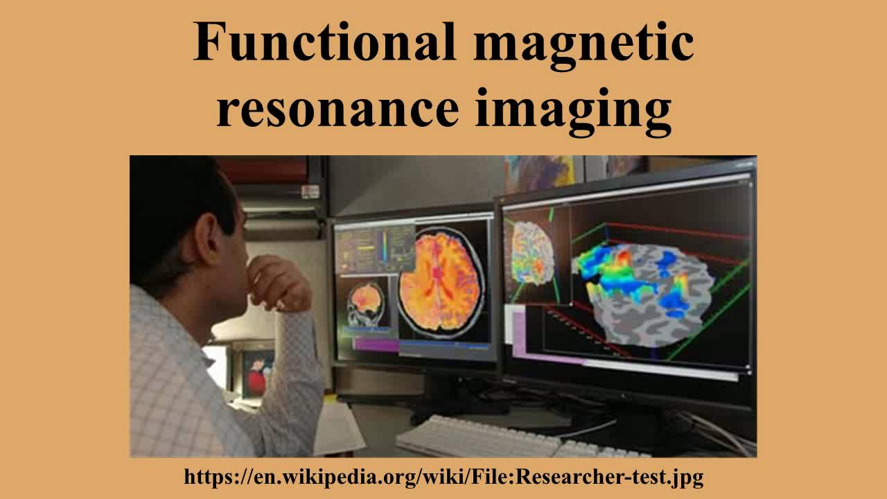
Functional Magnetic Resonance Imaging (fMRI) is a non-invasive neuroimaging technique that measures brain activity by detecting changes in blood flow and oxygenation levels, based on the principle that cerebral blood flow and neuronal activation are coupled; when brain regions become active, blood flow increases, displacing oxygen-depleted blood with oxygen-rich blood approximately 2 seconds later, creating a measurable signal through differences in magnetic properties between deoxygenated and oxygenated hemoglobin, which allows researchers to map neural activity with spatial resolution down to millimeters and temporal resolution spanning seconds to minutes, though it cannot distinguish between feedforward and feedback neural networks or provide direct measures of neuronal firing rates.
Click to watch →
Deep Dive
Bilingual Language Co-activation: The concept that a bilingual's two languages are constantly active and competing in the brain, even when using only one.

YLMP 2021: Prof. Robert Hartsuiker (Plenary)
106
Bilinguals show language co-activation at the lexical level during both production and comprehension. Evidence includes cognate effects in word recognition, false friend effects, and eye-tracking studies showing bilinguals activate both languages when processing words in one language. This raises the question of whether similar co-activation occurs at the syntactic level.
Click to watch →

Palestra de abertura: Psicolinguística e educação: uma interface necessária
395
The popular classroom advice to 'turn off' one's native language is scientifically incorrect—all languages remain co-activated simultaneously in bilingual individuals. Executive functions (attention control, inhibition, cognitive flexibility) play crucial roles in managing this co-activation and preventing interference. Linguistic similarity between languages creates particular challenges for acquisition, as learners struggle to distinguish between closely related languages. Research shows that bilingual experience shapes processing differently than monolingual experience, and viewing bilinguals as cognitively deficient fails to capture the complexity of bilingual cognition.
Click to watch →

Language Co-activation Influences the Development of New Implicit Linguistic Knowledge in Bilinguals
170
This research demonstrates that the constant activation of a bilingual's non-current language influences the development of new implicit linguistic knowledge, as evidenced by Cantonese-English bilinguals showing implicit learning effects of novel article-semantic category mappings only when their non-current Cantonese language remained highly activated, but not when it was suppressed through English-language video priming.
Click to watch →

The benefits of being bilingual
739
When bilingual people hear a word, both languages become active simultaneously. The brain's language system begins guessing what word might be heard before the word is fully processed. For bilinguals, this activation is not limited to a single language but extends to corresponding words in both languages. This phenomenon, called language co-activation, occurs because auditory input can map onto words in either language.
Click to watch →

LIF - Language in Focus Webinar with Mike Sharwood Smith
133
Once representations are associated across different cognitive systems, they become co-activated. This means that when one representation is activated, the associated representations in other systems are also activated simultaneously. In bilingual and multilingual contexts, this means that whenever a learner uses any of their known languages, all their languages will be activated in the background. For example, a Turkish native speaker who thinks their native language is irrelevant to English learning will still have Turkish activated in the background when using English.
Click to watch →
Deep Dive
Executive Function and Cognitive Control: Understanding the mental processes that enable goal-directed behavior, specifically task-switching and inhibitory control.

Lec 18: Executive control/function and its facets
450
Executive function, also called cognitive control, is a high-level cognitive process that regulates and guides cognitive processes in sensory, memory, and motor systems. It is considered the core of intelligent behavior in humans, interconnected with attentional mechanisms. The concept has evolved since the late 19th century with contributions from Bianchi and Luria, and the term 'cognitive control' has largely replaced 'executive function' in many domains, originating from cybernetics and AI research. Posner and his group established three key proposals: control processes are slower than automatic processes, they can suffer from interference from automatic processes, and they depend on a limited capacity mechanism. The Stroop task demonstrates this: reading a word is automatic and fast regardless of ink color, but naming the ink color is controlled and becomes impaired when the word conflicts with the ink color.
Click to watch →

Prefrontal cortex
2.2K
Executive function is the primary psychological term for prefrontal cortex functions. It encompasses abilities to differentiate among conflicting thoughts, determine good versus bad, better versus best, same versus different, predict future consequences of current activities, work toward defined goals, and make expectation-based predictions. Executive function also includes social control and supports concrete rule learning, with more anterior regions along the rostrocaudal axis supporting rule learning at higher levels of abstraction.
Click to watch →

STRONG Kids 2 Childhood Nutrition and Obesity in the Microbiome Brain Axis – Illinois
102
Executive function (cognitive control) is a set of processes vital for behavior regulation, including working memory (ability to hold and manipulate information), inhibitory control (ability to regulate attention and self-behaviors), and cognitive flexibility (ability to multitask and switch between rules). These functions support higher-order brain functions like reasoning, problem-solving, and planning. Executive function abilities predict long-term scholastic and vocational success, marital success, happiness, financial wellness, and lower risk for substance abuse in adulthood. These functions are proportionally dependent on the prefrontal cortex, which exhibits protracted development until the third decade of life, allowing health behaviors to impact cognitive function.
Click to watch →

Webinario “NEUROCIENCIAS & EDUCACIÓN ”
250
Executive functions are high-level cognitive processes that enable new behaviors, learning, and behavioral control. They include: inhibition (overriding automatic responses), working memory (holding information for goal-directed tasks), cognitive flexibility (adapting to new circumstances), autonomous behavior initiation (acting without external stimulation), application of strategies (planning to achieve goals), prospective memory (remembering to accomplish future tasks), and multitasking. These functions are located in the ventral prefrontal cortex and involve integrating multiple cognitive processes.
Click to watch →

Téorico 10 Cognitiva - Redescripción Representacional - Urquijo 2022
1K
Executive functions include processes for organization, planning, anticipation, and self-regulation. These functions were not included in earlier cognitive models and represent mechanisms that allow individuals to organize their behavior, plan future actions, and regulate their responses based on current goals and environmental demands.
Click to watch →
Prerequisite Knowledge
- Concept 01Basic Neuroanatomy of the Basal Ganglia: Understanding the location and general motor/cognitive functions of the basal ganglia, specifically the caudate nucleus.
- Concept 02Principles of functional Magnetic Resonance Imaging (fMRI): Knowing how fMRI measures brain activity through blood-oxygen-level-dependent (BOLD) signals.
- Concept 03Bilingual Language Co-activation: The concept that a bilingual's two languages are constantly active and competing in the brain, even when using only one.
- Concept 04Executive Function and Cognitive Control: Understanding the mental processes that enable goal-directed behavior, specifically task-switching and inhibitory control.
Subsequent Learning
- Step 01The Adaptive Control Hypothesis: Exploring Green and Abutalebi's theoretical model of bilingual language control and the wider fronto-basal-ganglia network.
- Step 02Clinical Implications in Bilingual Aphasia: Studying how damage to the left caudate affects language control, leading to pathological language mixing or switching.
- Step 03Neuroplasticity and Structural Changes: Investigating how long-term bilingualism structurally alters gray matter volume in the caudate and connected cortical regions.
- Step 04The 'Bilingual Advantage' Debate: Evaluating scientific literature on whether bilingual language switching enhances domain-general executive functions in everyday life.
Opening
Segment
Key Section
Opening
0:01Playing Section
- 1
Scene starts with musical introduction.
- 2
Sets initial atmospheric tone.
The Distributed and Domain-General Control Network Hypothesis
While the left caudate is often highlighted as the primary 'switch' for bilingual language control, many cognitive neuroscientists argue this perspective is overly localized. Opposing theories propose that language switching is managed by a broad, domain-general executive control network rather than a single subcortical node. This network relies heavily on cortical regions, such as the dorsolateral prefrontal cortex (DLPFC) and the anterior cingulate cortex (ACC), which manage conflict monitoring and attentional selection. Furthermore, critics point out that the left caudate's activation during language switching is not language-specific. Instead, it reflects domain-general cognitive processes, such as general motor response selection, habit routing, and goal-directed behavior. Under this view, the left caudate acts as a general-purpose coordinator of action selection rather than a specialized bilingual gatekeeper, and language control is achieved through the dynamic interaction of widely distributed cortical and subcortical pathways.
Deep Dive
The Adaptive Control Hypothesis: Exploring Green and Abutalebi's theoretical model of bilingual language control and the wider fronto-basal-ganglia network.

Lec 18: cognitive reserve, new developments
242
The Adaptive Control Hypothesis (2013) proposes that bilingualism itself does not guarantee executive control advantages; rather, the frequency and context of language switching determines cognitive benefits. Three switching contexts exist: single language context (one language per environment), dual language context (both languages used with different speakers), and dense code switching context (frequent mixing within utterances). Research on Singapore bilingual children and young adults found dual language context bilinguals perform better on task switching and inhibitory control tasks. This hypothesis explains why bilinguals in dual language contexts show better performance on non-linguistic cognitive control tasks.
Click to watch →

Treffers-Daller:Explaining individual differences in Executive Function performance in multilinguals
240
The Adaptive Control Hypothesis (ACH) proposes that inhibitory control adapts to contextual demands across three contexts: single-language contexts (exclusive language use), dual-language contexts (different languages with different interlocutors, requiring strong inhibition), and dense code switching contexts (mixing languages within utterances). ACH claims dense code switching requires less inhibition via opportunistic planning. However, evidence contradicts this: Hofweber et al. (2016) found South African English-German bilinguals, who engaged more in congruent lexicalization (intimate code switching), showed smaller conflict effects on flanker tasks, suggesting dense code switching may enhance rather than impair cognitive control.
Click to watch →

Associate Professor Christos Pliatsikas Keynote at the International Symposium on Bilingualism 14
318
The Adaptive Control Hypothesis identifies key brain regions involved in language processes and control that adapt in bilinguals. These include frontal cortex regions (particularly prefrontal cortex), anterior cingulate cortex (mid-brain), parietal regions (side of the brain), and the cerebellum. These adaptations relate specifically to linguistic contexts and are not uniform across all bilingual individuals; they depend on the intensity of bilingual experience and immersion level.
Click to watch →

Talk: Proactive and reactive control during language switching (Dr. Zlatomira Ilchovska)
104
The Adaptive Control Hypothesis proposes three main types of interactional environments affecting bilingual language control: (1) Single-language environment - where individuals speak one language at home and another outside without mixing them; (2) Dual-language environment - where speakers switch between languages depending on conversation partners; (3) Dense code-switching environment - where two languages coexist and are used simultaneously within sentences. Each context trains different aspects of cognitive control.
Click to watch →

IAS Lecture by Judith F. Kroll - Two sides of bilingualism - A lens to the cognitive neuroscience
351
The Adaptive Control Hypothesis proposes that language use is determined by environmental demands. Research comparing Spanish-English bilinguals in Granada (separate contexts), San Juan (integrated contexts), and State College (dominant English environment) found that proactive cognitive control correlated with better picture naming accuracy in Spanish only in the English-dominant context. This demonstrates that cognitive resources are recruited differently based on interactional context.
Click to watch →
Deep Dive
Clinical Implications in Bilingual Aphasia: Studying how damage to the left caudate affects language control, leading to pathological language mixing or switching.

Neurobiologia da Linguagem M5 A2 O Modelo clássico para a base neural da linguagem e as Afasias
27.1K
Broca's aphasia produces extremely deficient speech with few words per minute, mainly content words and function words. Wernicke's aphasia produces fluent but meaningless speech that cannot be understood. Conduction aphasia, caused by damage to the arcuate fasciculus connecting Wernicke's and Broca's areas, spares comprehension and production but impairs repetition. In bilingual individuals, aphasia typically affects the second language more severely when acquired later in life, as different neural circuits are recruited. Sign language users show similar deficits in both modalities, demonstrating that language circuits are modality-independent.
Click to watch →
NYCDCS Lecture 2: Aphasia in Multilingual Contexts with Aviva Lerman and Taryn Malcom
939
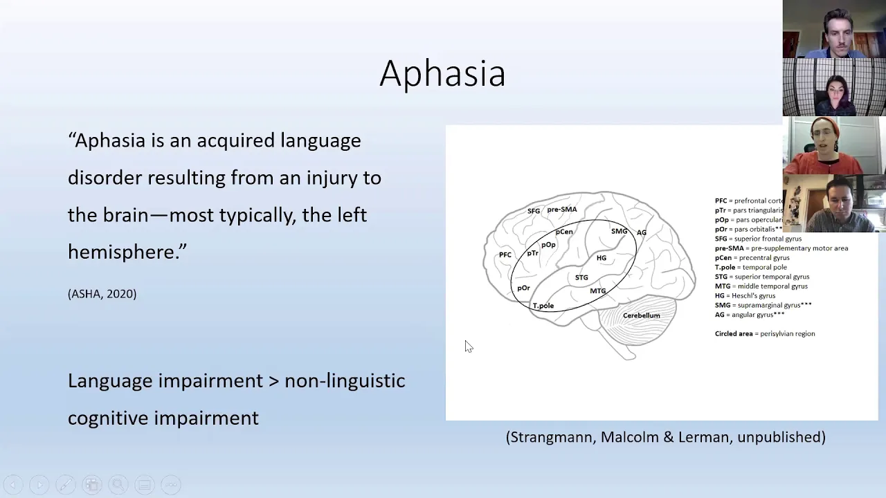
This section synthesizes sociolinguistic predictors of language performance and their clinical applications. Research identified key predictors: increased daily Jamaican Creole exposure correlates with better Jamaican Creole verb marking; speaking to equally fluent interlocutors reduces code-switching/mixing; greater enjoyment of a language correlates with better performance; higher value placed on a language correlates with better performance; lower perceived difficulty correlates with better performance. These findings highlight that language production reflects complex interactions between cognitive abilities, social attitudes, and contextual factors. Clinically, Jamaican Creole-English bilinguals should be treated like any other bilingual population. Key considerations include distinguishing normal language variation from disorders, recognizing that standardized assessments may not be valid for bilingual individuals, using narrative samples to compare language balance, being flexible about which languages to test/treat based on patient/family preferences, accessing cultural/linguistic brokers when needed, avoiding assumptions about patients' language backgrounds, and understanding that language differences can lead to misdiagnosis if not properly assessed.
Click to watch →

Language processing in the multilingual brain - Esti Blanco-Elorrieta
445
Understanding bilingual language organization has important clinical implications for aphasia rehabilitation after stroke. Aphasia results from brain damage disrupting language abilities, with recovery depending on which language elements remain intact. The unified mental lexicon model suggests that shared representations across languages may provide alternative pathways for recovery. Clinicians can leverage preserved language knowledge to help patients recover lost functions, and voxel-based lesion-symptom mapping can predict deficits based on lesion location and pre-existing language proficiency patterns.
Click to watch →

What Brain Damage Reveals About Language | Otherwords
155.1K
Aphasia is a language impairment caused by strokes, brain injuries, or neurodegenerative diseases. Broca's aphasia (expressive aphasia) results from damage to Broca's area, causing fluent comprehension but difficulty producing speech limited to single words or phrases. Wernicke's aphasia (receptive aphasia) stems from Wernicke's area damage, producing fluent but nonsensical speech with poor comprehension. Conduction aphasia involves damage to the arcuate fasciculus pathway, preserving expression and comprehension but impairing repetition. Global aphasia results from widespread brain deterioration, impairing all language functions. Treatment focuses on speech-language therapy and addressing comorbidities like depression and social isolation, which accelerate cognitive decline.
Click to watch →

"Syntactic processing in bilingual aphasia", Danielle Fahey
185
Bilingual aphasia presents unique challenges because grammatical structures vary significantly across languages, and the interaction between languages affects how aphasia manifests. Research indicates that bilingual individuals with aphasia typically show fewer deficits on grammatical structures that are fully overlapping between their languages (due to reinforced cognitive reserves), but greater deficits on partially overlapping structures that were acquired later in second language learning. This framework suggests that clinicians should assess bilingual aphasia patients in all their languages and consider cross-linguistic similarities when designing treatment interventions.
Click to watch →
Deep Dive
Neuroplasticity and Structural Changes: Investigating how long-term bilingualism structurally alters gray matter volume in the caudate and connected cortical regions.

Innovative Horizons in Neuro-Physiotherapy 2026
562
Neuroplasticity involves several structural changes: (1) Dendritic branching - increased branching patterns of dendrites; (2) Synaptic changes - formation of new synapses between neurons; (3) Axonal sprouting - axons can sprout like tree branches to create new connections; (4) Myelination changes - lipid layers surrounding axons that help in faster conduction of neural signals; (5) Neurogenesis - generation of new neurons, particularly in the hippocampus.
Click to watch →

What is Neuroplasticity?
682
True neuroplasticity involves structural changes such as anatomical changes in the brain, including changes in neuron shape and synaptic changes, especially when robust. This includes changes to the physical anatomy of individual neurons or whole brain structures that deviate from normal trajectories. Neuroplasticity often includes functional changes like new roles for brain areas and mechanisms like long-term potentiation (LTP) and long-term depression (LTD). Neuroplasticity can be induced by changes within the body (disease, injury) and outside the body, and often recapitulates developmental mechanisms.
Click to watch →

Neuroplasticity | Nervous system physiology | NCLEX-RN | Khan Academy
290.1K
Structural neuroplasticity involves physical changes in neuronal architecture, including dendritic branching, axonal arborization, and synapse formation. When neurons fire together frequently, structural potentiation occurs through sprouting—dendrites elongate and branch, and axons generate additional terminals to form new connections. Conversely, structural depression (pruning) eliminates unused connections, simplifying dendritic trees and removing axon terminals from inactive neurons. These structural changes occur across multiple timescales, from rapid synaptic adjustments to gradual morphological remodeling spanning years. Neuroplasticity underlies nervous system development, memory consolidation, learning acquisition, and recovery from injury by enabling the brain to reorganize and optimize its connectivity throughout life.
Click to watch →

Masterclass: Mapeo cerebral: cuando las redes neuronales cuentan tu historia
531
Neuroplasticity involves both the reorganization of connections between neurons (synapses) and physical changes in brain structure. The famous study of London taxi drivers by Eleanor Maguire demonstrated that extensive navigation experience could enlarge the hippocampus, with differences visible on MRI scans that could predict taxi driver exam success. This finding showed that learning and experience can produce measurable structural changes in the brain. Neuroplasticity encompasses changes in function, structure, and potentially even gene expression, demonstrating the brain's remarkable capacity for adaptation throughout life. This understanding has profound implications for rehabilitation and recovery from brain injury.
Click to watch →

Nöroplastisite (Sinir Sistemi Fizyolojisi)
24.4K
Structural neuroplasticity involves physical changes in neural connections. Potentiation leads to growth of new dendritic branches and axon terminals, increasing the number of synapses. Depression leads to pruning of dendritic branches and axon terminals, reducing the number of synapses. These structural changes can occur over different time scales.
Click to watch →
Deep Dive
The 'Bilingual Advantage' Debate: Evaluating scientific literature on whether bilingual language switching enhances domain-general executive functions in everyday life.

06h Bilingual advantage
424
While early research suggested bilingualism offers cognitive advantages such as enhanced attention control and delayed dementia onset, recent meta-analyses indicate no reliable evidence for a general bilingual advantage, with publication bias potentially inflating positive findings; the current consensus suggests bilingualism may not provide consistent cognitive benefits across all populations and tasks.
Click to watch →

M. Lehtonen ExLing tutorial
245
Executive functions are high-level cognitive abilities used to control and regulate behaviors, including focusing on tasks, suppressing interfering distractors, maintaining attention, and flexibly switching between tasks. Bilingualism may enhance these functions because managing multiple languages requires efficient executive functions—both languages are often constantly active, creating interference that must be suppressed, and switching between languages engages domain-general set-shifting processes. However, the bilingual advantage has been controversial despite many studies showing some advantage. The main reason is mixed results—sometimes advantages are observed in some tasks but not others. Publication bias is a significant problem: positive findings supporting bilingual advantage are more likely to be published than null findings, creating an overly optimistic view of the field. Meta-analysis statistically summarizes multiple studies to get a holistic picture. Researchers examined moderator variables that might explain differences in outcomes: age group, age of acquisition, proficiency, immigrant status, language pair, country of study, and study quality. The meta-analysis included 152 studies and almost 900 effect sizes from 27 different countries. Before bias correction, some positive findings existed for bilingual advantages in inhibition, shifting, and working memory, but effect sizes were very small. After correcting for publication bias using the PET-PEESE method, effects became close to zero or showed small bilingual disadvantages. Moderator analysis revealed no consistent patterns supporting the bilingual training hypothesis—age group, age of acquisition, and proficiency did not moderate the advantage as predicted. The conclusion was no general bilingual advantage in any domain of executive functions when correcting for publication bias.
Click to watch →

Autism and Bilingualism: What you know? What Do We Need to Know - Susan Fletcher-Watson, PhD
382
The concept of a 'bilingual advantage'—that bilingual individuals show enhanced cognitive abilities—is highly controversial in the research literature. One major problem is that most studies are cross-sectional (comparing groups at one point in time) rather than longitudinal (following individuals over time). Additionally, longitudinal data is scarce, and randomized controlled trials assigning children to bilingual versus monolingual conditions are ethically and practically impossible. Most research occurs in immigrant communities, creating atypical samples that make it difficult to find appropriately matched control groups.
Click to watch →

Shereen Sharaan - Evidence-Based Best Practices for Bilingual and Multilingual Children with Autism
2.5K
Bilingualism involves speaking two languages with varying proficiency levels based on exposure timing and duration. Language switching occurs in three contexts: single (different languages with different people), dual (both languages with same person at same time), and dense (mixing languages within sentences). Bilingual individuals experience slightly longer vocabulary retrieval times due to two active language systems. The bilingual advantage debate is highly controversial, with studies showing advantages in social cognition and executive function but limited by small sample sizes and inconsistent matching criteria across studies. No universal definition of bilingualism exists, ranging from knowing ten words to fluency in multiple domains.
Click to watch →

Emergentism, language control and the bilingual brain
974
The debate over whether bilingualism provides cognitive advantages has intensified since 2008, when media headlines claimed bilinguals are smarter. Researchers like Manolo Carreiras argue there is no general bilingual advantage, presenting evidence from interference tasks showing no differences between bilingual and monolingual groups across age groups. However, other studies suggest advantages in vocabulary learning and novel word processing. This controversy highlights the complexity of isolating bilingual effects from confounding variables like socioeconomic status, education level, and cultural factors that accompany bilingual populations. Methodological considerations include choosing appropriate languages for testing (avoiding cognates), using culturally sensitive measures, and carefully defining population groups.
Click to watch →
Opening
Segment
Key Section
Opening
0:01- 1
Scene starts with musical introduction.
- 2
Sets initial atmospheric tone.
The Distributed and Domain-General Control Network Hypothesis
While the left caudate is often highlighted as the primary 'switch' for bilingual language control, many cognitive neuroscientists argue this perspective is overly localized. Opposing theories propose that language switching is managed by a broad, domain-general executive control network rather than a single subcortical node. This network relies heavily on cortical regions, such as the dorsolateral prefrontal cortex (DLPFC) and the anterior cingulate cortex (ACC), which manage conflict monitoring and attentional selection. Furthermore, critics point out that the left caudate's activation during language switching is not language-specific. Instead, it reflects domain-general cognitive processes, such as general motor response selection, habit routing, and goal-directed behavior. Under this view, the left caudate acts as a general-purpose coordinator of action selection rather than a specialized bilingual gatekeeper, and language control is achieved through the dynamic interaction of widely distributed cortical and subcortical pathways.
Basic Neuroanatomy of the Basal Ganglia: Understanding the location and general motor/cognitive functions of the basal ganglia, specifically the caudate nucleus.
Principles of functional Magnetic Resonance Imaging (fMRI): Knowing how fMRI measures brain activity through blood-oxygen-level-dependent (BOLD) signals.
Bilingual Language Co-activation: The concept that a bilingual's two languages are constantly active and competing in the brain, even when using only one.
Executive Function and Cognitive Control: Understanding the mental processes that enable goal-directed behavior, specifically task-switching and inhibitory control.
The Adaptive Control Hypothesis: Exploring Green and Abutalebi's theoretical model of bilingual language control and the wider fronto-basal-ganglia network.
Clinical Implications in Bilingual Aphasia: Studying how damage to the left caudate affects language control, leading to pathological language mixing or switching.
Neuroplasticity and Structural Changes: Investigating how long-term bilingualism structurally alters gray matter volume in the caudate and connected cortical regions.
The 'Bilingual Advantage' Debate: Evaluating scientific literature on whether bilingual language switching enhances domain-general executive functions in everyday life.
36.3K views350likes2:02@AmericanMuseumofNaturalHistoryOriginal Release: 2012-06-25
[Music] [Music] [Music] e [Music] [Music] [Music]
Up Next
61:59
How Bilingualism Reshapes the Brain Across the Lifespan
@ladal2518
131 views•2021-09-02

21:55
Deciphering Neural Circuits Controlling Anorexia in Mice | Richard Palmiter
@AllenInstitute
2.3K views•2011-10-10

4:10
Vagus Nerve (CN X): Anatomy, Nuclei & Functions Explained
@Alilamedicalmedia
305.2K views•2022-10-31

13:03
How Exercise Benefits Your Brain: Science Explained
@TED
11.4M views•2018-03-21
Related Study Plans & Knowledge Roadmaps
Structured learning paths in Neuroscience
neuroscience
Appetite Regulation: Brain Circuits & Hormones
01Foundations of Endocrinology and Brain Anatomy
02Introduction to Energy Homeostasis
03Peripheral Signals: Leptin, Ghrelin, and Insulin
+ 2 more learning modules
14h5 modules
Explore Roadmap
neuroscience
Neuroeconomics: Value, Risk & Brain Mechanisms
01Foundations of Neuroscience and Brain Anatomy
02Introduction to Neuroeconomics and Subjective Value
03Dopaminergic Prediction Errors & Reinforcement Learning
+ 3 more learning modules
18h6 modules
Explore Roadmap
neuroscience
Spatial Navigation: Cells, Grids & Brain Maps
01Foundations of Brain Anatomy and Memory
02Place Cells and Head-Direction Cells
03Grid Cells: The Brain's Coordinate System
+ 2 more learning modules
12h5 modules
Explore Roadmap
neuroscience
BCIs: Neural Decoding & Prosthetic Control
01Neuroscience Foundations: How the Brain Signals Intent
02Signal Acquisition: Recording the Brain's Activity
03Neural Decoding: Translating Brain Waves into Data
+ 2 more learning modules
25h5 modules
Explore Roadmap