







![Биология поведения человека: Лекция #14. Лимбическая система [Роберт Сапольски, 2010. Стэнфорд]](https://i.ytimg.com/vi/jM_nP8gLBXM/sddefault.jpg)










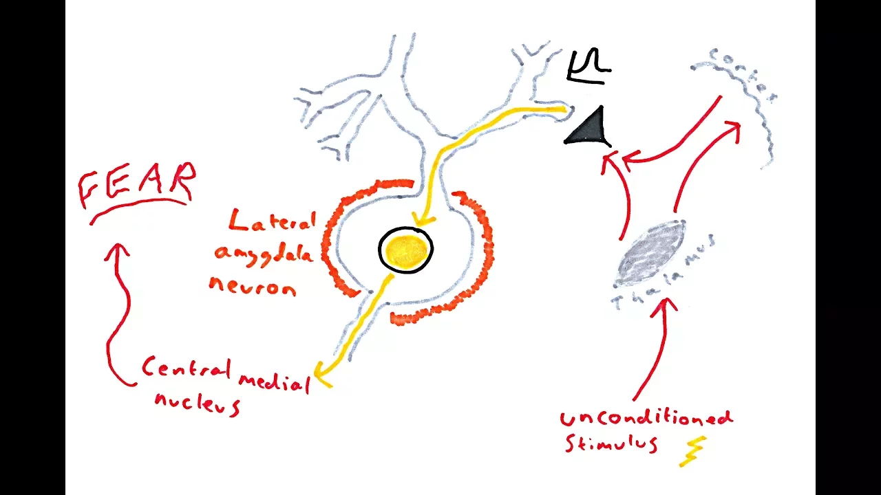





Fear conditioning specifically causes potentiation of synaptic connections between learning-recruited neurons in the lateral amygdala, forming autoassociative excitatory networks that enable a small number of pyramidal neurons to encode multiple memories through strengthened recurrent connectivity within neuronal ensembles.
Fear Learning Strengthens Amygdala Ensembles | Journal Club Review
Added:Basic principles of Classical Conditioning (Pavlovian learning), specifically how a neutral conditioned stimulus (CS) associates with an aversive unconditioned stimulus (US) to elicit a fear response.
The concept of synaptic plasticity, particularly Long-Term Potentiation (LTP) as a primary cellular mechanism for strengthening neuronal connections during learning.
Anatomy and functional organization of the limbic system, with a specific focus on the role of the lateral amygdala in threat detection and emotional processing.
The theoretical concept of a 'memory engram'—the physical or biophysical representation of a memory trace within a specific network of neurons.
The neurobiology of fear extinction and its clinical implications for treating anxiety disorders, phobias, and Post-Traumatic Stress Disorder (PTSD) in humans.
Advanced neuroscientific techniques used to identify and manipulate engrams, such as optogenetics, chemogenetics (DREADDs), and in vivo calcium imaging.
The role of hippocampal-amygdala pathways in contextual fear conditioning, spatial memory integration, and memory generalization.
The downstream molecular and genetic cascades (such as CREB activation and local protein synthesis) that stabilize and consolidate potentiated synapses over time.
161 views2likes1:02:37Original Release: 2025-04-26
Okay. Uh, everybody ready? I'm going to assume that is a yes. Somebody give me a yes.
Yes. Yes. Great. Okay. Uh, so welcome to another uh aspirational neuroscience journal club. Uh this time we are covering this paper uh entitled fear learning induces synaptic potentiation between engrams neurons in the rat lateral amigdula by Abatus. Uh it was published in uh nature neuroscience in 2024. Uh the authors used a very technically challenging technique uh simultaneous 12 cell patch clamping to determine the functional connectivity between lateral amydala uh fear memory ingra cells. And what they find uh uh is that fear conditioning specifically causes potentiation of synaptic connections between learning recruited neurons. These findings of synaptic plasticity in an autoassociative excitatory network of the lateral amygdala may suggest a basic principle through which a small number of parameal neurons could encode a large number of memories. I chose this paper because I think it reveals some general principles regarding how memories are formed. uh namely that synaptic connections onto and between Ingram cells are strengthened uh forming an ingram engram ensemble that is both selective to and uh to a particular input stimulus and that is um uh effective in amplifying that stimulus to drive later behaviors.
I also chose this because I think it suggests a possible connetoics experiment which could demonstrate the decoding of a non-trivial memory from a static conneto and uh I'll discuss this at the end of the talk and I'm looking forward to uh feedback on whether this would actually be considered a non-trivial enough uh to for example win the uh aspirational neuroscience memory decoding challenge which which is of course what these uh uh uh journal clubs uh should be kind of gearing toward.
board. Uh, okay. So, before going on to the main paper's results, I'm going to review what I believe to be the main theory of how auditory fear memories are formed uh to help put this paper's findings in in context. This is probably dramatically oversimplified, but that's that's where we are. Uh so first we have the auditory phalamus the medial geniculate nucleus um and the auditory cortex uh which I'll just summarize as auditory cortex for the rest of this talk uh which represents distinct sounds by distinct patterns of activation over their neurons and the axons from these auditory regions project to the uh uh lateral amygdala as shown in this diagram. Uh the lateral amydala cells also receive input from pain circuits.
I'm not showing that here but they just receive input from pain circuits directly. And the lateral amydala axons uh project to downstream areas central amydala and later uh to instigate freezing behavior of the animal. The lateral amydala cells also connect to each other recurrently as shown here. These connections are the main focus of today's journal club paper. Um, for the description of learning that I'm about to lay on you guys, uh, uh, I want you to assume that there's a lot of silent synapses, uh, uh, in this box here. So, silent synapses are synapses that have NMDA receptors so that they can sense coincident activation and undergo heavy and strengthening, but they don't provide a significant signaling prior to learning. So just assume that there's some small synapses there. Now uh during an auditory fear event, the sound produces a unique pattern of activity across the auditory cortex as shown here while simultaneously a random unique subset of lateral amydala cells becomes active due to the uh the pain signals. uh this subset of lateral amydala cells is believed to be chosen at random due to the cell's uh intrinsic excitability uh which fluctuates on a day-to-day basis.
So it's a random subset. Uh the previously silent auditory cortex to lateral amydala synapses that I just showed there that right there uh undergo LTP and strengthen. And at the same time according to today's journal club paper these lateral amydala recurrent synapses also strengthen these here. And so to summarize this oversimplified picture, it is thought this is what is thought to be the neural substrate of a fear memory. Number one, an ensemble of engram cells in the lateral amydala.
Number two, uh synapses among these engram cells that endow the engram with autoassociative amplification powers.
And number three, uh, synapses from auditory cortex to the lateral amydala that endows this engram with specificity for one sound in particular. And if that particular sound occurs again, uh, let's say the next day, uh, these synaptic path pathways will amplify that sensory input signal and cause the mouse to freeze even without a pain signal.
Now there have been many papers that have shown the strengthening of these auditory cortex the lateral amydala uh synapses that I'm highlighting here. uh but I think the the best one the best one that I found is this paper this 2014 paper by uh Nabavei uh at all and I'll take a few minutes to go through its results uh before I go through the main uh Abetus 2024 paper that really researches these recurrent synapses in the lateral amydala everybody I can yeah I was just going to ask you if you were able to hear when when I raised my hand and if you want to be interrupted at all because yes, go ahead and interrupt me.
Uh so this refers to this slide, the previous slide, the one before. So um I guess what I'm wondering is um and I also wondered while reading the paper is um what previous knowledge or models or theories are there uh about how it is that you choose or that the amygdala can choose that this is actually something that should be remembered as something to be afraid of. Say for example achieving uh strengthening of the syninnapse in the presence of pain and sound but only for pain that is above a certain level or uh auditory stimulus plus the pain above a certain level versus say just a very strong auditory stimulus or uh or pain just by itself without any particular uh correlation to anything. So so the short answer is I I don't know. uh but uh I've I've uh in the process of this I read a review article on the lateral amydala in general and there are a bunch of modulating um uh circuits also that go into this uh there's dopamine circuits um and so uh I think I think what puts it over that threshold is probably um uh probably just simply not in this paper and not in these diagrams.
Okay. Yeah. This was u I guess you said modulating circuits. So this is in line with another concern I had when reading the papers that because of the way most of the studies were done, you know, XVivo, in vitro. Yes. Um that that a lot of the say rhythmic modulation and other factors that have to go into a proper description of the system, its modes and when it does something and how it switches are not always included.
Uh that's right. In fact, I I touch on that later in the um uh in the talk.
Okay. Fantastic. Thanks. I'll highlight that. I I think I I think there's a couple of things that I uh that you're bringing up that I'll touch on later in the talk actually. Awesome.
Okay. So uh the the NAVO 2014 paper uh what what they did is they injected uh the auditory cortex and the auditory phalamus uh with an optogenetic virus to tag random subset of the cells. So that's what's shown here. Uh this this caused optogenetic channels to be expressed in the axons of these uh cells that terminate in the lateral amydala.
Uh that way they could shine blue light with a little fiber optic cable onto just the lateral amydala and uh uh while the rat was being uh shocked and this created a fear memory to this optogenetically activated fake sound condition stimulus and presumably uh it is strengthening these synapses. That's that's what I'm going to be talking about here. And so in that in that paper here's uh uh here's the uh the plot that they show that shows that the memory was was stored. So the uh the y-axis here is lever presses. They could see that the rat was freezing if it stopped a le lever pressing task. And so when they shine the blue light uh it it froze. Now the where the authors really show that this memory is encoded in these particular synapses uh they is here they used a laser light uh the same laser light to perform a LTD a long-term depression protocol. So that's when they um slowly activate the uh uh the they have a slow one hertz action potentials um that specifically shrink those synapses. Uh and when they do that uh behaviorally the memory is erased.
Then they could go and do a long-term potentiation experiment where they rapidly uh activate these um auditory cortex axons to strengthen those synapses back up and they show that lo and behold the memory comes back.
uh and they can do that multiple times and they have a quite robust result that they could erase and restore uh this behavioral memory um uh simply by uh by shrinking and regrowing these or re-expanding these uh these synapses from the auditory cortex onto the lateral amydala. So, uh, I wanted to I wanted to make clear that there's a bunch of cool results, uh, uh, that talk about those key synapses, but right now we're going to go to the main paper uh, which talks about these lateral amydala to lateral amydala synapses and uh, as shown here. So, uh, this uh, abatus at all 2024 paper, I'm breaking it down into about uh, six uh, experiments. uh the titles they have uh I' I've numbered them though. Uh there's actually just one experiment here, this Xvivo connectivity after fear conditioning that I think has the main clear results. So I'm going to spend most my time on that. Uh so in in experiment one, the authors determined the baseline connectivity statistics of the lateral amydala. Uh so what they did is they they they took a rat, chopped its head off, uh sliced a 400 micron thick uh slice out of its brain containing the lateral amigdula, put that in artificial cerebral spinal fluid, so it was still alive and did uh 12 pipet whole cell patch clamp recordings uh on cells in the lateral amydala.
uh uh as shown in this diagram, current was injected in one of the cells uh and then they listened to the other uh so that it produced a train of action potentials while they were listening to the other 11 cells uh monitoring them for microexitatory postsaptic potentials uh which is going to happen whenever there's a uh um a glutamate vesicle that is released because of that action potential. So if they saw any of those, then that's a sign that the cell that they're driving is connected to the one is synaptically connected to the one that they're uh listening to. They use 34 rats in total 90 slices uh to test um a total of 637 excitatory neurons uh with 4,157 possible connections. So they they listened to all of those pairs uh through all those experiments of which they found 89 actual connections which implied a 2.1% probability of synaptic connections between any two excitatory uh lateral amydala cells. Uh they did a whole bunch of other things that are really cool but are probably not so relevant. They calculated the quantal uh release size. Uh they um uh 460 microvolts per uh uh per um vesicle released. Uh they calculated the um uh uh five release sites on average per functional connection. Um uh also what they did is from that is relevant to a connetoics potential connetoic study is they uh because they re they backfilled all these neurons they knew where the cell bodies were uh and they found that uh cells that are closer to each other like within 100 microns uh had a higher probability of connecting with each other. So it maxed out at about 3.5%.
Um and uh and so that was kind of a a basic Can I ask a question about the previous experiment, please? Um can you go back to it? I I want to see the the traces.
So before these two images. Yeah, this one. Okay, this one here because there's something I I didn't quite get and that's probably just because I'm not an ephist person. Um so the red traces obvious what's happening. Also, we should keep in mind that these here are the titanic pulses where you're getting like a whole series of APS that are just not really giving you all the natural results that you would want to see for your um your EPS, but you're getting that on the next slide then because that's where they do the single AP stimulation and show you what it is. But I'm wondering about the gray here. So, the cell one to cell two, these are patch clamp, right? So when you're doing extracellular, it's very normal that you're also stimulating a variety of cells that are nearby. But is this supposed to indicate that cell 2 is very strongly connected to cell one or is this an I was wondering that myself. Uh I I don't know. Uh why are there off diagonals here? It's it's a great question. I I don't know. Okay. If there's anybody on the call who's an eiz person who does patch clamping, maybe you can chime in and and you know clarify for us because this is uh I was wondering about that. Yeah. Yeah. Sorry.
Uh um uh and there's a whole bunch of other stuff that I'm not going to be able to answer on these electrophysiology experiments as well.
Um uh so in experiment two they did more electrophysiology on the same slices. Uh basically what they were doing is uh uh they were uh putting in action potentials on one and uh and now that they knew that there was a synaptic connection they were trying to uh uh quantify uh the type of of a synaptic connection. So they could classify it as a facilitating or stable or depressing synapse. uh from this they calculated the overall temporal summation that could be expected at 183%. I'm not exactly sure what that means but uh the main meaning is that if multiple input action potentials arrive simultaneously then the synapses have the uh are more effective. Okay. uh uh they uh did current injection experiments and showed that uh the average threshold for spiking I think this is a delta uh was 28 molts. Uh again this is laying the groundwork for um uh a statement that they're making later. Uh so they put these together and they estimated that an average of 34 of the synapses out of the total 230 synaptic inputs uh from lateral amydala to lateral amydala neurons um for for each neuron uh would have to be activated together to cause an action potential. And then they uh uh they note that previous studies have shown that 10 to 30% of the excitatory neurons in the lateral amydala are recruited into a fear memory ingrain. Uh uh and they say this corresponds to a certain number of synapses. Uh actually let me just read their thing here. uh based upon previous findings uh fear conditioning recruits 10 to 30% of lateral amydala neurons uh and on the premise that fear conditioning can indeed induce local synaptic potentiation that reaches a similar level of this temporal summation this should allow for reliable signal propagation across the lateral amydala so what they're really setting up here is they're saying look from all the electrophysiology that we do uh it it makes sense that a signal isn't going to be propagated through uh if you don't have kind of a local ensemble that's going to increase the uh um uh uh the uh sensitivity if you will. Uh but if if you do have that local ensemble then uh and it's got lateral amydala to lateral amydala potentiated synapses then it might very well amplify a signal uh down the line.
Uh okay. So that was actually one of the places where I was thinking oh yeah and this is where modulation is probably going to play a pretty big role as to whether you can achieve postsaptic spiking or not. So um those numbers since they were done I think that was in vitro. Yeah. So all these are almost everything here is in vitro. Yeah. Okay.
Yeah. Yeah. uh or X Xvivo slice I guess is what they call it. Uh this actually so I'm moving on to experiment three.
Remember experiment four is the one that I I consider to be the main uh one that has meat. Uh so uh in experiment three uh the authors performed a set of simple uh heavy and LTP experiments in these acute slices. So uh this makes sense that they would do this, right? They've got the slices there. They've got neurons that are connected. um that they found they can uh give uh you know I don't know 100 hertz uh stimulus to one and see if it um strengthens its synaptic connection and they did find that in this but it was a super weak result uh at least if I'm reading it correctly they they say that um uh they I if you look in this figure the first spike um I can't show my Do you see my mice my mouse?
Yes, we do. Oh, you do? Okay. So, this first spike here uh this this is uh the facilitation of the first spike. So, uh if they gave this LTP protocol, then there's a little bit of a significant uh rise in um uh in connectivity, but all the other spikes uh uh spikes two through eight of a train uh didn't show any. And this was only on accommodating synapses and stuff like that. So I I feel like even though they got a result here and they showed LTP, I it's it's super weak. And I think uh that's because this was a slice and they weren't squirting um uh dopamine on it or it wasn't uh it didn't have the the the right things to actually uh I was wondering. I didn't read it carefully enough to catch this, but maybe you did. So, when they were testing this, um, did they repeatedly provide the stimulus or did they only do it once or how what exactly were they measuring here as the uh this is this is in consequence of what?
So um uh my understanding is what they did is they um uh they it probably says here we use a standard Hebian association protocol uh to associate 15* 10 presinaptic action potential uh action potentials with a post synaptically evoked action potential at a delay of 10 milliseconds.
And so the result was after 10 uh after 15 sequences of 10 stim 10 10 10 APs in a sense or um am I reading I I think so I think so I see okay yeah in that case it does seem kind of weak yeah it does seem kind of weak and they get much much stronger results uh for the next one so that happened in the animal so I would just ignore this one uh so this is uh paper uh this is experiment number four. Uh I think this is the main experiment here. So um in experiment number four, the authors injected rats with a virus to mark all the cells in red and uh in the lateral amydala and uh those cells that were highly active within the last 90 minutes uh would produce green fluorescent protein. Uh so they would be marked yellow. Um uh uh and they they did this be by using uh a degradable form of green fluorescent protein that would degrade over an hour I guess. Uh so if a neuron is active, it's going to be producing green fluorescent protein, but uh uh that green fluorescent protein is going to be degraded. Uh so uh they're only going to see green fluor they're only going to see these yellow um uh neurons uh uh when they sacrifice the animal uh 90 minutes after having undergone something that would activate those neurons. Okay. So the uh they used um uh so the experiment that they did is uh uh they injected the virus then on on uh they had three conditions. They had a home cage uh condition, a paired condition, and an unpaired. Uh on week two, day one, on day one, essentially, they would either just leave the rat in the home cage or they would um in the paired condition, they would um uh give the rat a tone and then uh overlap that with a shock.
uh and in the unpaired condition they would give the rat a shock and then they'd wait like 10 minutes and and then have a tone. So the idea is that uh only in the paired condition should these be uh should the tone be associated uh with the with the shock. Uh uh then on day two they tested it. Right? So they they gave a tone and uh the images up at the top here, H, U and P uh home cage unpaired and paired.
Uh uh and the is images of a slice 90 minutes after they tested with a tone. So only those neurons that were active when they gave a tone on the second day are going to be yellow and and so that's why only the paired condition has a bunch of um uh of yellow engram cells. So those are the engram cells. Okay. Um now why did they do that? They did this because uh they wanted to do electrphysiology. So they did electrophysiology on the slice, but they could tell whether they were in a yellow neuron or a red neuron. And so this is the key result of the paper here. And it's it's funny, it's the key result of the paper. So of course they put it in extended figure 7. Uh so they found significantly more connections between Ingram cells. Uh uh 7.2% 2% of Ingram to Ingram cells uh engram cell pairs were connected uh versus only 1.4% of non-engram cells. Uh not only that, the connections between engram cells were much more uh uh strong. So uh and and it showed up not just on the first spike but all the spikes. So this this answers your question Randall about I I think this is way stronger result of LTP. Um so they they had a uh micro EPSP response of 1.67 millolts versus 2 molts uh24 millolts uh for non-engram uh connections. And they also uh in the same experiment they uh they tried to increase they did an LTP again on these uh on these cell pairs and uh the they use this to show that this really is LTP because if you try to do LTP on something that has already done LTP then you don't get any LTP and uh and that's what they show they they only get LTP and the ones that were non-ening connections uh uh the the the ingram to ingram uh synapses are already LTPD out that make sense um so let me um uh so since this is the most important one I'm going to read this uh this is how they put it this suggests that through a heavy mechanism fear conditioning leads to synaptic strengthening between recruited lateral amydala neurons and may thereby ensure after fear conditioning a strengthening of local synaptic connections that can pro promote reliable signal propagation across the lateral amydala. All right, we're we're getting there. Any questions on that one? That was the kind of crucial one.
Anybody still there? That one was very clear. Um I should say though it for me you know it feels like um because theory tends to run ahead of experiment and models do a lot of things where we're already assuming what they're trying to show in detail here.
It feels like yeah I don't know I mean it feels as if they're showing things where you go duh of course but that's because I buy into the I buy into like the standard model of how this works or how how memories work in a sense. Yeah.
Yeah. Yeah. Although I I haven't seen a paper that actually goes to these recurrent connections in lateral amydala before this one. Yeah. No, that's what I mean. Like uh the you know actually doing that empirically and testing it in great detail by actually looking at the individual synaptic sites and stuff comparing with patch clamping that takes a lot of work and so it you know it's like experimental physics always lags behind theoretical physics and it's the same here. Yeah. Yeah. Okay. So uh the last two experiments are pretty quick.
Um uh so the the previous experiment one problem with the previous experiment is that uh it could mean that those connections were already there. Okay. Uh uh maybe the that ensemble of cells was already connected together and that's one of the reasons why it became the engram in the first place. Just for clarity when you say the connections were already there you mean strong connections, right?
Exactly. Not making new synapses here.
Okay. Absolutely. That's good point.
Good point. So, it could be that there was already really strong connections the um and there didn't have to be LTP uh between these cells uh in order to strengthen those. They've just kind of picked out a strongly connected subset. Um so in experiment five, what they did is they they tried to address that. They used uh 32 fine wire electrodes mounted on little micro drives to do single unit recording in the living rat. Uh and this allowed them to record neurons both before and after fear conditioning. So they could essentially essentially retroactively ask the question were these neurons that did become more connected were they already more connected? where they are already at that level of of uh functional connectivity before uh before the fear condition. Now, unfortunately, of course, this means because they're this is invivo and they're using wire electrodes, they're not doing patch clamping. Uh this means that they can only record spikes from neurons. So, they can't do the type of direct electrphysiology that uh verifies that there are actually a synaptic connection and to measure its strength. Instead, they had to rely on Granger causality.
Uh, which my understanding is it's just a simple way of saying if neuron A spikes after neuron B, then you have some slight evidence that neuron B is is That's right. And you don't know if it's a direct connection or an indirect connection through something else and all that. Yeah, exactly. So, so uh this is why I uh you know this this adds something uh but it's not as good as experiment number four. So, uh in any case, what they what they did is they uh they uh did the fear experiment and uh they could see what neurons were um uh spiking to say these are the ones that were recruited for the for the engram.
Uh and then they did Granger causality uh of the spike trains both before and after. And what they found is that uh uh there was uh an increase in the functional connectivity between ingrained cells but only after the fear conditioning. And experiment six essentially does exactly the same thing except using optogenetics uh to drive the recruited cells instead of um uh uh instead of just relying on their own uh firing.
Um okay so to summarize the whole paper uh uh this is the paragraph that I I I picked out. Uh these findings reposition the lateral amydala from a passive relay station into an active hub where synaptic plasticity strengthens uh lateral amygdala recurrent connections within neuronal ensembles following fear learning. Thus, fear memory encoding involves not only recruitment of intrinsically more excitable uh LA neurons and the potentia potentiation of their external aference but also stronger binding of these neurons together in a local network that after fewer learning can promote signal processing across the lateral amydala. And that's all I have for the paper. The rest is a memory decoding experiment that I want to throw at you guys. um uh uh based upon this. So is this is a good place for Sorry, Ken to be the one who keeps on clicking the hand up button. Um so the thing that the hand up button so the thing that isn't in the paper but that I was very curious about because I haven't studied amiga in as much detail as hippo campus. If you remember last time around, last paper, we had conversations about remote memory independent of hippocampus after a certain amount of time, hippocampus acting as a kind of cache when you're building new uh updating memory and things like that. I'm wondering in this version of okay, the amygdala is not just a passive pass through, but instead it's doing learning. uh it has sparse connectivity just like you see sparse connectivity in CA3 uh as opposed to what you might find in sensory cortex neoortex. Um is there do you know is of papers and experiments showing um fear memory becoming independent of amydala the same way that you know contextual memory becomes independent of hypocampus?
That's a great question. Looks like Ariel might have an answer to that. He's got his hand up. Yeah, I'm gonna throw that to anybody else that does. I I do not have an answer. I have a a followup to Randall's. I'll give anyone else a moment to jump in if they have an answer.
Not know this because I read Lau's uh book on um uh the synaptic self and I'm sure that he talked about this. Um uh there are a lot of fear uh memory systems and uh the frontal cortex is in is has its own fear memory. Uh so there is at very at the very least there is um a cortical other side that can be dissociated but what you don't know is whether it becomes offloaded to another site from amydala after a certain period of time.
That's right. I I don't I don't know.
Yeah, I'm basically curious about the same stuff. So, like with uh non necessarily fear memories, other memories that have been recorded, uh there's been some cool work where you see like simultaneous formation of engrams in the hippocampus, but also in cortex. And slowly what you see over time is for like long-term recall. So, like days to weeks later, uh the cortex becomes more important. the hippocampus starts to wipe the original memory. Uh I'm curious whether the same thing happens for the amydala or a different thing happens where like do those lateral amydala neurons store the memory forever or just temporarily until it becomes cortex dependent. Um and like it's not part of this paper, but I'm curious if anyone's seen whether they've done uh studies trying to see if those lateral amigd lateral amydala neurons are still important weeks later.
Well, I know one thing that they've done is they've done a bunch of studies on um uh I forget what they call it, uh but when they u when you give the same stimulus but without a shock multiple multiple times, uh and you essentially the animal learns to not freeze.
uh they they've done studies to ask where that is. And if if you've forgotten a memory that way, there's a technical term for it, but I'm blanking on it right now. If you've forgotten a memory that way, then if you get one more shock uh with a tone uh uh with the original tone, then that memory comes back much more quickly than you would think. So, these are definitely extended periods of time. And the research that I've seen on that is all also based in the amygdala. And they make make an argument that uh the amygdala is actually learning not to forget the original memory but to learn a new memory that cancels out the original memory.
Interesting. Yeah. Another thing called extinction by the way. Thank you.
Another thing that's in the in kind of along the same lines where I'm wondering about you know how much of this is happening in amydala and and what amigdula can be responsible for is I'm wondering about any tests involving the capacity and also the the ability to distinguish like how many different kinds of fear memories can one distinguish and how many fear memories can one learn in what span of time like can you learn a thousand different fear memories that you can distinguish in one day for instance. or something like that. Yeah, I have no idea. Uh, and it's it's interesting that this paper in the um in the abstract makes it seem like there's a whole bunch of memories, but they never unpack that to uh to to say how many. I I have no idea. It's it's a it's a good question. and and uh given that there's 10 to 30% of neurons in an engram, that's the kind of thing that you could potentially calculate uh and then test. All right. So, um uh if there's no more uh pressing questions, I wanted to throw this at you and then we can um have a back and forth on this uh if that's okay. Any other Nope. Okay. Okay. So, uh I propose a a memory decoding experiment. Okay.
Based based upon this. Here we go. Uh I'm going to have some diagrams later, but I'm going to give most of it in text right now. So, create two distinct optogenetic memories like the NAV uh paper that I just showed. um uh one would be with one channel rodopsin uh that has one excitation frequency and crimson r with which is another optogenetic channel that has a different excitation frequency. So you could basically have two fake tones and the axons associated those with those two fake tones could one could be labeled in green fluorescent protein the other could be labeled in a red fluorescent protein.
Uh this could also potentially be replaced with optogenetic tagging uh with uh immediate early gene type stuff but I I don't know if that would work or not. Um uh so the other thing same animal uh would undergo uh immediate early gene labeling of the ingram cells associated with both of those fear memories. So two fear memories um uh two different tones, two different fake tones, right?
uh uh this paper that we covered a while back uh Abdu 2018 uh showed that you could label two separate uh lateral amydala engrams uh one labeled using COS TTA system and the other using COS antibbody uh labels after and then sacrifice the animal uh okay that puts a bunch of fluorescents in that labels the engram cells and the um and the auditory cortex neurons. Then you take a subset, a relatively small subset, maybe a couple hundred microns on a side uh uh electron microscopy volume that allows you to get all of the connections uh within the lateral amydala uh that you need. And then you attempt to decode the memory and I'll show you how to do this uh based upon only that EM connectivity.
And you do this by sorting and stuff.
I'll I'll I'll show you that. Uh and then you compare that to the ground truth of those labels. And so this this is a diagram that kind of diagrammatically shows uh what I was uh what I was talking about.
So, um, let's see. You've got these two labeled, uh, uh, fake auditory signals that are associated with, uh, you know, the green is associated with the yellow, uh, engram cells and the red is associated with the blue engram cells. Um, and then you do a an a an electron microscopy volume that uh incorporates, let's say, all of those connections. And you would get something like this. So this would be if you took all the lateral amydala to lateral amydala connections that you get in this electron microscopy diagram, you get some connection matrix like this. But the idea is that you could sort that and if you sorted that there would be clusters of highly inter interconnected ones. That's what this paper is really really saying. It's saying that hey if you sort the conneto of the lateral amydala you will see uh ingram clusters and so you sort the neurons based upon those and then you uh uh then you um this is showing the connections from the auditory cortex axons to those neurons that are sorted and and so what I hope you see is you see stripes that are kind of uh saying, "Hey, this axon was active for this memory, but this axon wasn't."
If you thresholded those, you should get something like this. And so this would be the predicted memory, the predicted sound, right, for in one. And this would be the predicted sound for engram 2. Okay? Now that's all based on electron microscopy so far.
Ken, can I ask a quick question? Yes.
Yes, please. So, uh, this looks super familiar somehow and I'm trying to remember if it's familiar to me from modeling or from experiment. Have you seen have you checked to see if there are experiments quite like this perhaps in hippo campus?
Uh, I I mean I'm I' I was kind of hoping that something like this would pop out of your um of the paper that you're doing next time apparently. Oh, okay.
Well, then I can chime in on that because I mean uh it it sounds like a good plan and it aligns with a thing that I was thinking as I going through the paper. I was thinking, okay, wonderful experiments. What I would love to see is I would love to see some high throughput images of all of this um just just to actually see something in the circuitry. But yeah, so this is going definitely in the right direction.
This is a pure electrophysiology paper.
Yeah. Yeah. Yeah. You want to learn the circuits a bit. So definitely but it's just that this this sorting and then um seeing how your input connections are being activated along with that and all this this is it just looks like I've seen this in hippocample paper somewhere and I'm just don't remember if that was modeling or experiment. We should definitely look that up. Um yeah I would love I I haven't seen this. I' I've seen um uh uh Sebastian Song had a decoding memory uh kind of theoretical paper once that had diagrams like this. Um that's that's one place where I've seen it.
Okay. Um in any case, the the idea is that uh all of that prediction was based only on the electron microscopy. But if we were to overlay the fluoresence, this is what you should get. All this hypothetical, right? I mean you know and it's it's you know this would be beautiful if it happened this way right but uh the idea is that the um uh the the ingram uh the the lateral amydala ingram number one should be um uh should have the yellow uh immediate early gene labels in it right and it should be preferentially connected to the green uh fluorescently labeled axons uh that that there are ways to um uh to get that information into the electron microscopy. So this is a correlated electron microscopy and and light microscopy study. Um but in any case the idea is that uh you would you would literally have the ground truth and so the predicted vectors obviously they won't be exactly the same as the ground truth vectors uh but you could compare the two and you could compare the two uh based on let's say the dotproduct or something right uh and uh compare it to a bunch of random vectors that you chose. And if only one out of let's say two to the 10 random vectors were as close to the ground truth memory vector as the decoded predicted vector that we got then we could say that 10 bits of information was successfully decoded. And so uh to to end this up because this is the end uh I'm I I I propose to the group that this would meet some kind of qualification for being a non-trivial uh memory decoding as long as the number of bits of information uh decoded was itself non-trivial. Let's say 10 bits. And that's it.
Thanks. Awesome. Awesome presentation and also awesome proposed experiment. Um I guess we can debate whether it's non-trivial. you already had mentioned to me earlier that you thought some of us might think it's potentially trivial and and I think it might be in the sense that say the kind of information you could pick up in an MRI to distinguish in you know Jack Gallen's kinds of experiments between whether someone is seeing one image or another will also have a certain number of bits and they may not overlap by much and then you could say we're really is just distinguishing two things but still I I like your approach better because of the random vector um part of it. And I could imagine that, you know, you can easily take this from having two engrams that you're comparing to three or four if if there were enough ways to color things. I guess that's the question. There there's one paper that I saw that does three uh labels for um these immediate early gene stuff, but that's pushing the very limit. Um you know, most most papers just do one and they're very uh difficult at that. Yeah.
uh a question, a slightly technical question that might just reveal my uh computer science information theory ignorance, but it's 10 bits because you're comparing like how many let you know which of the two engrams it's from uh versus how many neurons are just corresponding to like completely disconnected random other neurons. If you increase the like population of neurons you were sampling from, does that mean that the bit number would go up or what's the ultimate determinant of how many bits I I I was I I was asking some people around here if that is a proper way of thinking about information. Uh I I don't know if this is the proper way of thinking about information. I I I'm I'm thinking of it from the point of view of I have a lack of information and I'm narrowing down between possibilities and if I narrow it down uh like uh one out of a thousand then that should be 10 bits.
Yeah, the bit measure is typically a question of how many different things can you distinguish. So for example, that's why say you know if we have a computer bite with eight bits in it, you can distinguish 256 different things and that's eight bits of information.
Yeah. But yeah, I mean this is a crucial this is a very crucial point, right? Because it's kind of like uh I think this gets to the heart of what non-trivial would be. So go ahead Ariel. Yeah, I was just trying to clarify whether there's like three possibilities. it's like engram one, engram two or like random neuron or whether we're considering it's like more possibilities that we're detecting out of I mean I mean essentially this is um if you were thinking about this as uh uh sounds right uh if you knew which we don't okay I tried to think of an experiment that could do this and I I couldn't really if if you somehow knew the uh the exact um uh uh receptive field properties, you know, frequency tunings, let's say, of the auditory cortex neurons. And there were a thousand of them. And uh that that um pallet was uh pallet anyway that that that set of neurons could could give all manner of different sounds, right? Then if you said, I've looked at this conneto and I know exactly which sound you were playing when the mouse was shocked, right? And it was yeah, Beethoven's fifth or something. You played Beethoven's fifth for this mouse when you shocked it. Uh uh right right out of like a clock or orange or something. uh and uh that that would be more than just saying there was a memory or not. That would be a non-trivial uh decode, right? Yeah, that would be non-trivial. That's just like the bird song experiment in a sense, right?
Trying to determine different songs.
This Oh, why did you have to mention Bethovven's fifth? Because now I'm thinking temporal and now I'm thinking so what evidence evidence do we have in amygdala that it can do hetereroassociative things and ext distinguish you know temporally extended things rather than a single tone and all of that it's it's a good question uh I mean one thing is this is not the hippocampus right it's uh this is a uh I think it's an older structure whatever that means uh maybe not maybe not but you know it's got similarity to um uh straight But remember the last paper right? So the the context remembered by hippocampus which can be a sequence gets associated with something in amigdula gets a basically one label or something like that or at least that's the theory then absolutely but but I I would I would argue that uh without any evidence whatsoever uh is that uh uh we have areas in the brain that are uh uh that turn temporal information into a flat representation. Uh the auditory cortex does this, right? Uh uh the hippocampus does this perhaps. Okay. For sequences or whatever. Yeah. Yeah. And and so the the amydala doesn't have to do that. It could be relying upon a flattening of a temporal sequence that is happening in the auditory cortex.
And that way it could be uh sensitive to a temporal sequence but it doesn't have to do the computation itself.
Yeah. That that having said that I don't know I mean you know it it's like if we were talking about the hippocampus you would expect not just you know like a single engram this why I think the amydala is is simpler uh than than the hippocampus. Yeah. And I think that doesn't really matter. I think I was just distracted by the thought about temporal whatever. But since we just assume that whatever it is that amydala depends on beyond that coding in sensory cortex coding in hypocampus whatever as long as you have x distinguishable things then you can strive for an experiment where you do something non-trivial by being able to identify a specific memory out of a set of possible exper possible memories.
Yeah. Wasn't there a previous maybe it was just an invivo paper where they did a uh distinguishing high versus low auditory tone uh in the auditory cortex that has some analogies to what you're describing here. Um um the details papers that we did um one is this uh Abdu paper um that I that I referenced, but I think the one you're talking about is uh Tony Zedar uh his group uh that was distinguishing a high and a low tone um uh going into the stridum and they were saying the did the mouse learn or the rat learn to lick right or left?
Yeah. Yeah, that was the one high tone or low tone. Yes. And they and they marked uh so it's a good point. Um uh they used I believe a they could have done two ways. I don't remember how they did. They could have injected um the auditory cortex. Uh the auditory cortex has a bit of a tontopy. So they they could have injected to the high frequency part of the auditory cortex, but I don't think that's what they did. I think they used some kind of um immediate early gene labeling uh to have uh uh optogenetics be expressed only in those auditory cortex cells that were active for a particular tone that they played the animal. I'd have to I'd have to review the paper.
Yeah, I mean in so far as you have an experimental setup where yeah, as like Randall was saying like how many memories, how many like distinct zone tone uh like associations can you teach the the rodents? Um, but I certainly feel I mean with two maybe it's already enough to be non-trivial, but if you're getting to like five or 10 or 20 or something like I think it'd be very hard to argue that uh if you were pulling the memories out that it was still non-trivial. I mean for me the key things are like is it distinct to like this animal? Is it like something from its learned experience uh as opposed to like a generic evolutionary style instinctual memory? Um, and how like specific is it and like arbitrary is it?
Like it's that it's like paired this arbitrary sound with this arbitrary experience. I mean, it seems like a reasonable candidate to me. Who who would argue this is still trivial?
I wouldn't argue that um as in the context of the whole memory decoding challenge since you're also trying to get people to express whether or not they are satisfied with the idea that engrams are represented by synaptic strength or connection strength expressed for example through LTP those people who have different ideas about what else is required to encode a memory they're not necessarily get from this particular proposal what they're looking for, but I wouldn't know how to stuff that in there as well because you can't test everything and and also you don't know what other recommendations they would have. Yeah.
Andy, what do you uh you think this would be trivial? No.
No, no, I think it'd be I think it'd be non-trivial.
Yeah. The other reason why I was um I I I think any anything that we gave a non-trivial um uh moniker to uh would have to say something about it. It's it's like there's if you're given a conneto, where do you start to um uh to get a handle on in order to get information? And the and the idea is that this experiment is saying look you you need to look for the for the engram ensembles. Uh if there wasn't a connections between those that you could pull out of the uh of the EM conneto then you wouldn't have any place to start you wouldn't be able to distinguish these two. Uh and so uh it it leads to other experiments. I think this is what you guys are saying that you know uh uh how many of these ensembles could you pick out reliably um from any particular area including like the hippocampus or or something where where these should the same type of principle should be at work. Yeah.
And there's another level of course that I'm interested in when it comes to memory decoding because you know I always think of it as say a whole pipeline of encoding and decoding and then you're taking a snapshot here right at the amigdula and you're looking at engrams in there. Now if those engrams themselves are not sufficient to describe exactly what you're what is encoded there. They're only able to distinguish between different things that are encoded. That still leaves us with, you know, looking forward to brain preservation and whole brain emulation and all that leaves us still with the question of if you had a slab of brain and you want to reconstruct something in there and get back what was this what did this person learn? What is in there?
What did this animal learn? Being able to distinguish isn't enough for that. At some point you have to get to to to mapping out that whole pipeline. So all these connections so that you can then represent oh yes so this you know or or at least it has to work in the context even if you can't tell what is in someone's memory that they heard Beethoven's fifth right because it's not just distinguishing between things but oh this is Beethovven's fifth then even if you can't figure out how to do that at least as a whole it should work together it should if you then ran the entire pipeline it should function it should be able to recognize Beethoven's fifth, right? Because just the slice of Amigdala can't can't do that obviously. So I I know I'm just rambling now about like thoughts on my mind, but it's a great experiment.
Anyway, my proposal and I I offered up as just an example because I I think and we're we're over time, so I'm I'm this is my attempt at uh at at a wrap-up. Um uh we really should be talking about the memory decoding challenge and uh getting uh a bunch of ideas on uh what would eventually win something like that. And so I offer this up as a concrete example that um uh is not uh may not have all the stuff that we're looking for, but it's it's kind of it's kind of getting in that direction.
And so um uh so with that any uh any uh any other points? Um what I would add to that is I I like that you have there an example that tells people concretely what you're looking for in the memory decoding challenge. And then what would be nice if you, you know, if we distribute this, if you show this around, if this, if Ariel puts it out there, um, is if people come back with a list of things they think are missing or that one would still need to know or critiques, etc., so that you can kind of build up more that explains, okay, all of these things fit into the memory decoding challenge. Kind of a almost a, yeah, what falls within, what falls without kind of Yeah. lists.
Yeah.
All right. Thanks. Thanks everybody.
Thanks all of you. Thanks for the greatation, Ken. Thanks. Thank you.
Thanks everyone.
Up Next
58:50
Amygdala Inhibitory Neuron Dysfunction in Schizophrenia | Neuroscience Seminar
@faujupiter4909
377 views•2020-10-07

21:55
Deciphering Neural Circuits Controlling Anorexia in Mice | Richard Palmiter
@AllenInstitute
2.3K views•2011-10-10

4:10
Vagus Nerve (CN X): Anatomy, Nuclei & Functions Explained
@Alilamedicalmedia
305.2K views•2022-10-31

13:03
How Exercise Benefits Your Brain: Science Explained
@TED
11.4M views•2018-03-21
Related Study Plans & Knowledge Roadmaps
Structured learning paths in Neuroscience
neuroscience
Appetite Regulation: Brain Circuits & Hormones
01Foundations of Endocrinology and Brain Anatomy
02Introduction to Energy Homeostasis
03Peripheral Signals: Leptin, Ghrelin, and Insulin
+ 2 more learning modules
14h5 modules
Explore Roadmap
neuroscience
Neuroeconomics: Value, Risk & Brain Mechanisms
01Foundations of Neuroscience and Brain Anatomy
02Introduction to Neuroeconomics and Subjective Value
03Dopaminergic Prediction Errors & Reinforcement Learning
+ 3 more learning modules
18h6 modules
Explore Roadmap
neuroscience
Spatial Navigation: Cells, Grids & Brain Maps
01Foundations of Brain Anatomy and Memory
02Place Cells and Head-Direction Cells
03Grid Cells: The Brain's Coordinate System
+ 2 more learning modules
12h5 modules
Explore Roadmap
neuroscience
BCIs: Neural Decoding & Prosthetic Control
01Neuroscience Foundations: How the Brain Signals Intent
02Signal Acquisition: Recording the Brain's Activity
03Neural Decoding: Translating Brain Waves into Data
+ 2 more learning modules
25h5 modules
Explore Roadmap