

















![Tomographic Image Reconstruction: Introduction (Part 1) [L28]](https://i.ytimg.com/vi/KjYnjlFLzvA/maxresdefault.jpg)


![PET Imaging: Important Technical Advancements (Part 3) [L35]](https://i.ytimg.com/vi_webp/udgz7zmFp1c/maxresdefault.webp)
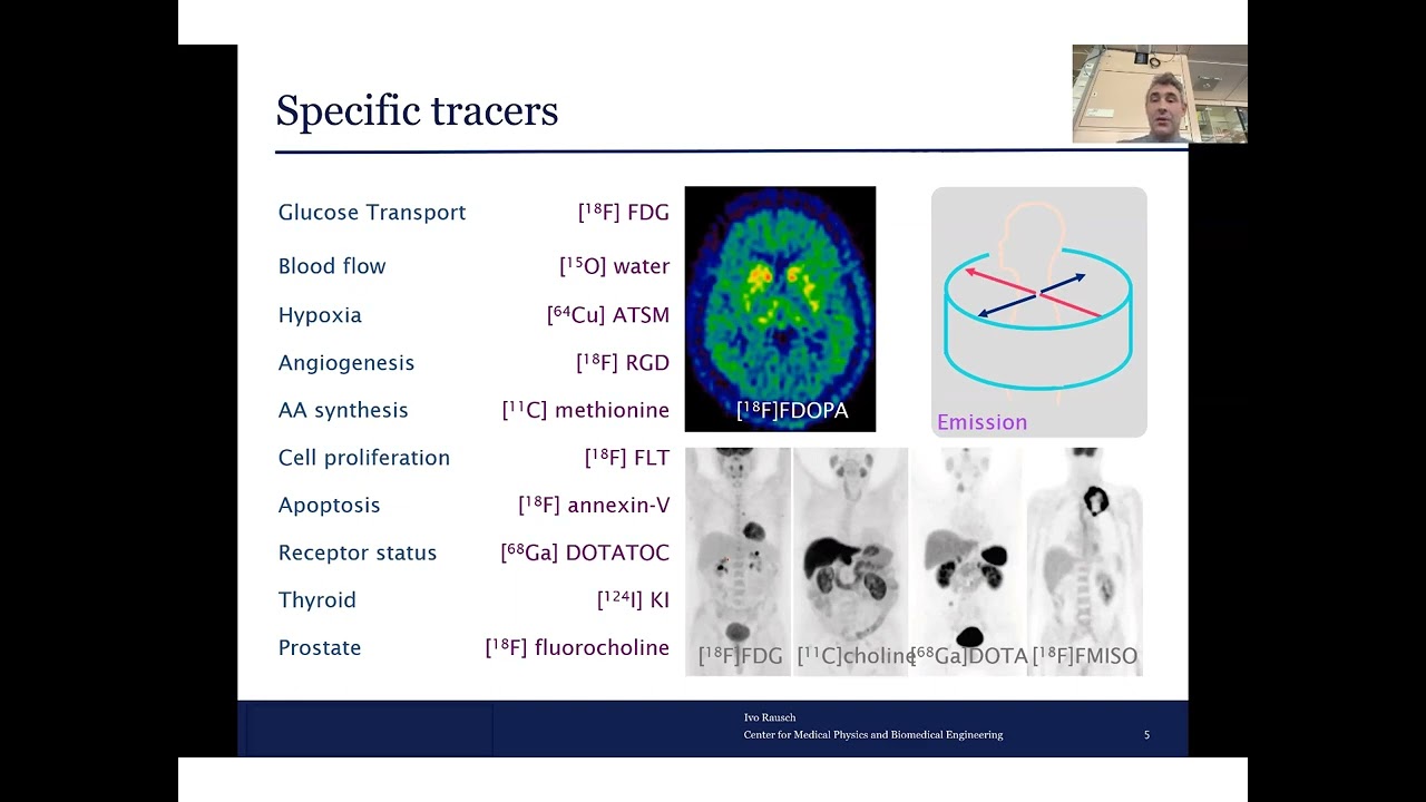

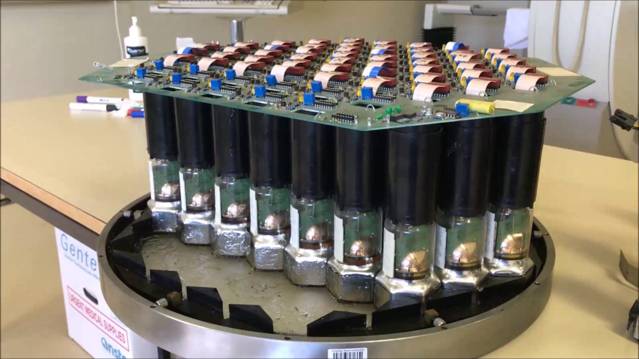


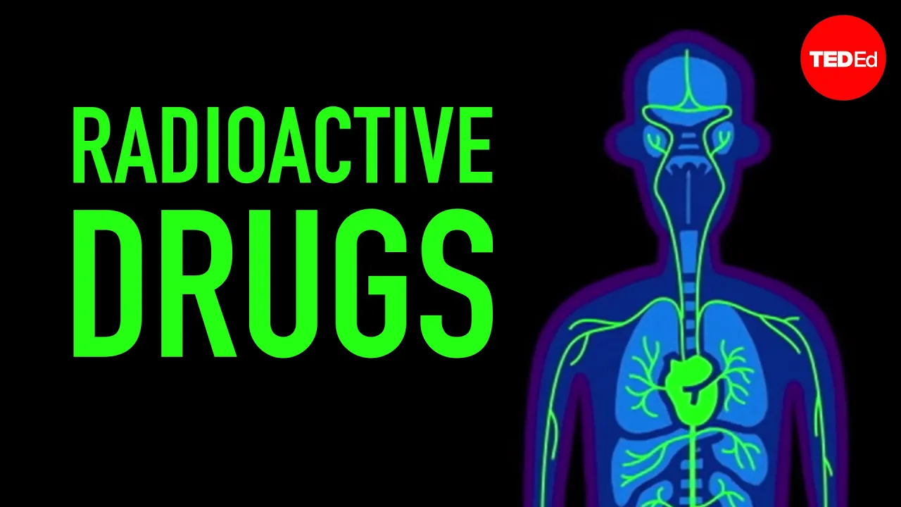
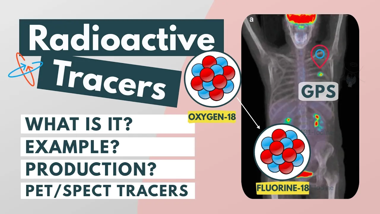


A PET (Positron Emission Tomography) scan works by injecting a custom-designed radioactive tracer (such as FDG - fluoro-deoxy-glucose) into the patient's body, which is preferentially absorbed by active tissues and cancer cells; the tracer contains fluorine-18 that decays to produce positrons, which annihilate with electrons to generate two gamma photons traveling in opposite directions; these gamma photons are detected by a ring of detectors surrounding the body, and the computer identifies the intersection points of multiple lines of response (LOR) to create detailed 3D images showing areas of high metabolic activity, such as tumors or brain regions with abnormal glucose metabolism.
PET Scan Working Principle | Cambridge A Level Physics (9702) A2
Added:The concept of antimatter, specifically the positron as the antiparticle of the electron.
The mechanism of beta-plus (β+) radioactive decay, where a proton decays into a neutron, a positron, and a neutrino.
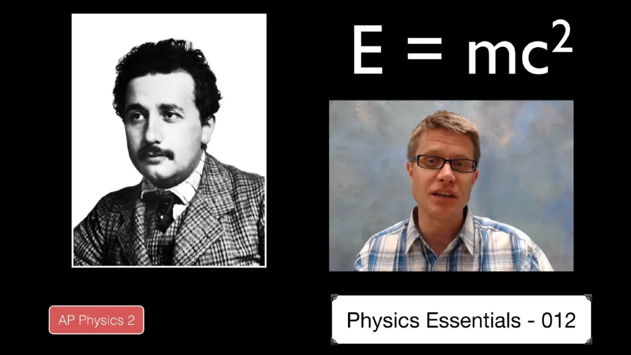
Einstein's mass-energy equivalence principle (E=mc^2) and how mass can be converted into energy.
The laws of conservation of momentum and energy, which explain why gamma-ray photons are emitted in opposite directions during annihilation.
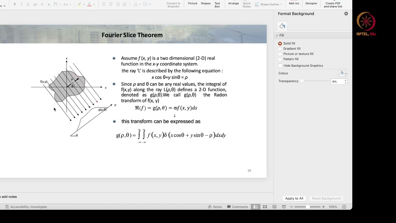
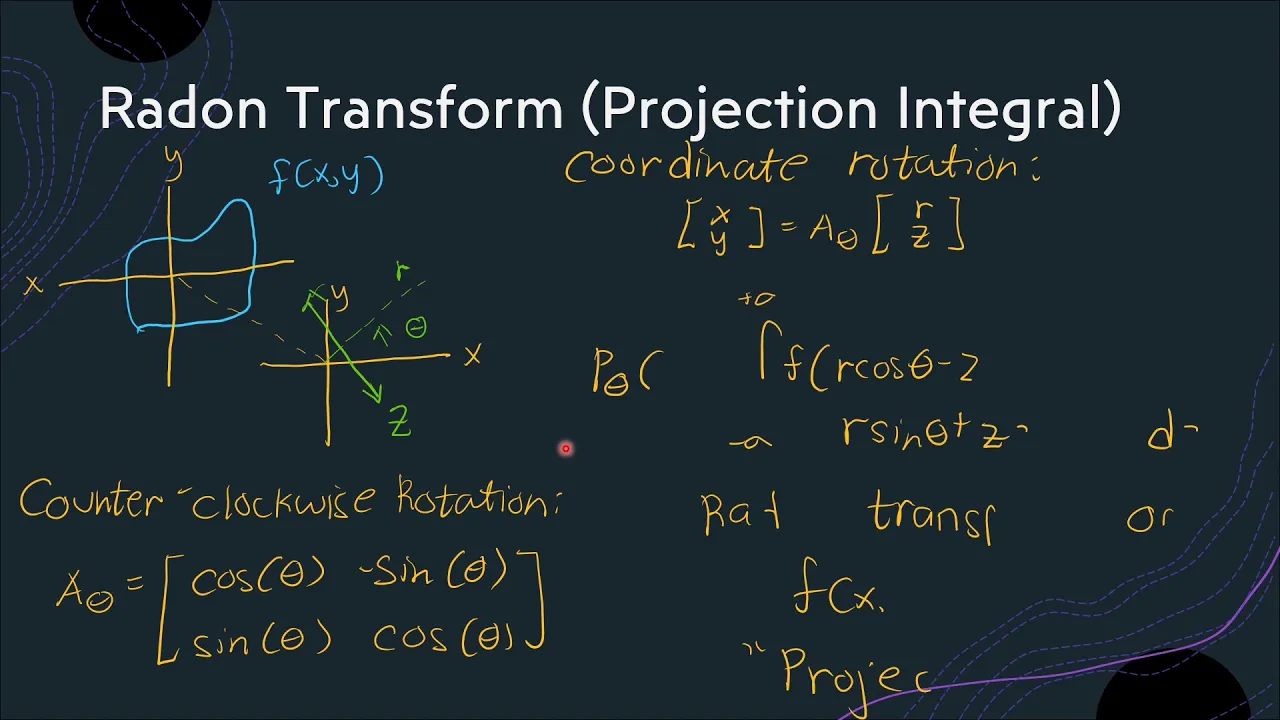
Mathematical principles of tomographic reconstruction, such as Filtered Back Projection, used to convert lines of response into 3D images.
The integration of PET with other imaging modalities, such as PET-CT and PET-MRI, to provide simultaneous anatomical and functional details.
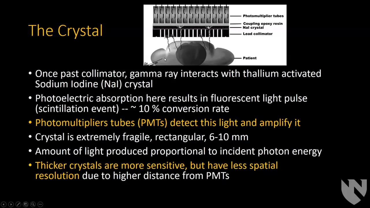
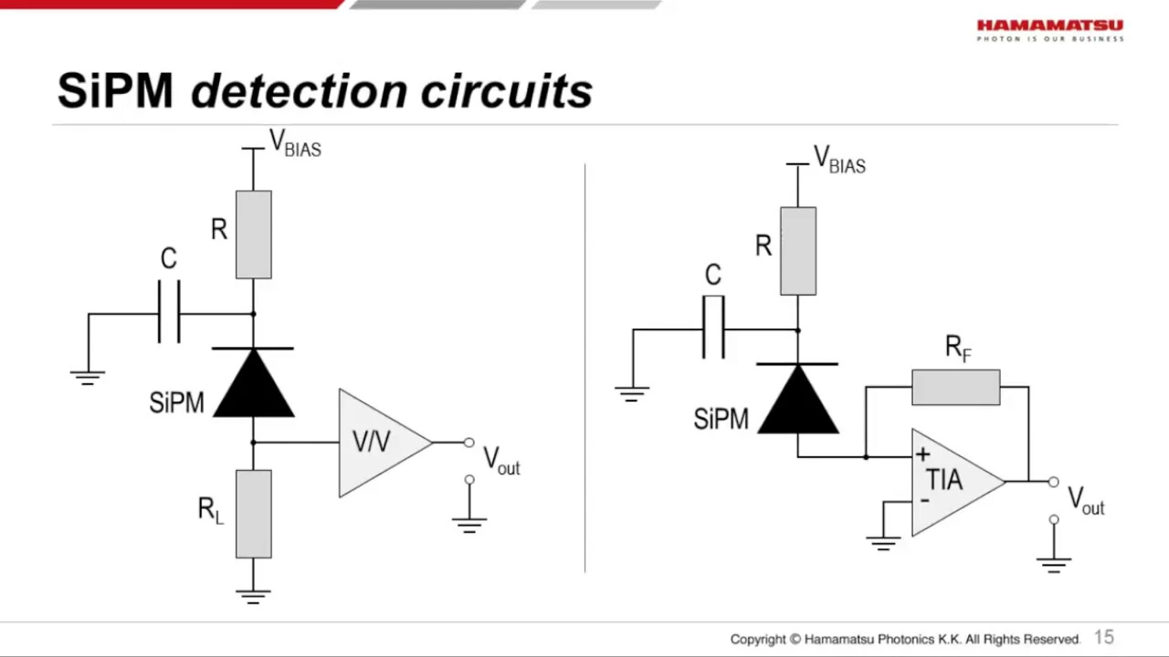
The physics and operation of gamma-ray detectors, including scintillation crystals and photomultiplier tubes (PMTs) or silicon photomultipliers (SiPMs).
The cyclotron production of short-lived positron-emitting isotopes (e.g., Fluorine-18) and the synthesis of radiopharmaceuticals like FDG.
9.7K views196likes18:21Original Release: 2022-03-16
so in the previous video we looked at the two physics principles are very important in pet scan now we are going to see what actually happens when you go to hospital to get a pet scan okay mislea what's happening yeah so if you are going to the hospital for a pet scan first thing they'll do is they will design the tracer for your body they will actually take certain measurements about your mass your weight you know your age i guess and also how you metabolize certain compounds so after some testing the custom design a radioactive tracer because remember the radioactive tracer have a very very short half-life today you go scan or today they make a special one special so when we introduce the radioactive tracer into your body most of the time by injection you can see our friend here poor dude is going for brain scan so when we inject the radioactive tracer let's go with the classic old fdg fluoro dioxide glucose for your brain and once this tracer is absorbed because it is after all glucose or sugar absorbed by the brain we get that annihilation process the gamma ray in the opposite direction okay so when it comes to writing working principles for your passive question what we want to see or what the examiners want to see is a basic understanding of the processes that has happened starting with the tracer so let's look at how it could look like in the form of a sentence so we need to first talk about how we would inject this tracer into the body okay injected into the human body and then you can say that it is taken up the fdg or the floral dioxide glucose taken up preferentially by your active tissues or your rapidly growing cancer cells let's say our patient has a brain tumor okay and the important thing here is also to mention the annihilation process okay so the annihilation process here is when the fdg basically not dj actually the fluorine 18 the spy compound decays to produce a positron we've learned this in the previous video okay this positron will annihilate on contact with the electrons producing your two high energy gamma photons traveling in the opposite direction so remember you can be asked to explain a little bit more about the annihilation process for example why do they travel in the opposite direction how where is the energy of the gamma photon coming from but the focus of you writing a working principle essay is to showcase that you understand one there is a need for a tracer number two cancer cells and active brain cells really like this tracer and it will absorb this number three there is an f18 inside the tracer that would decay to produce a positron upon annihilation two gamma photons traveling in the opposite direction so this first part is basically describing the trees so just now we looked at the tracer and your tracer the main idea is during the annihilation process there is two gamma photon that will be traveling the opposite direction now we're going to look at the detection how do we pick up these gamma photons and what do we do with the data so you can see this fancy ring here once the patients once we know that the patient the tracer has reached the patient's brain we're going to put the patient into a machine and the machine basically consists of a ring this looks like a ring right it's a ring of gamma ray detectors duh so gamma ray detectors is to detect gamma rays so during the annihilation process we'll have a gamma ray traveling this direction and another gamma ray traveling this direction so it will activate these two detectors it's the activated gamma ray detector and when it activates this is when we know a hey hey hey hey somewhere between these two lines lie an annihilation event somewhere between these two lines lie and annihilation event which also happens inside the tissue that we want to study your brain tissue has absorbed the fdg inside the fdg there's a fluorine 18 fluorine 18 causes this gamma to simultaneously hit and ping this two activated detector so when we reverse back the reasoning definitely on this line there is a tumor but teacher this line is a very long line is the tumor here is the tumor here on the forehead is the tumor here at the back of the brain well in order to get accurate data what do we do uh in experiment we take more readings another line we need another line and see where they intersect ah so the thing is of course there's only one positron right there like many many positrons because you know these are all atoms so probably very soon after that we will get an opposite ping in the opposite direction maybe something like this because the gamma will shoot out this way so somewhere along the intersection we will get the position of the tumor all right so let's write that down inside our working principle as regard we got to sentence this all right so as mentioned just now we are talking about gamma photons and gamma photons they penetrate the body very easily it's as if your body is not there okay and right now what will happen is we will put a pet scanner positron emission tomography scanner of circular ring detectors so because this design is so important you must mention in your explanation or in your description that we have a scanner of circular ring detectors don't just say put it inside a detector it's not it's ain't no normal detector yo it's a circular ring that looks like a fancy crown okay so we will ping but don't use the word ping we are going to detect the gamma rays photons simultaneously okay so we are going to be able to activate two simultaneous one and this is known as the coincidence detection all right incidence detection and uh you might be thinking teacher you know when it comes to coincidence detection right let's say we look at this picture we have this patient here and then we ping sensor number one and sensor number two it feels like sensor number one will ping slightly earlier than sensor number two because the distance traveled by the first gamma photon let's say i call this gamma 1 is a little bit shorter than the distance traveled by the second gamma photon let's say i call this gamma 2.
then i'm like well speed of light right it's a photon it's traveling so fast the extra few cm of human tissue doesn't really register but we do have a tolerance window very short of five to ten nanosecond okay so the important thing to mention is since it penetrates the body easily we have a ring detector around the body part that we want to scan and if there is simultaneous detection meaning we hit them in the same direction okay this is coincidence detection all right so then we draw that straight line so the line between two simultaneously so the simultaneous word is very important because you know sometimes random things can happen and certain detectors will be activated because there's background radiation all sorts of things are happening inside the detector inside your body at the same time that we don't care about because there are different tissues there's also digesting glucose so we only care about the pain that happens or the activation that happens at the same time so you will call this the line between two simultaneously activated please don't use the word ping i say ping because i'm a millennial cie no no activator don't don't be like the activator i am the student that never writes the keyword and lose marks don't be like me write the keywords so simultaneously activated gamma detector is known as the line of response sometimes i use the short word short term lor okay so this uh gamma ray detector gamma photons that do not arrive in pairs are ignored okay i want you to look at a few other types of detection that can happen inside the ring detector of course the first one is the normal coincidence this is 180 degree this one is this is the actual data that we want this is yes okay but sometimes due to scattering so if you remember in quantum physics whenever we have a gamma photon and then it hits the electron and then one will go in this direction the gamma photon will go in this direction and then the other electron will go down in this direction so due to scattering sometimes it will change direction this one we have to ignore because if we take the line of response you know we do some coordinate geometry and we find the the straight line that connects this coordinate and this coordinate it kind of makes no sense because i don't even have a patient on this line okay second one we also ignore so the interesting thing about the second one is we have two nucleus decay at the same time so basically we have two annihilation events happening at the same time so two annihilation occur together and this is because of the random nature you know if you study the previous chapter in nuclear physics you understand that normally all nuclear reaction is random so this one is normally due to random nature random nature of the radioactive decay so there's going to be a lot of activation of all kinds of detector and i guess in real life it's quite a messer you got the actual one that you want which is the left one on the left side and you got all these other nonsense that can appear due to scattering or just wrong coincidence that you do not want so we need to ignore those when we do some data filtering that the computer have to do let's go write it down on top there in our notes so gamma photons that do not arrive in pairs are ignored we try our best to clean up the data other simultaneous detection that are made at different angles uh which is wrong not the it's not the the coincidence we're looking for okay where the position of the source of tumor okay where is data is at the intersection of two lines of response the correct intersection that's the one we want okay so we go on to the picture again you can see a little bit the first one like misny mentioned is 180 degrees apart the path of the two what you call it the two gamma ray photons 180 if you draw an angle for diagram number two you will see that that's a bit lesser already so a little bit like maybe i don't know what angle is this uh 8 120 i feel like 120 can 120 degrees and the third diagram is i don't even know what you just cannot it's just called two completely different line it's like emission thinks that these two is on so they draw the line here yeah but it's not the correct one never mind it's okay so that's when the detector has confusion so that's what i mean when we write the sentence where they are at different angles that one we don't want so when we have lots of lines of response and of course not just one location where this occurs you just draw many many many many many many many many lines that is just for one position in the brain because obviously we'll draw a few more lines just for that one spot just one smaller knee yeah because that is where the tumor is where we have high activity lots of glucose the cancer cell is just eating the glucose so all that information i mean the more lines we get the better we can pinpoint who who is the one releasing so much uh gamma radiation okay so here missy joe i don't know how many lines ready that is for one spot but your brain is big there are many spots in the brain so we have to take many many many many analyze many many points and across the entire body so there's a combination actually of many images from different different angles different different pairs different different location everything all combined to form a 3d image you can form a 3d image if you want to yeah then we can rotate and view at different angles so here are some 3d images we found on the internet that you can look at to see how a 3d pad scan image would look like so as you can tell there's a lot of brightness that's happening here and this is a 3d image because we can flip and rotate it depending on what we want to stare at so this is a case of a patient with liver cancer so i don't know whether you can tell you can see the triangle shape of the lever here and this red color shows many many annihilation event a lot of line of response gamma gamma ray detection coming from this part and this part and all these red color spots of the body showing that you know this patient has a level of the cancer that spreads here are some other images as well also 3d this is an example of breast cancer all right so you can see there's a really hot spot right here on the shoulder because a lot of again if you do bio or you understand a little bit about the human anatomy a lot of your lymph nodes and your memory glands is connected underneath your armpit so it's like right here hot spot so there's a lot of you imagine this person that the ring is around his his the ring is around her shoulder and then you will be able to see that a lot of line of response is coming from this part and the interesting thing is you also probably notice there's some colder responses around the body because this is injected into into your tracer the tracer actually travels throughout your body you will notice that the brain is also very hot because we are thinking people so whenever we are awake we are conscious our brain is actually very active and you will also notice that the bladder is also a bit full because you know will pee out a lot of the tracer it won't stay in your body as you should not this is another one this is ovarian cancer that has been metastasized or basically quite bad it starts from the ovary a lot of activity red color is large activity so normally on a pet scan we will expect a lot of activity in the brain but we're not we're not necessarily always looking at the brain any kind of abnormal growth in the body will absorb a lot of glucose causing that high activation red color so let's go back to where we started our pet scan journey so the pet scan is porcitron it's in the name my dudes and mission tomography so of course the positron here is to represent your beta plus annihilation event and then the tomography is to show the 3d image so we started this journey by looking at certain pictures of the brain and when we can build pictures of the brain this is another 3d image that we can slice at different different places and you can even see for a coma comatose patient there is almost no brain activity normally you watch the drama video then the doctors say oh no the patient got no brain activity coma this is how it looks like in a ct scan someone that is minimally conscious probably asleep deep sleep that's very green no activity fully conscious your brain is moving of course we can adjust the sensitivity to pick up different detection and because of this we can actually we have actually done a lot of research and scanning onto the human brain to understand mental illness better okay so as you can see someone with ocd is plagued with ocd the prefrontal cortex which is the front part of the brain that is very involved in organizing stuff and being very particular and meticulous about detail is fully on fire whereas a normal brain is just somewhat on oh yeah i should probably put this back in the right place whereas the ocd bridges i need i need this to be like that and you can see that it mentions here that this shows that there is a very high glucose metabolism so a lot of sugar all right the anxiety brain is also have also have a lot of activity because people with anxiety are normally people who have a lot of fear driven things and constantly feel unsafe in this world so alzheimer's is another one you can see that there is a it's not that there's no brain here okay there is brain tissue but no activity or very little activity in the tissue because very little glucose is metabolized so the normal brain this one that's mild cognitive impairment and then as alzheimer's progress you will get the brain actually darkens all right so we actually can see a lot of mental health uh problems with the brain and we can study the brain better when we can do pet scans and also all the other medical scanning in the journey to understand the least understood organ in the human body which is your big brain so don't forget in this pet scan journey besides describing the role of the radioactive tracer the annihilation event and the working principle about the ring scanner around the parts of the body that you want to scan this is basically the focus of our pet scan that's it how's your brain today take a break if you need to bye-bye yes go and think how your brain might look like okay i think that's all for this video then see you in the next one
Up Next

5:36
Gravity and Falling Objects Explained by General Relativity
@ScienceAsylum
167.6K views•2018-03-06

12:53
21cm Hyperfine Transition in Neutral Hydrogen: Radio Astronomy Basics
@AaronRobertParsons
12.4K views•2011-10-13
114:06
A2 Medical Physics Review: MRI, Ultrasound, X-Ray & CT | 9702
@ETphysics
13.6K views•2021-04-30

27:14
Entropy and the Second Law of Thermodynamics Explained
@veritasium
27.5M views•2023-07-01
Related Study Plans & Knowledge Roadmaps
Structured learning paths in Physics
physics
Nuclear Physics (Decay, Fission & Reactors)
01Atomic Structure and the Binding Force
02Radioactive Decay and Half-Life
03Nuclear Fission Mechanics and Chain Reactions
+ 2 more learning modules
12h5 modules
Explore Roadmap
physics
Soft Matter (Polymers, Colloids & Rheology)
01Foundations of Soft Matter & Thermal Forces
02Polymer Physics and Macromolecules
03Colloids, Surfactants, and Self-Assembly
+ 2 more learning modules
38h5 modules
Explore Roadmap
physics
Analytical Mechanics: Lagrangian & Hamiltonian
01Mathematical Foundations: Calculus of Variations
02From Newtonian Mechanics to Constraint Dynamics
03The Principle of Least Action & Lagrangian Mechanics
+ 3 more learning modules
28h6 modules
Explore Roadmap
physics
General Relativity (Spacetime & Cosmology)
01Foundations of Relativity: Special Relativity
02The Geometry of Spacetime: Equivalence Principle & Curvature
03Einstein's Field Equations: The Mathematics of Gravity
+ 2 more learning modules
55h5 modules
Explore Roadmap