






















The cerebellum maintains adaptive coupling with the forebrain (cortex, basal ganglia) through reciprocal neural pathways, and when basal ganglia dysfunction occurs (as in Parkinson's disease, dyskinesia, or dystonia), the cerebellum compensates through plastic changes in its output pathways; however, this compensatory adaptation can be therapeutically enhanced through targeted cerebellar stimulation, which normalizes aberrant neural activity in the cerebello-thalamo-cortical and cerebello-striatal circuits to alleviate motor symptoms.
Adaptive Cerebello-Forebrain Coupling in Health and Disease
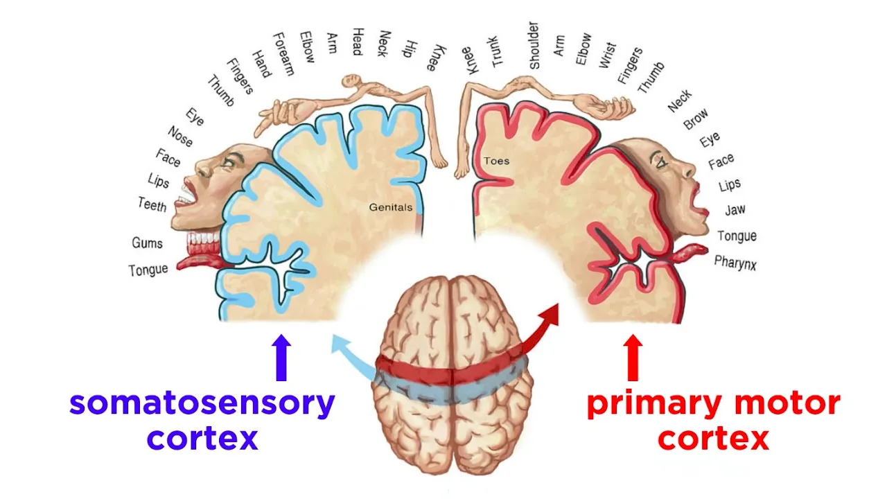
Added:Functional neuroanatomy of the basal ganglia, cerebellum, and cerebral cortex, and how they interact to regulate motor control.
The fundamental mechanisms of neural and synaptic plasticity, specifically how neural circuits adapt to changes or damage.
Basic pathophysiology of movement disorders, including the role of dopamine depletion in Parkinson's disease and abnormal circuit activity in dystonia.
Core principles of clinical neuromodulation, such as Deep Brain Stimulation (DBS) and its therapeutic applications.
Closed-loop and adaptive neuromodulation techniques that target multi-structure brain networks dynamically.
The role of cerebello-forebrain pathways in cognitive and psychiatric disorders beyond classical motor symptoms.
Advanced computational modeling of the interactions between the basal ganglia and cerebellum during sensorimotor integration.
Recent clinical trials and experimental protocols evaluating non-invasive cerebellar stimulation (e.g., cerebellar TMS or tDCS) for movement rehabilitation.
438 views4likes51:23Original Release: 2023-03-07
hi everyone welcome uh to our Tuesday seminars today I'm so happy to uh hear from Daniella Popa Daniella she received her MD from in Bucharest and then did a PhD in University of Paris in neuroscience and pharmacology then went uh to New Jersey to Rutgers uh to do a postdoc with Dennis faray and she's now back in Paris a group leader at uh equal normal Superior and she's probably one of the very few people in the world who studies uh cerebral cerebellar interactions and I this talk in particular is of particular interest to me to listen to about how pathological conditions that affect the basal ganglia result in changes that uh take place in the cerebellum and potentially therapeutic Ways by which that one can engage the cerebellum so Daniela thank you so much look forward to hearing your work okay so hello everybody so thank you very much Reza for this invitation and introduction so I came later into the cerebellar community but I'm really very happy to be part of it and I'm also very happy to be with you online today so our team here in Paris is focusing on the study of neural land interactions between the cerebellum and other brain structures in Behavior and today I will present you our data um on the Adaptive cerebellum for brain coupling in normal and pathological conditions uh so notably motor disorders such as Parkinson's disease dyskinesia and dystonia so the secrets that I will discuss today involves the cerebellum the basal ganglia and the cortex which are the main players of the turbine motor circuits and they do not work independently one from another so they can have reciprocal connections and for example here there are the cerebello cortical reciprocal connections with the layer 5 neurons from the cortex pipe Beamer from the cortex projecting to the pawn time and then to the cerebellar cortex and to the cerebellar nuclei so this word is descending pathway and then for the ascending pathway the cerebellar nuclei projecting to the motor Thalamus and then back to the cortex we also know that there are also basal ganglia cortical loops and in this case the basal ganglia projects to the motor talamus to another part of this motor Thalamus that projects to the cortex and also more recently it has been shown that there are cerebellum basal ganglia loops and in this case the cerebellum for the ascending pathway projects to the impalaminar talamus that protects to the basal ganglia so inflicts such as parapro circular or Central lateral in rodents so we see that overall we have a heavily interconnected Network but we also know that all these structures um are used by the brain in order to learn and to complete efficiently motor actions so each of them has the ability to learn in order to optimize the success of a movement and um yes these functions in each structures for example here uh uh in the cerebellum or basal ganglia is different neurological disorders so Ataxia for cerebellum Parkinson's disease for basal ganglia which can affect the other structures so we are interested to to see what goes wrong into the cerebellum when something goes wrong in the rest of the motorcycle and we uh uh we focused on the basal ganglia so our working hypothesis is the plasticity in the cerebellum continuously adapts the motor function including when the basal ganglia are dysfunctional and our objectives are to identify the plastic events involving the cerebellum in pathologies associated with the basal ganglia so we looked at Parkinson's disease at dyskinesia and dystonia and we did not only describe these plastic events but we wanted to use the plasticity in order to correct the problems and for this we induced plasticity in the system through cerebellar stimulation to to treat the disorders so we explored the benefits of the cerebellar stimulation to reverse the motor dysfunctions so we know already that there is something happening in the cerebellum in Parkinson's disease and if we look at the circuit in the normal State the cerebellum excites the cortex through the motor Thalamus and then the basal ganglia inhibits the cortex to the motor thalamus so a pathology developing in the basal danglia can also show abnormal activities in the cerebellum and this can be the case of Parkinson's disease where we know that the pulmonergic depletion is in the basal ganglia and we know there is a disruption in the basal ganglia function so here you can see a resting state study and here you see an upper activation of the pallidum in Parkinson and also we know that there is a disruption of the cortical function so in this case here we see in blue a productivity of the posterior parital cortex however increased metabolic activity in the cerebellum here in red is consistently observed so this was a resting state study but in the last year more and more studies shown that there is abnormal activities in the cerebellum also during movement so I will introduce you to some of these studies notably emerging studies so here for example you can see in red and hiper activation of the cerebellum during the automatic execution of a learned motor sequence here in this study you see two moments of the execution of the movement and you can see here that in the control patients there is an activation of the basal ganglia but this activation disappears in the Pakistanian patient but there is evidence for an over increase activity in the cerebellum compared to the control subject at the same moment of the movement as if it compensates for the lack of the activation of the basal ganglia so we can see that this is transient because we do not see this anymore a little later so it must be linked to a movement Unlimited in time for that type of movement so we see increased cerebral activity in the parkinsonian patients that may reflect redistribution of cerebral computations so we saw a normal activities in the cerebellum during movement but there are also during learning so in these studies the author's perform a learning trial and error sequence learning and they look here at the number of correct movements to the Target in parkinsonian patients so they are in black and then in Contours in Gray and we can see that at the beginning there is a difference between the two but then the parkinsonian patients can learn and then the authors look at the activities the brain activities when the performance was equivalent between the parkinsonian patients and the controls and here they saw this over activation bilaterally in the cerebellum so this over activation helps the parkinsonian patients to perform as well as as the subject the control subject so we see that there are abnormal metabolic activities in the cerebellum but not only also in the thalamus and in the sensory motor cortex and in Parkinson disease we know that there is this dopaminergic depletion in the basal ganglia but it was not clear if this is sufficient to induce the brain activities that we see in Parkinson and we have the answer in a nominal model here a neurotoxin mptp monkeys with dopaminergic depletion where we see the same patterns as in patients so this shows that the dopaminergic lesions in animals and PTP monkeys here induce similar metabolic anomalies in cerebellum sensory motor cortex as in patients and also we have a cerebellum or cortical covariance that can be suggestive of a functional Network so Timmerman in his study he looked at the dynamic of this cerebellar cortical uh thalamo cortical Network in parkinsonian patients with tremor and he found that there is an oscillation that is synchronous at around eight hours so a Tremor related abnormal coupling in the cerebellar Talamo cortical group Loop so this circuit can also be a therapeutical Target and this is seen here in this study by gross where they implanted electrodes in the cerebellocortical thalamic relay which is called Vim and they found cells related with the tremors and then stimulation in this Zone will reduce the tremor so this shows that targeting the cerebellum cortical pathway with different stimulation can alleviate parkinsonian tremor so now I showed you that there are a modification in all these structures in the motor circuit but uh how about the cerebello cortical functional connectivity so these in humans is studied with the non-invasive transcranial magnetic stimulation so how does it work we can stimulate the motor cortex and then we look at an AMG response so the motor revoked potential and we can see here a response and then if we stimulate the cerebellum before stimulating the motor cortex we see a decrease in this response and this is called the cerebellar inhibition the CBI and this decrease depends on the um delay between the two stimulations and the cerebellum has to be always stimulated before the motor cortex in order to obtain the cerebellar inhibition and this can be explained um um and it's interpreted as a decrease in the acceptability in the motor cortex following the stimulation and probably it's due to the fact that we stimulate Burkina cells that are inhibitory so we have a decrease in the cerebellocortical drive now what's happening in Parkinson's disease with this CBI so in this study career they look first at the normal control so you see the same CBI as before and then they look in parkinsonian patients of medication and they see a decrease in the CBI and then parkinsonian patient on medication so we levodopa so levodopa was not able to restore the CBI so we see that we have this cerebellar CBI that is disrupted in Parkinson's disease and suggests that functional impairment of the cerebellar cortical pathway so um for this part I showed you anomalous activations and activities in the cerebellar cortical Network in Parkinson's disease and also evidence for the cerebello cortical functional disconnection in Parkinson's disease now what is the nature of the alterations of neural activity and functional connectivity in the cerebellum cortical Loop we decided to study this phenomena in animal models where we have more possibilities of manipulation so we use an experimental Parkinson's disease model called six hydroxy dopaminergic animal mouse so what we do is we inject a dopamineergic toxin the six hydroxy dopamine in the medial forwarding bundle and this bundle links the substantia Negro to the striatum and then we can look at the impact of the pulmonergic lesion and to do so we we do the immunostaining of the tyrosine hydroxylize which is an enzyme that is essential for the pulmonergic sentences and it's seen in dopaminergic terminals so here we can see that in the case of collision we have a diminution of the the paminergic terminals in this triatum where we inject the toxins so we do this unilaterally and this has an effect on the motor activity so for the motor activity we use as a test called the use of the purple so what we do is that we put a mouse in a cylinder and then we look at how it uses the four limbs and we can see that there is an increase in the use of the ipsilateral for limb because the contralateral is impaired because of the lesion so we see this over weeks this changes in the use of the four pole and we also see changes in Locomotion so for Locomotion we do the open field so in this case we put a mouse in an open field and then we look at his activity and we can see here trajectories for the normal mice so here in Black in the open field over weeks and here for the lesient mice so we see that we have a total decreased travel that it's decreased um in in the legion mice over weeks now if we look at the gate so here in red and blue you see the four Limbs and the Hem limbs and here is the same Mouse before and after the dopamineergic region and you can see that after the pulmonergic lesion they acetate initiating movements so this is interpreted as akinesia and also they have smaller steps that is are called fascination so all this is consistent with the literature and confirms that we have a classical model of Parkinson's disease now what is the nature of the changes in this cerebellar cortical Pathways in this animal model of Parkinson's disease where we know that we have a degeneration in the dopamine in the basal ganglia so to answer this question we set up electrophysiological recordings in the cortex and in the cerebellum so here uh we perform Electro theoretical Electro physiological recordings in the cerebellar clay and in the motor cortex in fully moving mice so implanted electrodes that are fixed and we can follow the the mice over weeks and here you have examples in the motor cortex and in the cerebellar nuclei so the interposing clause and if we look at all the data we can see here um the classical decrease activity in the motor cortex that is observed normally in Parkinson's disease over weeks but we see an increase activity uh in the interposed nucleus so we look at a distance if we can look at this as if the cerebellum was trying to compensate for the low motor activity in the motor cortex by increasing its excitatory Drive so we saw decreased activity in the cortex increased activity in the cerebellum plane but we know that the cerebellar nuclei can be influenced either by the cortex through the excitatory projection to the pump time but also they can be influenced by the poor kidney cell activity in the cerebellar cortex that inhibit the cerebellum play so we look at the cellular booking cells activity in this model and to do so we are we did this in l7genic mice that we developed here in France with colleagues and this allows us to specifically stimulate sets of porchineal cells so we see that each time when we stimulate we have an action potential and then we opt to identify the Burkina cells and this animal model with the lesion uh in the dopaminergic system and here you can see a classical bikini cell from the cerebellum so in the normal animals High firing and we see that it hires even more if we are stimulating optogenetically the protein cell and now in the animal the Legion of Parkinson's disease we see a decreased firing rates for some cells we see cells with slow and irregular firing rates and we even see uh cells silent leukemia cells that Fabian was a recording when he was using the intermittent light stimulations while he was moving down with his uptrot so we see this decrease in the Pokemon cell inhibitory action on the cerebellar will play that can also show participate to the increased cerebellar activity that we see in the cerebellar clay interposed so we saw this decrease activity and also decrease activity in the motor cortex how is the functional state of the cerebello cortical pathway in this model so to answer this question so in humans I show you that to answer a such a question we can use a transcranial magnetic stimulation and CVI and here we took advantage of the L7 child adoption frangelic mice so we specifically stimulate the pukino cell in the cerebellar cortex while recording in the motor cortex so in the previous study in the lab we already showed that we can stimulate spokaneous cells and look at the motor cortex and have a waves that will uh be uh transferred uh through the water circuit so in that study we stimulate booking a cell so here we see that increase in in blue is the stimulation and then when we moved to the first synapse in the dental nucleus here we see that we have a tight inhibition and then the start the cells restarts firing and when we move to the event nucleus we also see that some parameter planes the clay cells requires cerebellum in order to fire and then in a motor cortex we could find this excitatory response in the motor cortex in the layer 5 of the motor cortex where the motor Thalamus projects and then we also look at the local field potential in the motor cortex so here we don't see much during the stimulation but then we see developing the way that is linked with a current sink here in the layer five so it's a local phenomena so this means that um when we stimulate working a cell we can have this inhibition excitation sequence in the ascending pathway and that stimulating protein cells are recording the local potential Immortal cortex it's a good way in uh and looking at the cerebral cortical functional connectivity so it's what we did in our animal model of Parkinson's disease so stimulating for kidney cells recording the motor cortex so here you can see examples of the motor cortex local field potential during the cerebellar stimulations so in in black for the controls in red for the lesient mice so there is not much during but then after the stimulation you see this wave as I showed you in the normal case just four and if we look at all the animals we see that we have a decrease in the amplitude for the lesion animals so a depression of the cerebellar cortical pathway which is consistent with the change that I showed you in the CBI in patients so overall we found that there are decreased activity in the pukemia cells but then fast firing in the cerebellar nuclei a slow firing with the motor cortex despite this past firing in the cerebellar McLean and also we found the reduced motor response that is evoked by cerebellar after stimulations so we see the presence of this compensatory adaptation here in the cerebellum following the basal ganglia lesion and the benefit potentially limited by the depression of the cerebellar cortical pathway so the service activity and its coupling to the forebrain are modified by pathological alteration in the basal ganglia now we asked if we can use cerebellar perturbations and their impact on the forebrain to improve the condition of patients and two groups look at the effect of cerebellar transcranial magnetic stimulations in levodopa-induced dyskinesia in parkinsonian patients is the gold standard in Pakistan and it works very well but after a few years many patients have abnormally voluntary movements that are also called the lipodupa-induced dyskinesia and we can look at this with the score so here you can see at the administration of the level of buying patients in Black you see an increase in this abnormally voluntary movements over time and then the authors perform a protocol of theta burst cerebellar stimulations less than two minutes a day during two weeks and they could show that they have a decrease in the dyskinesia score and even better that this decrease can stay after the end of the stimulation so they show that there is a functional impact of cerebellar stimulation on levodopa-induced dyskinesia so now with better knees during her PhD we ask if we can reproduce this therapeutic effect in rodents and for this we focused on a side effect of the levodopa the oral lingual dyskinesia and this type of dyskinesia is also seen in mice after the administration of the lobodopa had mice for nine weeks and first she induced the toxin the lesion with the 60 drugs toxin for three weeks and then she gave the level dopa to have the dyskinesia so the dyskinetic mice and we performed the stimulation the double stimulation like in the um patient study but this time specifically in L7 Child Of synchronic Mice are specifically on porchinia cells in the cruise 2 so the auto lingual zone of the cerebellum and um we performed this in two groups the correctivity called corrective cerebellar stimulation so we have the increase in the dyskinesia and then we start these stimulations and we also have another group that we call preventive cellular stimulation and in this case we start the simulations in the same time with the levodova so now we can look at the data so here is the total oral levodupa induced dyskinesia score and here you can see so the for control uh mice with levodopa there is no effect but then in parkinsonian mice with the levodopa we have this increase in the oral levodopa into this in Asia and it's stable over weeks and then with the cerebellar stimulation on the cruise tool and protein cells we have this decrease almost down to zero of the level of induces kinesia and we have this effect even after the end of the stimulation and even better if we start the stimulation in the same time with levodoba we can even prevent The Apparition of this abnormal involuntary movements so we see that we have this beneficial effects of the cerebellar stimulation on the levodopa induced disc in Asia and the effect it's even stronger than in patients because here we are almost down to zero with the oral dyskinesias now what is the effect of the protein cell data stimulation on the cerebellar output the first synapse so for this we recorded in the interposed while stimulating with the Theta burst protocol the protein cells and here you can see an example in the interposed nucleus and here the normalized responses and you see here that we have this powerful rhythmic inhibition of the cerebellar clay during the perkinia cell data stimulation magnetic stimulation in patients is unknown but here we show that stimulating selectively workingia cells is sufficient to have an effect and we also revealed that preventive stimulations accept a protective protective effect now is this Behavior effect reflected in neural activities so to answer this question parentis during her PhD recorded from the motor circuit and then to start with the interposite levels of the cerebellar so here the firing rate and here the first one corresponds to the pre-level dopa phase and then the other results correspond to the levodoba treatment and one more time controls we see that levodoba does not induce changes in the firing rate but then in the level dopa induced this in Asia we see this small decrease in the firing rate in the interpersonal players over weeks and we see the correction of this decrease with the corrective group and then the prevention of this decrease in the preventive Group by the cerebellar stimulation we also look at the firing irregularity so the CB2 of successive Isis and here you see examples in the level Dubai in this dyskinesia group and in a control group of traces and waves and if we look at all the data so we don't see much on the levodopa activity for the control group but then we see this increase in the firing irregularity in the interpersonal flows um in the level dopa induced dyskinesia and then uh with the cerebellar stimulation we see this decrease and we have corrective effect and we even prevents this abnormal increase in the firing irregularity when we started the preventive stimulation in the same time with levodoba so we have this normalization of the aberrant pattern of the activity that was observed in these kinetic animals in the interposed nucleus using these cerebellar simulations so we also look at the powerful secular Thalamus and on the motor cortex so for the paraphrocular thalamus here in the control group we don't see an effect of the lepodopa but then in the level dopa induced the skin Asia group we see the small decrease of the activity over weeks and once again we see that we can prevent this with cerebellar stimulation now in the motor cortex so uh here we see this decrease in the level dope at the beginning so the first is the one before the levodopa so it corresponds to Parkinson's disease so um it's the decrease activity in the motor cortex that I present you in the study just before compared to the controls and then we see that once we give levodopa we have this increase activity in the firing rate in the motor cortex and here again in a motor cortex we can prevent this abnormal increase in the motor cortex firing by cellular stimulations so yes the neural activity in the cerebellum paraphasicular nucleus and the motor cortex is normalized after cerebellar stimulations no we think that the stratum is at the origin of the dyskinesia so we ask it Australia several markers of this in Asia can also be normalized by cerebellar stimulation so in this case we look at the straddle marker called Force B Delta hose B which is causally linked to dyskinesia and this is a transcription factor with a long lifetime in the cells and here it has been shown by others that this accumulates industriatum during this kinesia and also that there is a correlation between the first video tapos B and the dyskinesia score so we look at this first B Delta could be in our animals and here you can see in control minus 100 and then we see this increase in levodopa induced dyskinesia so on the lesion side compared to the control side and then we also see that we have cerebellar stimulation can normalize this Triathlon expression of the marker of the cellular activity that accumulates and participates to this kinesia so this shows that the plus b in the striatom so the basal ganglia activity can also be normalized so because we know that the parapascicular thalamic nucleus is projecting to the striathome we ask if the effect of cerebellar stimulations can involve the cerebellar fascicular pathway and to answer this question first we look with Ximena at the cerebellar nuclear anatomical projection to the thalamic parasicular nucleus and for this so Ximena injected the Retro AV in the parafascicular nucleus and then she looked in the cerebellar clay and then here we can see the dental the interpose and the vestigial so we see that all of them project massively to the parapascicular thalamic nucleus so we looked at the effect of porkiness cerebellar data stimulation coupled with a chemogenetic inhibition of fibers between the cerebellar nucleus to the paraphasicular nucleus so to do so we use CNL so chemogenetics CNO and inhibitory dreads specifically in the cerebellum nucleus to those paraphasicular nucleus and we look at the behavior so here if you remember when we started a preventing group when we started stimulation in the same time with the levodopa we can prevent The Apparition of these abnormal movements and then we did the same so we did a Pokemon cells laborator stimulations but this time we inhibit in the same time the projections from the cerebellar nucleus to the paraphasicular nucleus and in this case we see levodopa in this dyskinesia so we cannot prevent any more the operation of this abnormal voluntary movements and this so this shows that the specific projections from the cerebellar nucleus to the paraphasicular nucleus are required for the level of induced dyskinesia alleviation by the porchineal cell stimulations is important for the therapeutic effect of cooking a cell stimulations so we showed um during Bernie's disease that the cerebellum is an actor in The normalization of the behavior so we see the effect on the oral lingual dyskinesia and also on the cerebelloportical pathway and on the cerebellar stratal pathway in Parkinson's disease so now to end I would like to to say a few results about Estonia so dystonia is this neurological pathology characterized by the simultaneously contraction of antagonistic muscles leading to involuntary movements and postures and we know that this is a circuit Disorder so it involves basal ganglia termocortical and cerebellum cortical pathways and here we see for example structural abnormalities so changes into gray matter densities in the cerebellum Paramus and the sensory motor cortex in one dystonia so this is the writer from dystonia so we also know that the cerebellaramic alterations are present in genetic forms of dystonia and they are predictive of the penetrance of the disease and dystonias are associated with abnormalities in synaptic plasticity in the motor 63 and may result from pathological learning so we studied a genetic animal model of basal ganglia disruption Estonia and in this case this is a new familiar monogenic dystonia so deep 25 called genome that calls for G alphaol so the gel file it's important for the signal transduction in the striatom so this is in collaboration with the nearby and we participated to the validation of this model so this model is a symptomatic model when the mice are young adults they do not they do not see the show or any historic movements or postures but then we can inject oxotremorin so this is a muscarinic Agonist so in in the striatal or systemically so this will mimic the hyperkol energy that is present in the stratum in dystonia and with these injections we can have an abnormal motor score and also we can see that there are dystonic movements and postures and now we've hint Roma and Laura so we ask what are the changes in the cerebral server coupling in dystonia and to do so we implemented a optical fiber antenna adoption in the cerebellar plane and then electrodes in the thalamus and we recorded the mice while stimulating in the thalamus while stimulating the cerebellar cortex so we did this in what we call pre-symptomatic so as I told you before uh spontaneancy there is now dystonic posture and then we induce dystonia and then we wait later when there is no more dystonic postures and we call this asymptomatic post-induction so here you can see the effect in the thalamus of the stalamic cells when we stimulate the dentite nucleus of the cerebellum and we see this in the pres-intomatic and also in the asymptomatic post-induction of the dystonia so these are examples but when we look at all the data of the cerebellar excitation of thalamic neurons we can see here that in the pre-symptomatic already we have this increased excitability in the cerebellar dynamic circles in the dystonic mice and this aberrant excitability it's Amplified post-induction of dystonia so we see that we have this abnormal potentiation in the genome mice that suggests a functional impairment of the cerebellar dynamic pathway in dystonia so we also looked at cerebellar stimulation Theta ball stimulations in this model so this time we use the same protocol as we studied before but this time we applied this protocol in the cellular plane while recording in the thalamus the activity and we look at the responses of the cerebral ceramic pathway before the Theta burst and after the data burst and it's what you can see here so here are examples so responses in the talamus in the Predator burst in the genome oxotremarine and then post Theta burst and when we look at all the data we see this depression in response to cerebellar stimulation so we have a depression that occurs on this over excitable pathway so it normalized the exitability of the cerebellar thalamic circuits so we see that we have this abnormal depression in the cerebellator pathway after cerebellar stimulations in the geneal mice so this suggests that we have this normalization of the exitability of the cerebellar Palam of silky in the auxotremoline-induced dystonia in general mice so can this normalization also play a role in the behavior so to answer this we look at the dystonia score in the wild type on General mice after the Theta burst so we did this over three days and here you can see so in the general mice you see that the cerebellar stimulations decrease dystonia scores in the general mice suggesting an improvement of the condition of the animal so as a general conclusion for the cerebellum programming Pathways in Parkinson's disease dyskinesia and Estonia I showed you that the basal ganglia pathology is Alter cerebellothalamic function and that cerebral stimulation can correct the basal ganglia dysfunctions and now we'd like to figure what and where are the plastic phenomena taking place during motor disorders so I would like to thank all the team that we are co-directing with clemony now so I present you the data from the physio pathology but we also work on motor and emotional learning and special processing and if you want to come to Paris to work with us we have two postdoc positions available so write us I would like also thanks the founding uh to thanks Reza again for the invitation and thank you all for your attention Daniel that was what a lovely convergence of human work fmri stimulation non-invasive stimulation and animal models thank you so much for that let's uh let's see if we have open it up for some questions for Daniella sure maybe I can start up um by asking you about how the loss of dopaminergic input to the striatum might result in changes in the Baseline activity of purkinje cells and nucleus neurons so do you think that dopamine by itself might have an effect on the property of the neurons in the cerebellum or is it or is it the input to the cerebellum has somehow change from the mossy fibers or other things so yeah this is this is a very interesting question so it will be really nice to know this so it's hard to say that it's really direct because there is evidence sure of some of the pulmonary receptors in the cerebellum but it's not so clear um if we really have um dopamine energy role in the cerebellum so we wanted to to put the canola and to inject the dopamine into the cerebellum and see exactly what's happening or maybe these days we should with the Delight look at the dopamine while we are doing this to really see if something is happening in the cerebellum um but I would say that this is more a compensatory effect so I I don't think it's really directly linked with the dopamine at least in in our hands what we know for now but I think it would be really nice to follow this and to yeah to follow the dopamine know that we can do this online uh while we have the lesion to see exactly what's happening in the cerebellum also on the dopaminergic side Maria please go ahead do you have a question to ask Daniella sure thank you very much for the great talk uh I have a probably naive question considering the CBI data as you are showing that like in the decrease and CBI uh is quite you know not very stable in humans so I just wonder was it tested that it's really the same timeline and it's like it's a real decree so it's different timeline and also like using your wonderful approach with animals like may be tested it's actually real timeline of CBI and also it's the matter topi because this five milliseconds it's it really puzzles me all the time it's really short thank you very much yes thank you Maria for for this question so yes this is a this is important to know and sure with I'm not sure that we really look at the I mean in detail at the Timeline so what is what is what's really impressive for us because we look after to see uh once we saw that we really have a decrease in the cerebellar cortical functional connectivity in Parkinson's we look to see um if this is something that was seen in patients so there are not many reports on this and I guess that this is really hard also to uh to do all the time because it involves already um muscular response because we stimulate the motor cortex and we look at how the muscles are so it must also depend exactly where we look so but um it has been shown that if the delay is it's bigger there is no effect so which really means that I think it's a way to read the functional state of the cortical functional connectivity cerebellar cortical functional connectivity at least at one moment but now if we if we I think at the study where I show you with the samurai that in Parkinson's uh during the execution of the movement we can have um a moment when we have the over excretion of the cerebellum but then it just a few minutes after it's it's just not there a few moments after so probably we should really look better at the at the timeline and also for the somatoto P I think it's um it's not hard to do but I mean in humans uh we can also do this in uh in mice we didn't do it so for the somatotope we are very happy to to have the L7 Channel adopting plantaining mice because we could really uh in the study with the level of enthusiastic initially at the time could really stimulate cruise to or lingual look at orolingual movements so we really were in the in the same uh secret uh but for Parkinson's yes I um I'm not sure that um that this is uh this is now Aradia thank you very much Rich you had raised your hand I was actually a very similar question it was about trying to kind of hear your take on kind of reconciling why the you know resting stage show cervix and increased connectivity uh functional studies show you know increased recruitment of the cerebellum and yet then uh it's really striking drop in the uh CBI response in in the Parkinson's uh patients both on and off medication just trying to think you know hear your your thoughts about how you kind of reconcile the those those different observations because they're really quite striking yeah yes this is this is really striking because sure if we look at the one structure we can see the activity but then sure um if we see this hyperactivity in the cerebellum probably it's really linked as a compensatory activity what we show in the animal um so the cerebellum really tries to to move to to increase the tonus so then um will there will be um more activity in the motor cortis because in Pakistanian uh in pakistanism we know that there is this decrease in the water activity of the cortex that is League short with akinesia and the symptoms in the Parkinson's so I guess that the HIPAA activity is more linked with that is the fact that the cerebellum tries to to increase this activity but then uh probably there is a a limit in this because we have this uh disconnection between the cerebellum and the cortex and um even if the cerebellum it's it's really over activated maybe it can't be even more so then it goes to talamus and the cortex in the case of Parkinson's disease that it's quite I mean advanced in the case that we look at with the dopaminergic lesion we are in a very Advanced if we want to compare very very Advanced state of the of the Parkinson yeah I was wondering if um I mean obviously if the purkinje firing rate was uh elevated in the uh um Parkinson State than doing the uh stimulation might not you know they are right increased in a significant way but is there any evidence in your animal models of elevated uh purkinje self Baseline sort of firing rates change we we really thought also that um uh because the increased activity can also come uh from the cortex or from this uh from the pokenia cells so what we saw in the cerebellum play so we didn't know how to look at the prokines so we were um so we saw cells that are not changed so they do not change not all of the cells change but what we are surprised is because we had these are seven transgenic mice that we could have with the uproad we could go down stimulate and from time to time we had the silent cells that they did nothing we could just see some complex from time to time when we stop them record them but they all the time uh answered to the cerebellar stimulation so um probably it's it's really um uh what we tried then I mean it's really hard to see we didn't see us I mean in our hands increase booking Essence only see decrease or no change so that's why we said maybe if we try to to wake up these cells the protein cells that are silent something will happen and then we'll see an increase in uh in the wave in the cerebellar cortical talama wave but I guess that the increase that we already saw in the cerebellar will play it's maybe it's just already uh some limit and then it's very hard to to push the threshold so to to really get an effect yeah well thank you for a really nice talk really interesting thank you thank you very much for your question Violetta you had a question all right yes sir hi Danielle I'm nice to see you again and a wonderful talk uh I'm most interested in this cerebellar stimulation of studies and um as I'm studying the prostitial nucleus I know there are some recent reports on Direct projection to VTA and we come to substantia Negra from some um projections from FN and I was curious what's your take on this neural population because you only looked at or okay correct me if I'm wrong you look mostly sending signals to the FN and sorry to the vcn and further populations from the bcn thank you yes thank you Violetta so um we also looked at the other locally actually so I presented the the most important data that we had on the interpose which is really the main uh nucleus that projects to the motor cortex but we had a look on the fastidious under the dentates uh and um we saw some modifications also in this in this nuclei so then for the vestigial to the VTA or the substantial in English or uh colleagues I mean show that you can stimulate and have effects in the VTA in the substantia Negra you can even have effect on dopamine um if you stimulate cerebellar nuclear in general and then I can answer with this more with the second study that we did in dystonia because in in the Parkinson levodopa Industries in Asia we stimulated the cerebellar cortex because we really wanted to be specific or lingual dyskinesias we are lucky to have a symptom that it's really in front of it but is more complex we went already with the stimulations in the cerebellar nuclei so we did the dentate shore but we can really see that we have these effects on the um talamus so you can induce the same type of effects also stimulating directly in the cerebellum then for the fastidual in the lab we did a look um in this motor studies but we looked in link with emotional learning so and what we see is that if we stimulate for example pastel nucleus we have immediate response in the parietal gray matter for example and we even show that this pathway was important for pre-learning but this this is a separate subject so I will say that um stimulating this glutamaturgical Pathways from the cerebellar nuclei even from vestigial for sure they can have an effect on the um on the ascending Pathways um and talamus and VTA and sunshine thank you thank you Violetta for the question any other questions for Daniella Daniella that was just wonderful thank you so much it was a real pleasure to listen to this beautiful word thank you very much Riza for the invitation and thank you all for the attention it's really great to be here online with you bye-bye hi everybody
Up Next

5:04
How to Learn Skills Faster: 4 Neuroscience Steps by Dr. Andrew Huberman
@virtusan
118.9K views•2022-11-18

21:55
Deciphering Neural Circuits Controlling Anorexia in Mice | Richard Palmiter
@AllenInstitute
2.3K views•2011-10-10

48:20
Memory Consolidation and Intrinsic Plasticity of Purkinje Cells
@shadmehrlab1352
214 views•2024-06-12

13:03
How Exercise Benefits Your Brain: Science Explained
@TED
11.4M views•2018-03-21
Related Study Plans & Knowledge Roadmaps
Structured learning paths in Neuroscience
neuroscience
Appetite Regulation: Brain Circuits & Hormones
01Foundations of Endocrinology and Brain Anatomy
02Introduction to Energy Homeostasis
03Peripheral Signals: Leptin, Ghrelin, and Insulin
+ 2 more learning modules
14h5 modules
Explore Roadmap
neuroscience
Neuroeconomics: Value, Risk & Brain Mechanisms
01Foundations of Neuroscience and Brain Anatomy
02Introduction to Neuroeconomics and Subjective Value
03Dopaminergic Prediction Errors & Reinforcement Learning
+ 3 more learning modules
18h6 modules
Explore Roadmap
neuroscience
Spatial Navigation: Cells, Grids & Brain Maps
01Foundations of Brain Anatomy and Memory
02Place Cells and Head-Direction Cells
03Grid Cells: The Brain's Coordinate System
+ 2 more learning modules
12h5 modules
Explore Roadmap
neuroscience
BCIs: Neural Decoding & Prosthetic Control
01Neuroscience Foundations: How the Brain Signals Intent
02Signal Acquisition: Recording the Brain's Activity
03Neural Decoding: Translating Brain Waves into Data
+ 2 more learning modules
25h5 modules
Explore Roadmap