This video demonstrates how flame tests and atomic emission spectra reveal the unique electronic structure of elements; when metal salts are heated in a Bunsen burner flame, their electrons absorb energy and emit characteristic colors corresponding to specific electronic transitions, while discharge tubes containing gases like helium, hydrogen, and mercury produce distinct line spectra that can be used to qualitatively identify unknown substances.
Flame Test & Atomic Emission Spectra: Chemistry Lab Experiment
Added:Deep Dive
The Bohr model of the atom, specifically how electrons occupy distinct, quantized energy levels and can transition between them.

فيزياء السادس علمي || 1- الفصل الثامن || النظريات الذرية
300.8K
Niels Bohr resolved the atomic collapse problem by proposing that electrons orbit the nucleus in specific, quantized energy levels without radiating energy. Electrons can only occupy these discrete orbits, each with a specific energy value. Electrons closer to the nucleus have lower energy, while those farther away have higher energy. For an electron to occupy a particular energy level, it must possess the exact amount of energy and angular momentum required. When an electron absorbs energy, it transitions to a higher energy level (excited state). The atom naturally tends toward stability, so the excited electron eventually returns to a lower energy level, emitting a photon with energy equal to the difference between the two levels (E_photon = E_final - E_initial).
Click to watch →
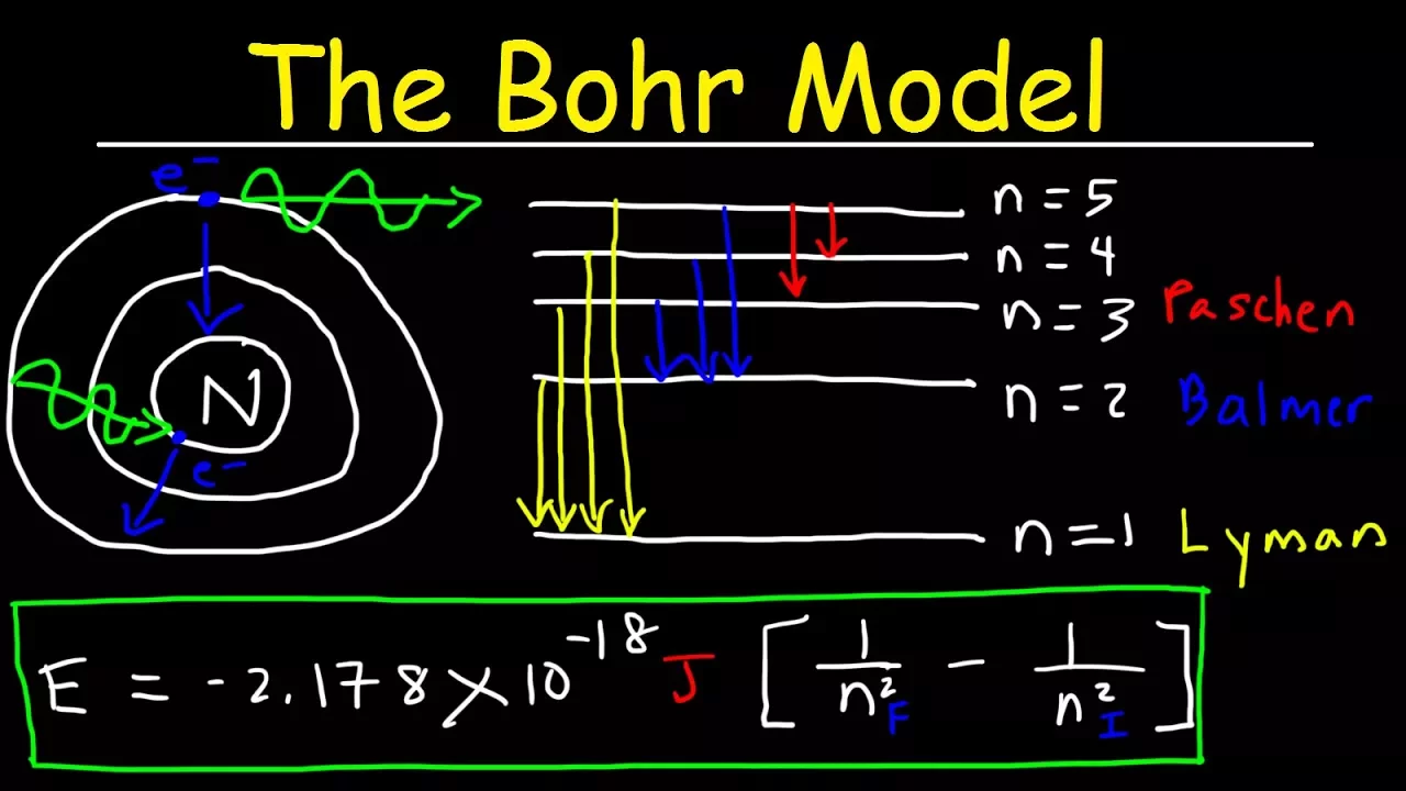
Bohr Model of the Hydrogen Atom, Electron Transitions, Atomic Energy Levels, Lyman & Balmer Series
1.6M
In the Bohr model of the hydrogen atom, electrons occupy quantized energy levels (n=1,2,3...), and energy is released or absorbed when electrons transition between these levels; the energy change is calculated using the formula E = -2.178 × 10⁻¹⁸ J × (1/n_final² - 1/n_initial²), where a negative value indicates photon emission during downward transitions, and the emitted photon's frequency and wavelength can be determined using E = hf and c = λf respectively; different electron transitions correspond to specific spectral series: Lyman series (UV radiation, n_final=1), Balmer series (visible light, n_final=2), and Paschen/Pfund/Brackett series (infrared, n_final=3,4,5).
Click to watch →
Structure of atom | Bohr's model of an atom | Part 10
95.6K
In Bohr's atomic model, electrons orbit the nucleus in specific circular paths called energy levels or shells, with each level having a fixed negative energy value (e.g., -13.6 eV for n=1, -3.4 eV for n=2 in hydrogen). Electrons can only transition between these quantized energy levels by absorbing or emitting electromagnetic radiation with energy exactly equal to the difference between the levels (ΔE = E_final - E_initial). When an electron moves from a higher to a lower energy level, it emits radiation; when moving from lower to higher, it absorbs radiation. The total energy of an electron in a hydrogen-like atom is given by E_n = -13.6 × Z²/n² eV, where Z is the atomic number and n is the principal quantum number.
Click to watch →

كيمياء الثالث متوسط / الفصل الاول / المحاضرة الثانية (منهج 2024)
538.9K
Niels Bohr proposed a revolutionary atomic model addressing Rutherford's limitations. Bohr suggested electrons occupy specific, discrete energy levels or orbits at fixed distances from the nucleus, rather than moving randomly. Each energy level has a distinct energy value and radius. Electrons can only exist in these quantized energy states. The first level holds 2 electrons, the second holds 8, and the third holds 18. Electrons transition between levels by absorbing or releasing energy. This model introduced the concept of quantized energy levels, where electrons jump between discrete states rather than moving continuously. The energy of an electron increases with its distance from the nucleus, explaining why outer levels hold more electrons.
Click to watch →

Atomic Structure L4 | Bohr Model of atom | #jee2024 #jee2025 #jeechemistry #monicabedi
71.6K
In the Bohr model of the atom, electrons orbit the nucleus in discrete energy levels without radiating energy, similar to floors in a multi-story building where the ground floor represents the lowest energy state. The total energy of an electron equals the sum of its kinetic and potential energies, with potential energy being negative and twice the magnitude of kinetic energy. When an electron transitions between energy levels, it absorbs or emits energy equal to the difference between those levels, with higher energy levels having less negative (or positive) total energy values.
Click to watch →
Deep Dive
The nature of light and the electromagnetic spectrum, including the relationship between wavelength, frequency, energy, and the visible color spectrum.
ATOMIC STRUCTURE # 6 || NATURE OF LIGHT || EMR SPECTRUM || CORPUSCULAR THEORY ||
222
The electromagnetic spectrum is the arrangement of all electromagnetic waves in order of increasing wavelength and decreasing frequency and energy. It includes cosmic rays, gamma rays, ultraviolet, visible light, infrared, microwaves, and radio waves. The visible light spectrum consists of seven colors: violet, indigo, blue, green, yellow, orange, and red, with wavelength ranges from approximately 400-750 nanometers. The relationship between wavelength and frequency is given by f = c/λ, where c is the speed of light (3 × 10^8 m/s). This fundamental relationship shows that as wavelength increases, frequency decreases proportionally.
Click to watch →

RSSB LDC 2026 Physics | Light (प्रकाश) Part- 2 | NCERT & RBSE Based | BK Pathak Sir
35.1K
This section covers electromagnetic radiation properties and visible light characteristics. From E = hν and E = hc/λ, we derive: (1) Energy is directly proportional to frequency (E ∝ ν), meaning higher frequency waves have more energy. (2) Energy is inversely proportional to wavelength (E ∝ 1/λ), meaning shorter wavelength waves have more energy. The electromagnetic spectrum from highest to lowest energy is: Gamma rays, X-rays, Ultraviolet, Visible light, Infrared, Microwaves, Radio waves. As wavelength increases, frequency, energy, and penetrating power all decrease. Visible light (VIBGYOR) spans violet (400 nm, highest frequency) to red (700 nm, lowest frequency). Green has the highest frequency among red, yellow, orange, and green.
Click to watch →

SSLC Physics | The World Of Colours And Vision | കാഴ്ചയും വർണങ്ങളുടെ ലോകവും | Full Chapter
69.8K
The electromagnetic spectrum is the complete range of electromagnetic radiation arranged by wavelength or frequency. It includes (from longest to shortest wavelength): radio waves, microwaves, infrared, visible light, ultraviolet, X-rays, and gamma rays. Visible light is the portion that humans can see, ranging from violet to red. Wavelength and frequency have an inverse relationship - as wavelength increases, frequency decreases. Energy is directly proportional to frequency. Infrared radiation is responsible for the heat we feel from the sun, while ultraviolet radiation helps the body produce vitamin D.
Click to watch →

Electromagnetic Spectrum
457
The electromagnetic spectrum consists entirely of light, with all radiation traveling at the constant speed of 3.0 × 10^8 m/s. The key differentiating property is wavelength—the distance between wave crests. Wavelength and frequency have an inverse relationship: smaller wavelengths (gamma rays) allow more waves per second, resulting in higher frequency. Energy is directly related to frequency, so gamma rays have high energy while radio waves have low energy. Visible light (ROYGBIV) is a small section of this spectrum, with red having the largest wavelength and lowest energy, and violet having the smallest wavelength and highest energy among visible colors.
Click to watch →
![Electron Configurations - Structure 1.3.1 [2025 SPEC] IB CHEM SL/HL](https://i.ytimg.com/vi/Wl0-OFVBfzk/maxresdefault.jpg)
Electron Configurations - Structure 1.3.1 [2025 SPEC] IB CHEM SL/HL
791
In the electromagnetic spectrum, wavelength, frequency, and energy are interrelated. Shorter wavelengths correspond to higher frequencies and greater energy. Conversely, longer wavelengths have lower frequencies and less energy. In the visible light spectrum, violet has the highest energy (shortest wavelength), while red has the lowest energy (longest wavelength).
Click to watch →
Deep Dive
The concepts of ground state versus excited state configurations of electrons within an atom.

Part 12: Structure of Atom | One Shot | Dr. Mandeep Dalal | 12th, NEET, IIT-JEE | Live Classes
102
Electronic configuration is the arrangement of electrons in atomic orbitals following Pauli, Aufbau, and Hund's rules. Ground state configuration is the most stable arrangement following all three principles. Excited state configuration occurs when electrons are promoted to higher energy orbitals, violating either Aufbau or Hund's Rule. For example, 1s² 2s² 2p³ is ground state for nitrogen, while 1s² 2s² 2p¹ 3s² is excited state. Excited states have higher energy and are less stable.
Click to watch →

TABLA PERIÓDICA PARTE 2
411
A ground state electron configuration has electrons distributed in the lowest possible energy orbitals. An excited state configuration has one or more electrons promoted to higher energy orbitals. The transition from ground state to excited state requires energy absorption (typically from a photon). The number of electrons remains the same, but their distribution changes. An excited state configuration requires less energy to remove an electron compared to the ground state because the excited electron is already in a higher energy orbital, closer to the edge of the atom.
Click to watch →
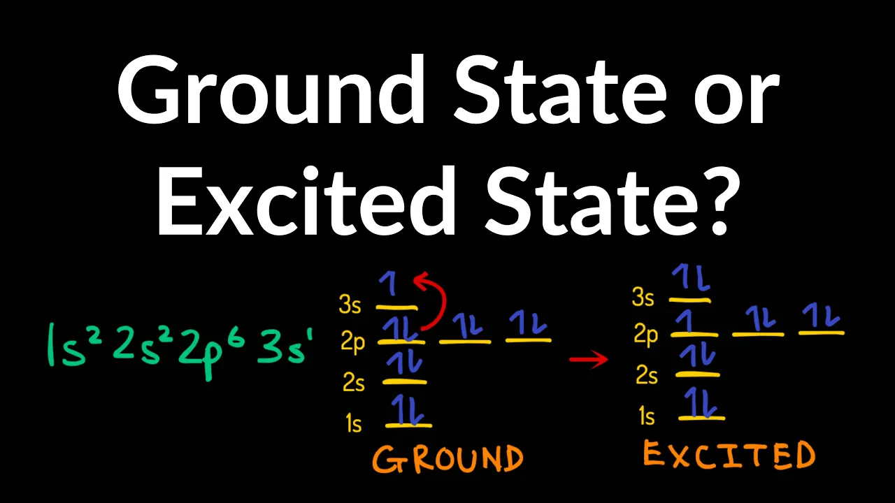
Ground State vs Excited State Electron Configuration Example, Practice Problems, Explained, Summary
303K
The ground state represents electrons in their lowest possible energy configuration, following the Aufbau principle where lower energy levels are completely filled before higher levels receive electrons. An excited state occurs when one or more electrons are promoted to higher energy levels, leaving lower levels incompletely filled. To identify these states, verify that the filling order follows 1s < 2s < 2p < 3s < 3p < 4s... and that each sub-level reaches its maximum capacity (s=2, p=6, d=10, f=14) before proceeding to the next higher level.
Click to watch →

CONFIGURACIÓN ELECTRÓNICA PARTE II
15.4K
The ground state (estado basal) is the lowest energy configuration of an atom, where electrons occupy the lowest possible energy orbitals following the Aufbau principle. An excited state occurs when an atom absorbs energy and one or more electrons move to higher energy orbitals. The ground state is more stable and has lower energy than excited states. When an atom absorbs energy (from light, heat, or electricity), electrons in the highest energy level can be excited to higher energy orbitals. For example, hydrogen in ground state (1s¹) can absorb energy and move to 2s or 2p orbitals. Excited state configurations do not follow the normal Aufbau order and are unstable, eventually releasing energy to return to the ground state.
Click to watch →

02- 03 MODERN ATOM TEORİSİ- 2 ve 3 | 2022 AYT KİMYA
2.1K
This section distinguishes between ground state and excited state electron configurations. The ground state is the lowest energy, most stable configuration of an atom, where electrons occupy the lowest energy orbitals according to the Aufbau Principle. An excited state occurs when an electron absorbs energy and moves to a higher energy orbital. For example, aluminum's ground state is 1s² 2s² 2p⁶ 3s² 3p¹, but an excited state could be 1s² 2s² 2p⁶ 3s¹ 3p². The excited state has higher energy and is less stable than the ground state. Excited states have different physical properties: the volume increases because electrons are farther from the nucleus, and reactivity increases because electrons are easier to remove. The chemical properties remain the same because chemical properties depend on the ground state electron configuration.
Click to watch →
Deep Dive
Basic laboratory safety protocols and the chemical properties of metal salts and ionic compounds commonly used in flame tests.
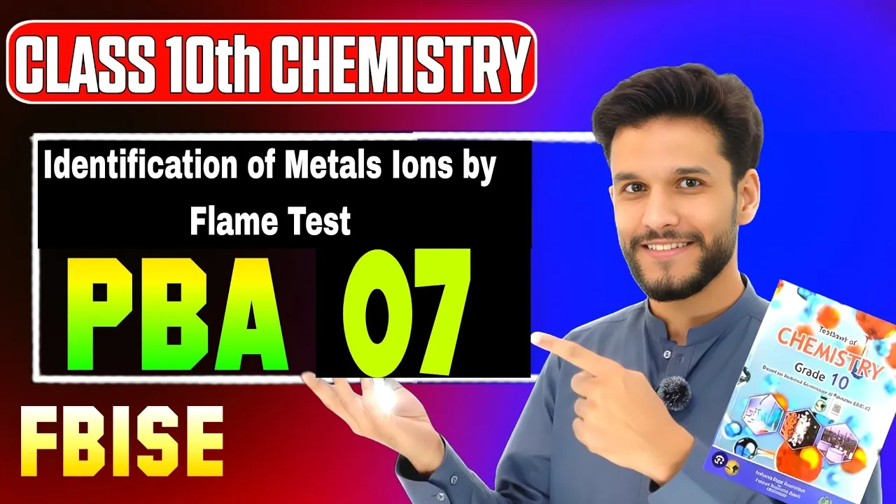
Identification of Metal Ions By Flame Test | Federal Board | PBA Chemistry Class 10
1.4K
A flame test is a qualitative analytical method for identifying metal ions by observing characteristic colors emitted when heated in a Bunsen burner flame. The technique relies on electron excitation: metal ions absorb thermal energy, causing electrons to jump to higher energy levels; upon returning to ground state, electrons emit light at specific wavelengths producing distinct colors. Required materials include metal salt samples (sodium, potassium, calcium, copper, barium chlorides), nichrome wire loop, concentrated hydrochloric acid, Bunsen burner, glass rod, and porcelain dish. Safety protocols mandate wearing gloves when handling corrosive HCl, working in well-ventilated areas, and using eye protection. The complete procedure involves: (1) preparing salt paste by mixing metal salt with concentrated HCl, (2) cleaning nichrome wire by dipping in HCl and heating until blue, (3) dipping wire into prepared paste, (4) holding in flame until characteristic color appears, (5) observing and recording the color. Each metal ion produces a unique color: sodium (golden yellow), potassium (lilac), calcium (orange-red), copper (blue-green), barium (apple green).
Click to watch →

Identify Sodium,Calcium, strontium, barium, copper & Potassium by Flame test | Che 10th | EXP.10.1
108.4K
This section details the laboratory equipment and chemicals needed for flame tests. Essential apparatus includes a platinum wire loop attached to a glass rod, a Bunsen burner with oxidizing blue flame, concentrated hydrochloric acid for cleaning, and watch glasses for holding samples. Required chemicals are metal chlorides: sodium chloride, calcium chloride, barium chloride, strontium chloride, copper chloride, and potassium chloride. The instructor explains that these chlorides are chosen because they dissociate completely in solution, releasing metal ions that produce characteristic colors when burned. Proper handling of concentrated acids requires safety precautions.
Click to watch →

Flame Test Lab | Chemistry Flame Test Experiment | Free Lab Report
7.3K
This comprehensive procedure demonstrates how to identify unknown metal ions using flame tests. The process begins with preparing aqueous solutions of metal salts by dissolving crushed chemicals in water and saturating popsicle sticks overnight. Students learn that when electrons absorb thermal energy, they jump to excited states and emit photons upon returning to ground states, producing characteristic flame colors. Each metal ion produces a distinct color (e.g., sodium = orange, copper = green/blue). Proper technique involves tapping off excess solution, holding sticks at the upper blue flame region, and adjusting burner settings for optimal observation. Safety protocols include using fire extinguishers and blankets. Students identify unknown samples by matching observed colors to reference charts, applying principles of atomic emission spectroscopy to determine elemental composition.
Click to watch →

গুচ্ছ রসায়ন সুপার সাজেশন | GST । গুচ্ছ পদ্ধতিতে ভর্তি পরীক্ষা ২০২৫ | Gst Update 2025 |guccho porikka
4.4K
Different metal ions produce characteristic flame colors: Copper (blue-green), Calcium/Strontium (red), Barium (apple-green). Sodium reacts violently with water, requiring kerosene storage. Halogenated compounds like DDT cause liver cirrhosis. Ammonium hydroxide irritates eyes by producing ammonia. Baking soda neutralizes acid burns. Benzene is toxic, so toluene is used as alternative. A desiccator dries chemicals using silica gel. Chromic acid (K2Cr2O7 + H2SO4) cleans glassware. MSDS provides chemical hazard information.
Click to watch →

Игротека с Барбоскиными | Выходные | Сборник мультиков для детей
57K
This segment demonstrates a chemistry experiment showing how different metal salts produce characteristic flame colors. The experiment uses potassium iodide (purple flame), copper sulfate with hydrochloric acid (red flame), and copper wire with copper sulfate solution (green flame). The video emphasizes safety precautions: wearing protective glasses and gloves, not leaning over flames, and not inhaling fumes. The experiment demonstrates that chemical reactions produce visual effects and that scientific experiments should focus on results rather than just visual spectacle.
Click to watch →
Prerequisite Knowledge
- Concept 01The Bohr model of the atom, specifically how electrons occupy distinct, quantized energy levels and can transition between them.
- Concept 02The nature of light and the electromagnetic spectrum, including the relationship between wavelength, frequency, energy, and the visible color spectrum.
- Concept 03The concepts of ground state versus excited state configurations of electrons within an atom.
- Concept 04Basic laboratory safety protocols and the chemical properties of metal salts and ionic compounds commonly used in flame tests.
Subsequent Learning
- Step 01The transition to the Quantum Mechanical Model of the atom, focusing on probability clouds (orbitals) and quantum numbers.
- Step 02Quantitative analytical techniques such as Atomic Absorption Spectroscopy (AAS) and Inductively Coupled Plasma Optical Emission Spectroscopy (ICP-OES) used in modern chemical analysis.
- Step 03Applications of spectroscopy in astrophysics, specifically how scientists analyze starlight to determine the elemental composition, temperature, and motion of distant celestial bodies.
- Step 04The industrial chemistry behind pyrotechnics (fireworks formulation) and the mechanics of specialized lighting technologies like neon, sodium-vapor, and halogen lamps.
Core Insight
Action Steps
Takeaway
Core Insight
1:04Playing Section
- 1
Identifies the central problem to be solved.
- 2
Lays out the high-level approach for resolution.
- 3
Explains the practical significance of the topic.
Limitations of Qualitative Flame Tests and the Bohr Model in Modern Spectroscopy
While flame tests and emission spectra of simple elements like hydrogen are valuable introductory tools, they present significant scientific and pedagogical limitations. Qualitatively, flame tests are highly prone to human error, contamination (particularly from sodium), and spectral interference, making them obsolete for precise analytical chemistry. Modern laboratories instead rely on quantitative instrumental methods like Inductively Coupled Plasma Mass Spectrometry (ICP-MS). Theoretically, these experiments often reinforce the Bohr model of the atom, which successfully explains the single-electron hydrogen spectrum but fails to account for multi-electron systems (such as helium and mercury) due to electron-electron repulsions. Relying solely on these classical observations can obscure the more accurate, probability-based quantum mechanical model of atomic orbitals.
Deep Dive
The transition to the Quantum Mechanical Model of the atom, focusing on probability clouds (orbitals) and quantum numbers.
Plus One Chemistry | Quantum Mechanical Model - Full Mark in One Video | Exam Winner
24.8K
The Quantum Mechanical Model replaced Bohr's fixed orbits with probability-based orbitals, where electrons are most likely to be found. This model emerged from the Heisenberg Uncertainty Principle, which states that we cannot simultaneously know both the exact position and momentum of an electron. Four quantum numbers describe electron location: (1) Principal Quantum Number (n) determines energy level and size, with values starting from 1; (2) Azimuthal Quantum Number (l) determines orbital shape, with values from 0 to (n-1) corresponding to S (l=0), P (l=1), D (l=2), F (l=3) subshells; (3) Magnetic Quantum Number (m) determines orbital orientation, with values from -l to +l; (4) Spin Quantum Number (s) describes electron spin direction, with only two values: +1/2 or -1/2. Each orbital can hold exactly 2 electrons with opposite spins. S-orbitals are spherical and hold 2 electrons. P-orbitals have a dumbbell shape with 3 orientations (px, py, pz) and hold 6 electrons total. D-orbitals have complex shapes with 5 orientations and hold 10 electrons. F-orbitals have very complex shapes with 7 orientations and hold 14 electrons.
Click to watch →

العاشر الدرس2 الحصة 6 النموذج الميكانيكي الموجي #العاشر #2010
7.3K
This comprehensive section covers the complete development of the quantum mechanical model of the atom: Planck and Einstein discovered electrons have dual wave-particle nature; Bohr developed a model for hydrogen (one electron) calculating energy levels and electron transitions; de Broglie confirmed wave-particle duality; Schrödinger then developed the quantum mechanical model using advanced mathematics to describe electron behavior as probability clouds. The section introduces the three quantum numbers: Principal quantum number (n) determines main energy levels (1,2,3,4...) with increasing size and energy; Azimuthal quantum number (l) determines subshell shape (0=s, 1=p, 2=d, 3=f) with values from 0 to n-1; Magnetic quantum number (m) ranges from -l to +l, determining orbital orientation. Subshell distribution follows: n=1 has s only; n=2 has s and p; n=3 has s, p, d; n=4 has s, p, d, f. Each subshell has specific orbital counts: s (l=0) has 1 orbital; p (l=1) has 3 orbitals; d (l=2) has 5 orbitals; f (l=3) has 7 orbitals. Orbital shapes are mathematically represented: s orbitals are spherical; p orbitals are dumbbell-shaped (∞) oriented along x, y, z axes; d orbitals have complex cloverleaf patterns.
Click to watch →
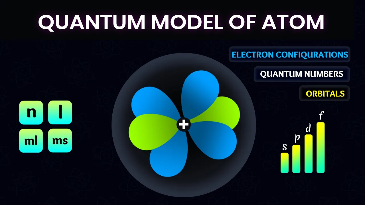
A Brief Guide to Quantum Model of Atom | Quantum Numbers
165.5K
This section develops the mathematical framework of quantum mechanics. Erwin Schrödinger's equation describes electrons as wave functions forming electron clouds rather than fixed orbits. The four quantum numbers characterize electron behavior: Principal (n) determines shell size and energy; Azimuthal (l) defines subshell type (s=0, p=1, d=2, f=3); Magnetic (ml) specifies orbital orientation; Spin (ms) indicates electron spin (+1/2 or -1/2). Shells divide into subshells: n=1 has s only; n=2 adds p; n=3 adds d; n=4 adds f. Subshells contain orbitals: s=1, p=3, d=5, f=7. Orbital shapes emerge from wave properties: s orbitals are spherical; p orbitals are dumbbell-shaped with one angular node; d orbitals have complex shapes with two angular nodes. Nodes represent zero-probability regions, while lobes show high-probability areas. This probabilistic distribution gives atoms their characteristic electron cloud appearance.
Click to watch →
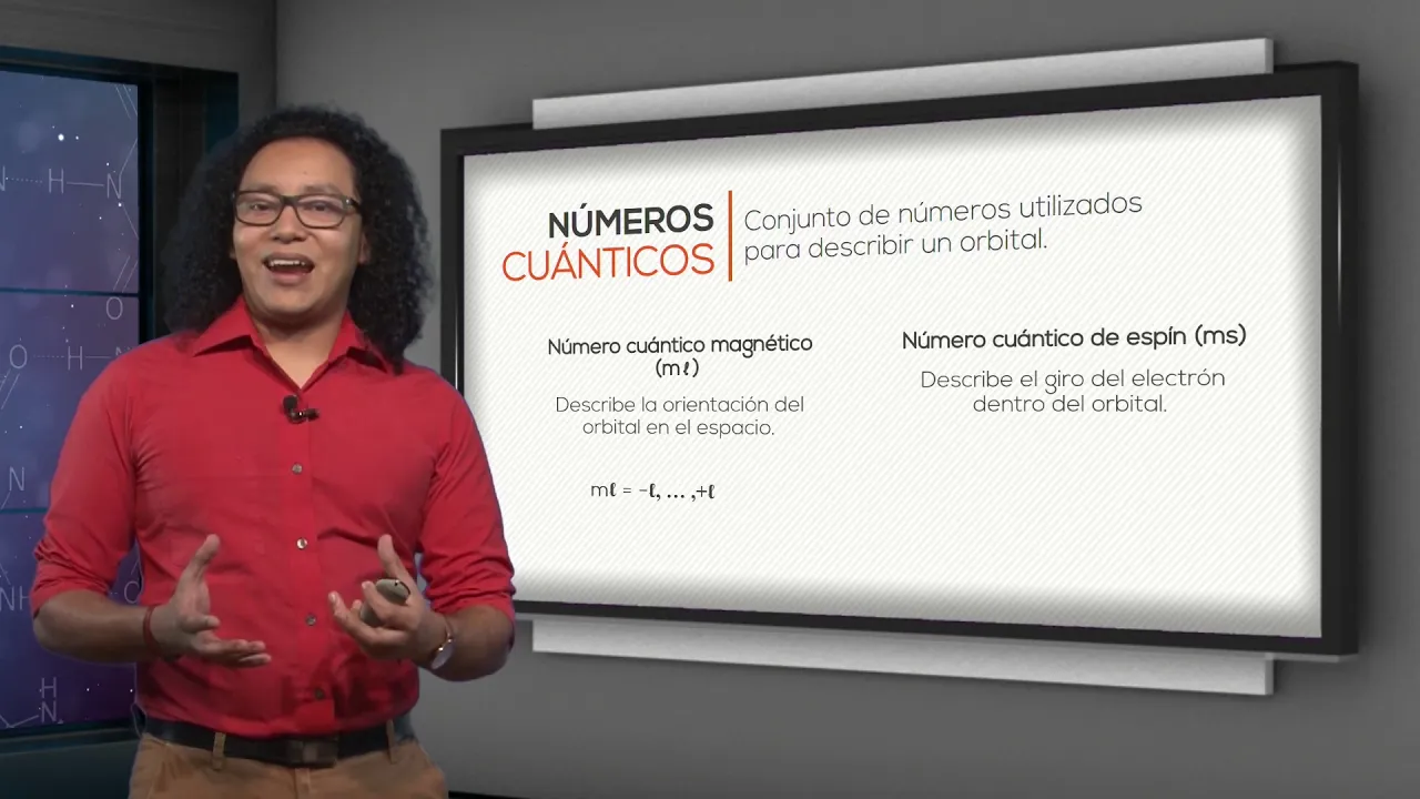
QI: Cap. 6.3. Orbitales y números cuánticos
4.5K
According to Schrödinger's atomic model, electrons do not have predetermined motion, and their position and velocity cannot be determined simultaneously. Orbitals are statistical representations of the probability of finding an electron in a specific region within the electron cloud. Four quantum numbers describe orbitals: the principal quantum number (n) describes size and energy (n ≥ 1); the azimuthal quantum number (l) describes orbital shape (0 ≤ l ≤ n-1); the magnetic quantum number (ml) describes spatial orientation (-l ≤ ml ≤ +l); and the spin quantum number (ms) describes electron spin direction (+1/2 or -1/2).
Click to watch →

Chemistry | Structure of Atom | One Shot Revision | Xylem Plus One NEET / JEE
1.1K
The quantum mechanical model replaces fixed orbits with orbitals - regions of space with high probability (90-95%) of finding electrons. Four quantum numbers describe electron states: principal (n) for shell, azimuthal (l) for subshell shape (s=0, p=1, d=2, f=3), magnetic (m) for orientation (-l to +l), and spin (s) for electron spin (+1/2 or -1/2). Orbital shapes: s is spherical, p is dumbbell-shaped, d has various shapes. Each orbital holds 2 electrons with opposite spins.
Click to watch →
Deep Dive
Quantitative analytical techniques such as Atomic Absorption Spectroscopy (AAS) and Inductively Coupled Plasma Optical Emission Spectroscopy (ICP-OES) used in modern chemical analysis.
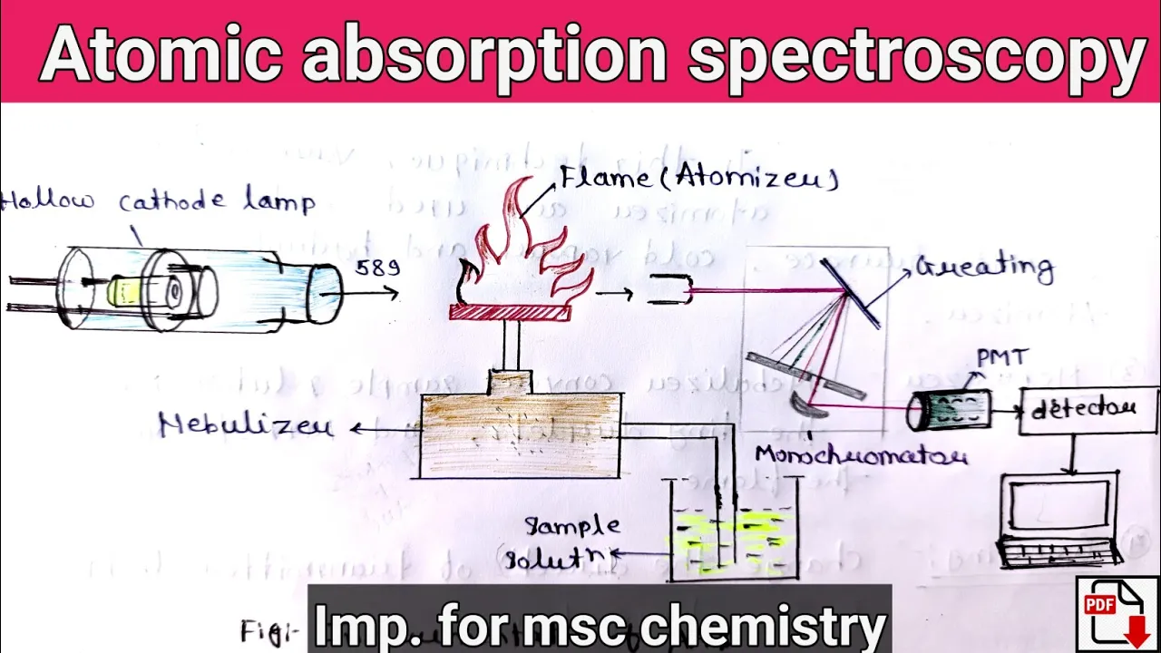
Atomic absorption spectroscopy | principle | instrumentation | applictions (@relatechemistry21 )
24.8K
Atomic Absorption Spectroscopy (AAS) is a chemical analysis technique for qualitative and quantitative element determination. The method relies on light absorption by free metallic ions following the Beer-Lambert Law. Four atomization methods exist: Flame, Graphite Furnace, Cold Vapor, and Hydride Generation AAS. The hollow cathode lamp generates characteristic element-specific radiation by sputtering metal atoms from a cathode. In Flame AAS, samples are nebulized into droplets that undergo evaporation, dissociation, and ionization in the flame, producing free atoms that absorb light. Quantitative analysis uses calibration curves comparing standard solutions to unknown samples, establishing linear relationships between concentration and absorbance for accurate elemental quantification.
Click to watch →

Los análisis de laboratorio en Minería
1.9K
Since the 1960s, modern instrumental techniques have revolutionized mineral analysis. Atomic absorption spectroscopy (AAS) quantifies metals by exciting atoms in a flame (air-acetylene or nitrous oxide-acetylene) and measuring light absorption. Modern systems include flame, graphite furnace, or dual configurations. Different metals require different flame types: air-acetylene for most metals, nitrous oxide-acetylene for refractory metals, and hydride generation for elements like arsenic and antimony. ICP (Inductively Coupled Plasma) offers greater sensitivity than AAS, allowing detection of trace metals at concentrations as low as parts per billion. ICP can analyze elements that AAS cannot detect. Modern ICP systems can be coupled with mass spectrometry for even higher sensitivity, detecting elements like vanadium, iron, cobalt, and platinum at microgram per kilogram levels.
Click to watch →

Espectroscopia de Absorção Atômica - Revisão por questões - Concursos de Perícia Criminal!
3.7K
ICP-OES represents an evolution over flame AAS, using argon plasma at 6,000-10,000 K versus flame at ~2,000 K. It enables simultaneous multi-element analysis with superior sensitivity and detects both metals and non-metals (except halogens). Graphite furnace atomization provides higher sensitivity than flame. Flame AAS is monoelemental with high specificity but requires extensive sample pretreatment and cannot perform multi-element analysis. ICP-OES is inherently multi-elemental with faster analysis but requires sophisticated equipment. The choice depends on analytical requirements—single-element precision versus multi-element screening—and factors including budget, sample complexity, and detection limits needed.
Click to watch →
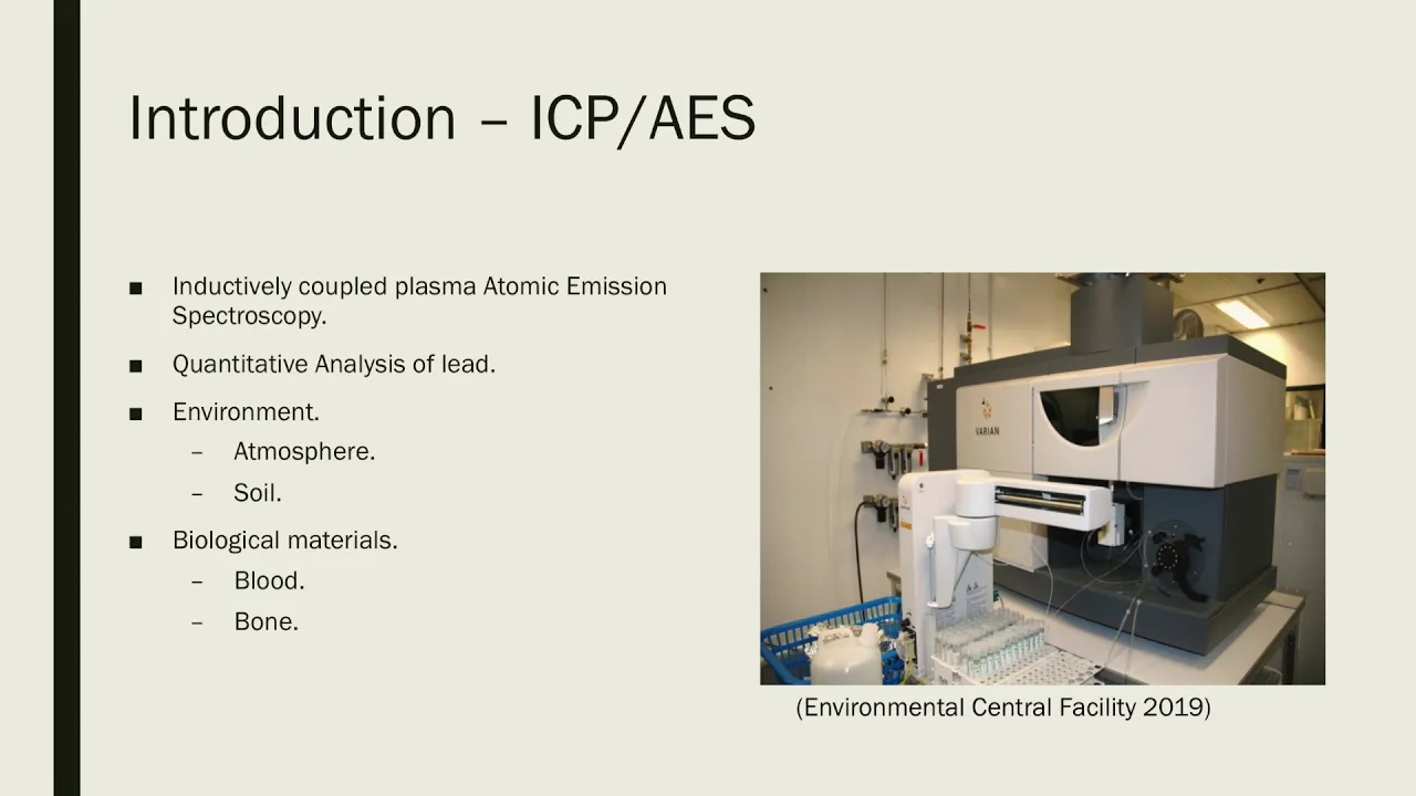
ICP AES vs AAS
7.9K
This video compares Inductively Coupled Plasma Atomic Emission Spectroscopy (ICP AES) and Atomic Absorption Spectroscopy (AAS), two analytical chemistry techniques used for quantitative analysis of lead in samples. ICP AES uses a plasma source to excite atoms, causing them to emit characteristic wavelengths of light that are measured to determine elemental concentrations, while AAS measures the absorption of light by ground-state atoms. Both techniques are widely used in environmental and industrial applications for detecting and quantifying lead contamination.
Click to watch →
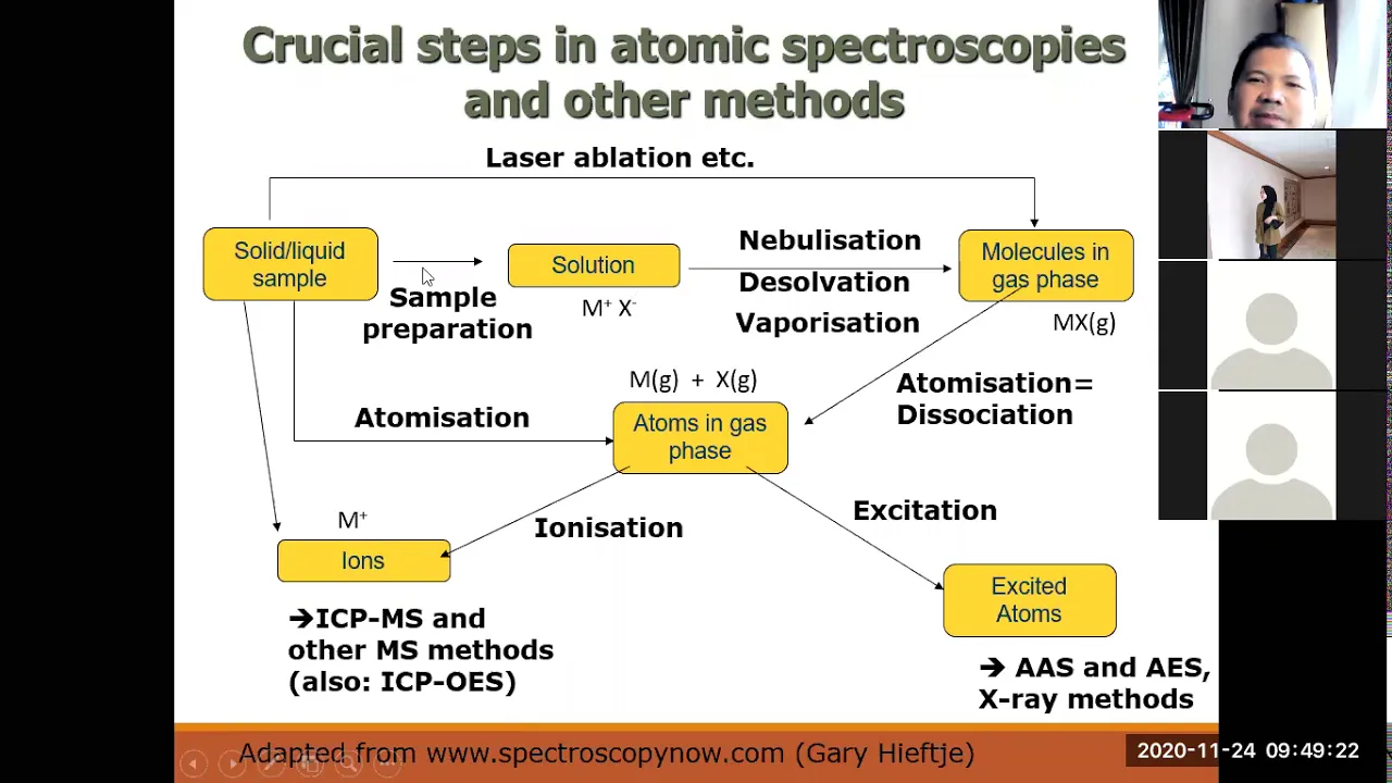
ICP MS
3.6K
Comparison of atomic spectroscopy techniques: (1) AAS - single-element analysis, detection limits 1-100 ppb, simple and inexpensive, (2) ICP-OES - multi-element analysis, detection limits 0.1 ppb, moderate cost, (3) ICP-MS - multi-element analysis, detection limits 0.001 ppb (ppt), highest sensitivity but expensive and complex. GFAAS offers higher sensitivity than flame AAS but is limited to single-element analysis. The choice depends on required sensitivity, number of elements, sample matrix, and available resources. ICP-MS is preferred for trace element analysis requiring ppt-level detection.
Click to watch →
Deep Dive
Applications of spectroscopy in astrophysics, specifically how scientists analyze starlight to determine the elemental composition, temperature, and motion of distant celestial bodies.

Light and Spectroscopy Lesson
408
Spectroscopy enables astronomers to study stars in three key ways. First, spectral fingerprints allow identification of chemical compositions—each element produces unique bright or dark lines at specific wavelengths, acting like fingerprints that reveal which elements are present in a star. Second, spectral type indicates temperature: hot gases emit light (emission spectra), while cool atmospheric gases absorb it (absorption spectra). Third, the Doppler effect reveals stellar motion—the Doppler shift causes spectral lines to shift toward blue (shorter wavelengths) when objects approach and toward red (longer wavelengths) when they recede. This allows astronomers to determine whether stars are moving closer (blueshift) or farther away (redshift), providing crucial information about galactic dynamics and cosmic expansion.
Click to watch →

Spectroscopy Cracking starlight's hidden code
505
Spectroscopy, the technique of splitting starlight into its component wavelengths, reveals the chemical composition, temperature, and motion of celestial objects through absorption and emission lines, making it a fundamental pillar of professional astronomy that allows astronomers to determine what stars are made of, how hot they are, and whether they're moving toward or away from us, with amateur astronomers now able to participate in this powerful technique using accessible equipment like star analyzers and LP spectroscopes.
Click to watch →

الفصل السادس - المحاضره ١- اطياف الهيدروجين - الفيزياء الحديثه | ٣ ثانوي | محمود مجدي٢٠٢٢
453.4K
Spectroscopy has many important applications: (1) Determining the composition of stars and other celestial bodies by analyzing their emission and absorption spectra, (2) Identifying chemical elements in unknown samples, (3) Studying atomic and molecular structure, (4) Measuring temperatures of hot objects by analyzing their blackbody radiation. The ability to determine the composition of distant stars is one of the most important applications of spectroscopy.
Click to watch →

Universe
139.9K
Astronomers study photographs of starlight split into spectra where each band has specific meaning. From spectroscopic photographs, astronomers can determine whether a star contains elements like iron, calcium, or carbon. By exposing the same photographic plate to both the star's spectrum and an iron arc spectrum, they can measure displacement between the two to tell whether a star is moving toward or away from us.
Click to watch →
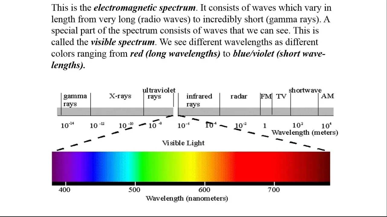
Redshift and BlueShift explanation presentation
7.7K
Scientists use spectroscopy to determine what stars are made of. The electromagnetic spectrum ranges from radio waves to gamma rays, with visible light being the portion we can see. When white light passes through a prism, different wavelengths separate into colors. Each element emits a unique pattern of wavelengths when heated, acting like a fingerprint. By attaching a spectroscope to telescopes, scientists analyze starlight spectra to identify elements present in distant stars, even thousands of light-years away.
Click to watch →
Deep Dive
The industrial chemistry behind pyrotechnics (fireworks formulation) and the mechanics of specialized lighting technologies like neon, sodium-vapor, and halogen lamps.
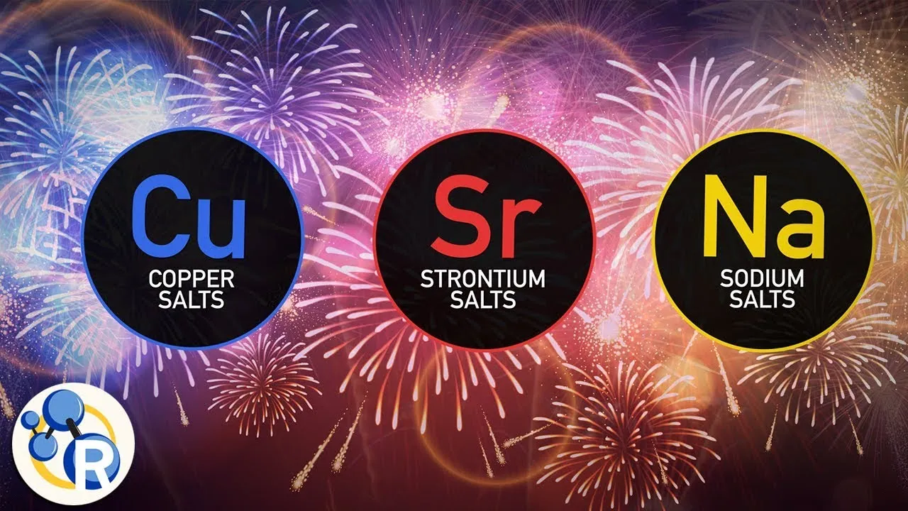
The Chemistry of Fireworks
607.3K
Fireworks are created through chemical reactions where oxidizers (like potassium perchlorate) combine with fuels (such as sulfur, charcoal, aluminum, or magnesium) to produce heat and light; different metal compounds emit specific colors when heated—strontium compounds produce red, copper compounds produce blue, sodium produces yellow-orange, calcium produces yellow-orange, and barium produces green—while the timing and bursting mechanism of aerial shells controls when and how these effects are displayed in the sky.
Click to watch →

Std 10 English Unit 5🔥PLAYING WITH FIRE | TOPPER BATCH Class FREE | Vidyakul
13.6K
Pyrotechnics is the science of fireworks, derived from the Greek words 'pyr' (fire) and 'technics' (art), which encompasses the physics and chemistry behind firework effects. The basic material used in fireworks is black powder (also called gun powder), invented in India over 2000 years ago, consisting of potassium nitrate, charcoal, and sulfur in a ratio of 75:15:10. This formula remained unchanged for centuries until modern chemistry introduced elements like aluminum, magnesium, and titanium to enhance brightness and color effects. Pyrotechnics extends beyond traditional fireworks to include devices ranging from safety matches to solid fuel rocket boosters used in space shuttles.
Click to watch →

1st Year : Illumination( I.T.I. ELECTRICIAN THEORY ) Mathur's Classes / Part-2
1.7K
Neon Sign Lamps use neon gas for reddish-pink glow in advertising, with different gases producing different colors (argon for blue, helium for yellow, chlorine for green). Xenon Lamps use xenon gas with 1.5-6mm electrode gap for cinema projectors and searchlights, producing intense white light. Halogen Lamps contain iodine and chlorine gases for decorative lighting during festivals. These specialized lamps serve specific illumination and decorative purposes.
Click to watch →

fireworks
146
Prior to the 19th century, fireworks were relatively dull until breakthroughs in chemistry revealed that oxidations with chlorates of barium, strontium, copper, and sodium produced intense bright colors, while metallic magnesium and aluminum enabled silvery light. Pyrotechnic stars contain four basic ingredients: fuel and oxidizer, color-producing salts, and a binder. Color compounds include strontium for red, calcium for orange, sodium for yellow, barium for green, copper for blue, and potassium/rubidium for violet. Aluminum produces silver and white flames, while barium stabilizes volatile elements. Common elements include chlorine in oxidizers, iron for sparks, and titanium for silver sparks. The brightest stars use aluminum fuel, with magnesium rarely used alone due to oxide layer issues. Common elements include aluminum for silver/white effects, barium for green colors, carbon in black powder, and various metals for specific colors.
Click to watch →

Химия. Удивительные эксперименты 🔥 Дома не повторять
144.2K
Different metal ions produce characteristic flame colors through electronic transitions: lithium (red), strontium (red), sodium (yellow), copper (green), barium (green), lead (blue-yellow), potassium (yellow-violet). These principles underlie pyrotechnics, where aluminum provides sparks and metal nitrates serve as oxidizers. Fireworks compositions combine specific metal salts with binding agents like starch paste. The hydrogen explosion demonstration illustrates explosive mixture formation and the critical importance of gas ventilation in laboratory safety protocols.
Click to watch →
Core Insight
Action Steps
Takeaway
Core Insight
1:04- 1
Identifies the central problem to be solved.
- 2
Lays out the high-level approach for resolution.
- 3
Explains the practical significance of the topic.
Limitations of Qualitative Flame Tests and the Bohr Model in Modern Spectroscopy
While flame tests and emission spectra of simple elements like hydrogen are valuable introductory tools, they present significant scientific and pedagogical limitations. Qualitatively, flame tests are highly prone to human error, contamination (particularly from sodium), and spectral interference, making them obsolete for precise analytical chemistry. Modern laboratories instead rely on quantitative instrumental methods like Inductively Coupled Plasma Mass Spectrometry (ICP-MS). Theoretically, these experiments often reinforce the Bohr model of the atom, which successfully explains the single-electron hydrogen spectrum but fails to account for multi-electron systems (such as helium and mercury) due to electron-electron repulsions. Relying solely on these classical observations can obscure the more accurate, probability-based quantum mechanical model of atomic orbitals.
The Bohr model of the atom, specifically how electrons occupy distinct, quantized energy levels and can transition between them.
The nature of light and the electromagnetic spectrum, including the relationship between wavelength, frequency, energy, and the visible color spectrum.
The concepts of ground state versus excited state configurations of electrons within an atom.
Basic laboratory safety protocols and the chemical properties of metal salts and ionic compounds commonly used in flame tests.
The transition to the Quantum Mechanical Model of the atom, focusing on probability clouds (orbitals) and quantum numbers.
Quantitative analytical techniques such as Atomic Absorption Spectroscopy (AAS) and Inductively Coupled Plasma Optical Emission Spectroscopy (ICP-OES) used in modern chemical analysis.
Applications of spectroscopy in astrophysics, specifically how scientists analyze starlight to determine the elemental composition, temperature, and motion of distant celestial bodies.
The industrial chemistry behind pyrotechnics (fireworks formulation) and the mechanics of specialized lighting technologies like neon, sodium-vapor, and halogen lamps.
4.5K views29likes4:51@emilyh-science-music-booksOriginal Release: 2021-12-11
[Music] [Music] [Music] [Music] do [Music] [Music] [Music] [Music] [Music] so [Music] [Music] [Music] [Music] [Music] [Music] [Music] [Music] do [Music] you
Up Next
3:21
Curved Arrows in Reaction Mechanisms: A Guide
@chegg
209.7K views•2024-02-15
83:56
Biocatalysis and Catalysis in Islatravir Synthesis | Merck Webinar
@SCIwheresciencemeetsbusiness
675 views•2020-10-08
10:06
1H NMR: Determining Number of Peaks from Structure
@MSJChem
59.2K views•2017-04-06

17:31
Edible Water Bottles: A DIY Guide to Sodium Alginate Spherification
@ryan
10.5M views•2019-06-21
Related Study Plans & Knowledge Roadmaps
Structured learning paths in Chemistry
chemistry
Nuclear Chemistry: Decay, Meds & Fuel Cycle
01Introduction to Nuclear Structure and Radioactivity
02Decay Dynamics and Kinetics
03Radiation Interaction and Detection
+ 2 more learning modules
24h5 modules
Explore Roadmap
chemistry
Supramolecular Chemistry: Forces & Machines
01Foundations of Chemical Bonding and Intermolecular Forces
02Introduction to Host-Guest Chemistry
03Thermodynamics and Dynamics of Self-Assembly
+ 2 more learning modules
16h5 modules
Explore Roadmap
chemistry
Photochemistry: Excited States & Reactions
01Foundations of Light-Matter Interactions
02Molecular Orbitals and Electronic States
03Photophysical Processes and Jablonski Diagrams
+ 2 more learning modules
18h5 modules
Explore Roadmap
chemistry
Coordination Chemistry: Bonding & Properties
01Introduction to Transition Metals and Ligands
02Crystal Field Theory (CFT) and Geometry
03Magnetic Behavior and Spin States
+ 2 more learning modules
14h5 modules
Explore Roadmap