This video demonstrates how to measure Trans Epithelial Electrical Resistance (TEER) using WPI's EVOM Manual instrument, covering setup procedures including file naming configurations (auto-indexed prefixes or unique names), meter parameter selections (plate size, resistance units, and range), blank subtraction methodology for accurate cell culture measurements, and the complete measurement workflow involving blank handling, data collection, and file storage to USB drives.
How to Measure TEER with EVOM Manual: A Step-by-Step Guide
Added:Deep Dive
Understanding of epithelial and endothelial cell biology, specifically the structure and function of tight junctions in forming cellular barriers.

BIOL 4610 Module 4 1 Part 1
133
Tight junctions are occluding junctions that maintain epithelial cell polarity by preventing apical and basolateral membrane proteins from diffusing between domains. They form a tight seal near the apical domain, creating a barrier to solute diffusion. Electron-dense tracer experiments demonstrate that solutes cannot pass through tight junctions from apical to basolateral side. Tight junctions are not completely impermeable; the paracellular pathway allows certain small molecules and ions to pass. The CFTR chloride channel is an example of a component that can cross tight junctions, and its mutation causes cystic fibrosis. Tight junctions are essential for transcellular transport, such as glucose absorption in the intestine, using sodium-glucose symporters and GLUT2 transporters. Tight junctions are composed of occludin, claudin (multipass transmembrane proteins), and JAM (single-pass transmembrane protein), forming tight bands that create the seal.
Click to watch →

Structural Organisation in Animals | Rapid NCERT Revision | Biology | NEET 2023 | Dr. Anand Mani
57.4K
Tight junctions are specialized connections between adjacent epithelial cells. They form a seal that prevents substances from passing between cells (paracellular pathway). Tight junctions help maintain the barrier function of epithelial tissues and prevent leakage of fluids.
Click to watch →

Cell Adhesion Molecules & Cell Cell Interactions - CSIR Dec 2025 | Emotion 6.0 | Lecture 14 | IFAS
8.9K
Tight junctions form continuous seals between adjacent cells to prevent passage of molecules and ions between cells. Key structural proteins include occludin (four transmembrane domains), claudins (four transmembrane domains), and junctional adhesion molecules (single transmembrane domain). Occludin and claudins associate with actin cytoskeleton to strengthen the barrier. Tight junctions are essential for maintaining tissue integrity and regulating substance transport across epithelial layers, preventing urine leakage from bladders and food particles from escaping intestines.
Click to watch →
4c-Summary of Intercellular junctions with animations
32.3K
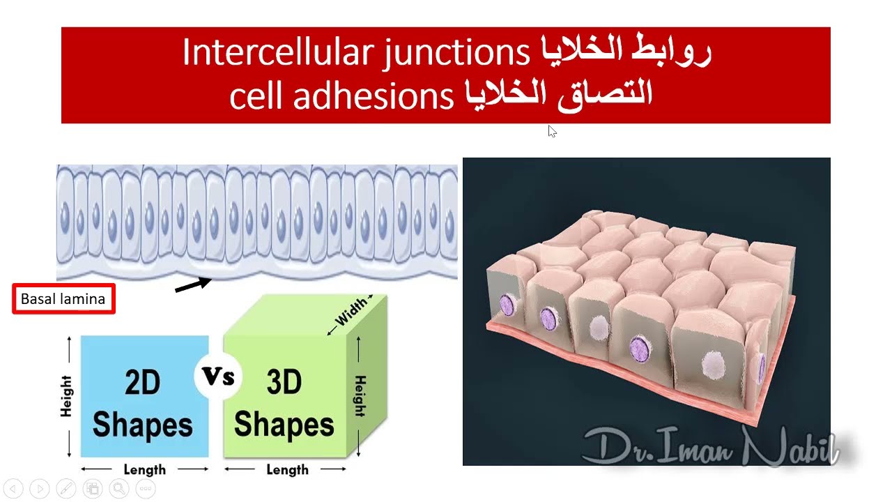
Tight junctions (claudin junctions) are formed when transmembrane proteins from adjacent cells fuse completely, sealing the intercellular space. This fusion creates a physical barrier that prevents substances from passing between cells. The barrier function requires that molecules and ions must pass through cells via pumps and channels rather than moving directly between cells. This mechanism is essential for maintaining distinct cellular compartments and controlling substance movement across epithelial layers.
Click to watch →
Histology lecture 3 week 3
148
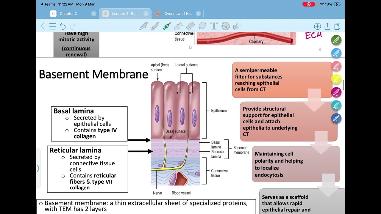
Tight junctions (zonula occludens) are the highest junctions located on the lateral surfaces of epithelial cells, forming a continuous belt around the cell like a belt (zonula means belt). They contain key transmembrane proteins including occludin and claudin. The primary function of tight junctions is to create a barrier that prevents the passage of substances between cells (paracellular pathway), forcing substances to pass through cells (transcellular pathway). This allows cells to control what substances pass through the tissue, providing a critical barrier function. Tight junctions also help maintain cell polarity by creating a distinct boundary between the apical and basolateral domains of the plasma membrane.
Click to watch →
Deep Dive
Basic principles of electricity, particularly Ohm's Law (V = I * R) and how electrical resistance is measured in conductive solutions.
Princípios Sobre Eletricidade Voltada a Automotiva
19.3K
Electrical resistance is the difficulty materials impose on current passage, measured in ohms (Ω). Resistance depends on material nature, temperature, cross-sectional area, and length. Good conductors have valence electrons weakly attracted to nuclei, facilitating current flow. Insulators have high resistance preventing current passage. Semiconductors have intermediate properties. Resistivity (Ω·m) is resistance of 1m length, 1mm² area at 20°C. Temperature increases resistance in metals but decreases it in semiconductors. Ohm's Law states current is directly proportional to voltage and inversely proportional to resistance: V = I × R. This allows calculating resistance from voltage and current measurements.
Click to watch →

11 Faraday's Law of Electrolysis and Ohm's Law For 12th NCERT IIT JEE NEET Entrance Exams 1
49.1K
Ohm's Law states that current (I) is directly proportional to potential difference (V) across a conductor (V = IR). Resistance (R) is directly proportional to length (L) and inversely proportional to cross-sectional area (A): R = ρ(L/A), where ρ is resistivity. Resistivity is a material property measured in ohm-meters. Conductivity (σ) is the reciprocal of resistivity: σ = 1/ρ, measured in siemens per meter. The cell constant (G* = L/A) relates solution resistance to conductivity. These principles form the foundation for understanding electrical behavior in both metallic conductors and electrolyte solutions.
Click to watch →
Лекция 1. Введение. Предварительные сведения
39.2K
Ohm's Law (V = I × R) describes the relationship between voltage, current, and resistance in electrical circuits. Voltage is the potential difference driving current flow, measured in volts. Current is the rate of charge flow, measured in amperes. Resistance opposes current flow, measured in ohms. A short circuit occurs when minimal resistance connects a power source, causing dangerously high current that destroys components. Resistors limit current to safe levels for components like LEDs and lamps. Ground (GND) represents a reference point with zero potential, and all other voltages are measured relative to this point.
Click to watch →

RRB Group D 2025 | Numericals Series | Electricity | Group D Physics + Chemistry #1 | Dharmendra Sir
192.9K
This segment covers Ohm's Law, which states that voltage equals current multiplied by resistance (V = IR). Students learn to calculate current when voltage and resistance are given by rearranging to I = V/R. The instructor demonstrates multiple examples with different resistance values, including decimal and large resistance values. Students practice decimal division and fraction-to-decimal conversion. The segment emphasizes that resistance is measured in Ohms and that students should be comfortable with various numerical formats.
Click to watch →

Electricity in 30 minutes 🔥 Class 10 Science One Shot NCERT Revision!
274.3K
Electric charge is a scalar, quantized quantity with symbol 'q' and SI unit Coulomb. The fundamental charge of an electron is e = 1.6 × 10^-19 C. Electric current is the rate of flow of electric charges, symbol 'I', measured in Amperes. Conventional current flows from positive to negative terminal, while electrons flow from negative to positive. Ohm's Law states that potential difference across a metallic wire is directly proportional to current flowing through it, provided temperature remains constant. The formula R = V/I defines resistance. Graphically, plotting V on y-axis and I on x-axis gives a straight line with slope equal to resistance. Resistance is directly proportional to voltage and inversely proportional to current. The SI unit of resistance is Ohm (Ω), named after Georg Simon Ohm.
Click to watch →
Deep Dive
Familiarity with cell culture techniques, specifically growing cells on semi-permeable membrane inserts (such as Transwell systems).
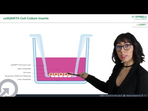
Introduction to Cell Culture Inserts – cellQART® at ASCCT 10th annual meeting
1.5K
Cell culture inserts are membrane-based devices containing semi-permeable membranes that split the culture chamber into apical and basolateral compartments, enabling cell polarization, co-culturing, and permeability assays. These inserts feature track-etched microporous membranes produced by breaking polymer molecular chains with accelerated noble gas ions to create defined pore channels, which are then chemically etched and treated with air plasma for optimal cell attachment. The quality of these membranes is critical for reproducible cell culture experiments, requiring uniform pore distribution, proper coating, and even welding to the plastic body. Membrane pore size selection depends on the application: 0.4-1 micron for epithelial and endothelial layers, while 3-8 micron sizes are suitable for migration and invasion studies. Clear membranes are preferred for phase contrast microscopy monitoring, while translucent membranes support faster cell growth. These membrane-based systems are widely used to model biological barriers including the blood-brain barrier, intestinal and respiratory epithelia, skin, hepatic and renal epithelia, and placental barrier, as well as for permeability and toxicity testing of drugs, chemicals, and cosmetics.
Click to watch →

Millicell® Hanging Inserts – How to use – Chapter 1: 2.5D Cell Culture
214
Millicell® Hanging Inserts enable 2.5D cell culture by providing a permeable membrane that allows cells to access media and nutrients from both apical and basolateral directions, promoting improved cellular growth and physiological relevance compared to traditional 2D plastic plates; proper setup involves peeling the insert from its blister pack, placing it in a cell culture plate with flanges flat and non-overlapping, ensuring even media levels on both sides, and using the apical assist rim for non-contact media exchanges during cellular growth.
Click to watch →

Inserts & Incubation
436
This video demonstrates the proper technique for transferring cell culture inserts to experimental wells, including rinsing with endal medium, adding progenitor medium, using sterile tweezers for transfer, removing old medium and discarding it in bleach, and placing the plate in a tissue culture incubator set at 37°C with 5% CO2 to maintain sterility and optimal cell growth conditions.
Click to watch →

How to Model the Human Airway at the Air-Liquid Interface: Expansion of HBECs
8.4K
This phase covers the transition from flask expansion to submerged culture in transwell inserts. Key procedures include: enzymatic dissociation using ACF solution (7-8 min at 37°C); cell counting with trypan blue and hemocytometer; centrifugation at 350g/1200 rpm for 5 min; resuspension in complete medium; seeding at 100,000 cells/cm² in apical compartment with 200 μL medium; adding 500 μL basal medium; performing full medium changes every 2 days until 100% confluence; then airlifting for mucociliary differentiation. Using PET membrane Costar inserts with 0.4 μm pore size achieves optimal differentiated morphology.
Click to watch →
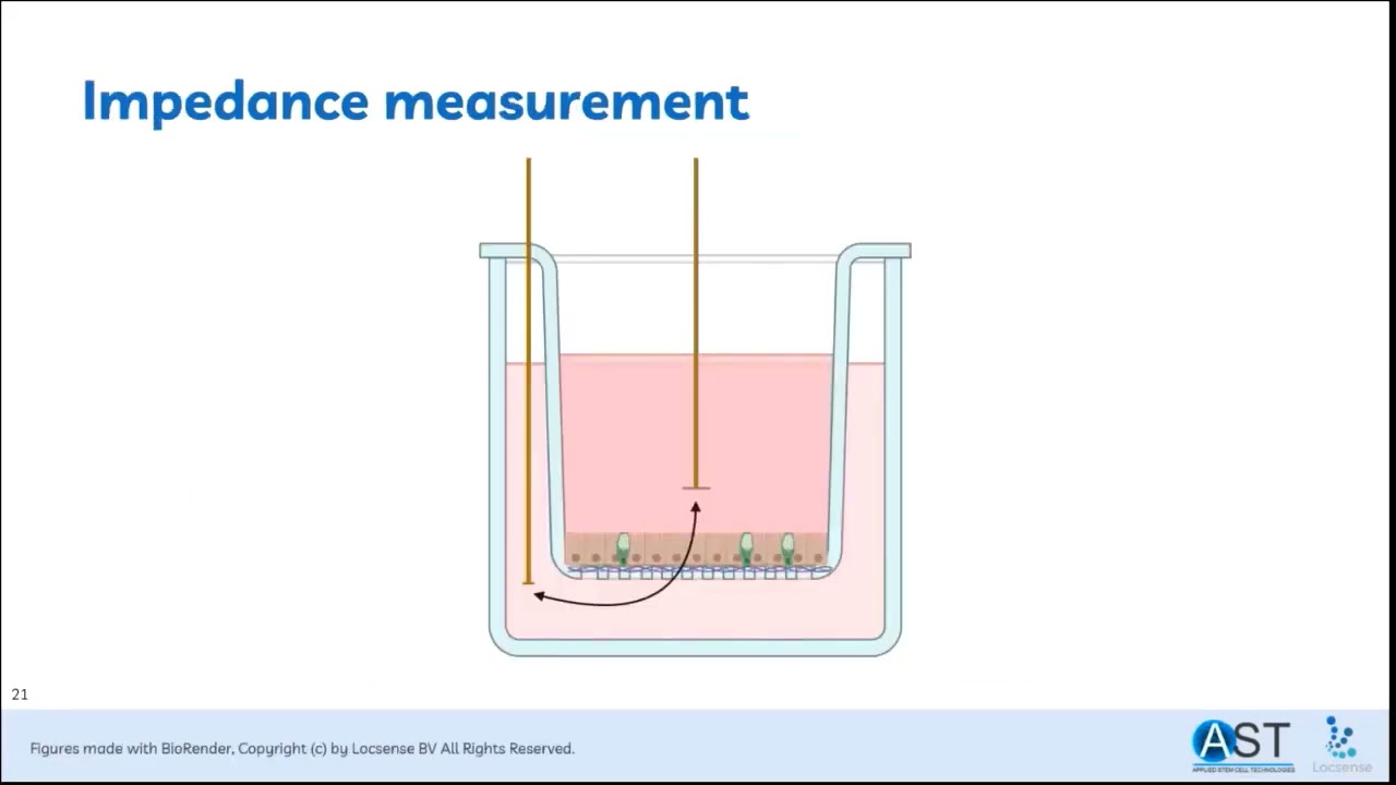
Does all Disease Begin in the Gut? Monitoring the Barrier Function of an In Vitro Gut Mimic
157
Transwell culture provides a 3D-like environment where epithelial cells contact wet layers on both apical (gut interior) and basolateral (blood vessel) sides. Collagen-coated membranes enable proper adhesion, with cells cultured for three weeks at 15% FBS in DMEM/F12 with supplements. Impedance spectroscopy applies alternating current across electrodes to measure electrical resistance. At low frequencies, impedance reflects resistors (tight junctions); at high frequencies, it reflects reactances (capacitance). TEER quantifies tight junction integrity—the electrical resistance caused by tight junction proteins like claudins and occludins. Higher TEER indicates better barrier function.
Click to watch →
Deep Dive
Knowledge of aseptic technique and contamination prevention when interacting with live cell cultures and instruments.
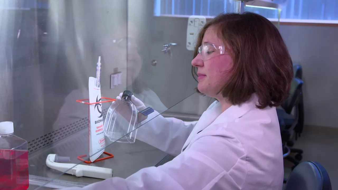
Reducing Cell Culture Contamination: Aseptic Technique
10.8K
Aseptic technique in cell culture involves systematic contamination prevention through personal protective equipment (gloves, lab jacket, glasses), thorough biosafety cabinet preparation with IPA cleaning, proper item handling (spraying and wiping fridge items, immediate spill cleanup), and strict pipetting protocols (never pouring, holding caps face-down on cleaned surfaces, avoiding vessel interiors) to maintain sterile conditions and protect cell cultures from microbial contamination.
Click to watch →

Cell Culture: Aseptic/Sterile Technique Dos & Don’ts | CST Tech Tips
330.6K
Proper aseptic technique in cell culture requires working within a biosafety cabinet using laminar airflow, disinfecting all items with 70% ethanol before use, avoiding lateral arm movements to prevent disrupting the air curtain, using single-use pipette tips, and keeping the workspace organized to minimize contamination risks from bacteria, fungi, and mycoplasma.
Click to watch →

NPTEL Week 2 Assignment Cell Culture Technologies
913
Aseptic techniques are essential for preventing contamination in cell culture laboratories. A laminar flow hood has three distinct areas: clean space for sterilized equipment, working space for hands and pipettes, and dirty area for waste. Hands must be sterilized with 70% ethanol and gloves worn. Contamination sources include: improperly sterilized equipment, contaminated cell stocks or media, cell culture reagents (serum, amino acids), and personnel with non-sterile hands or contaminated gloves. Common contaminants include viruses (hard to detect), mycoplasma (spreads quickly), bacteria (most common, easily visible), and yeast.
Click to watch →
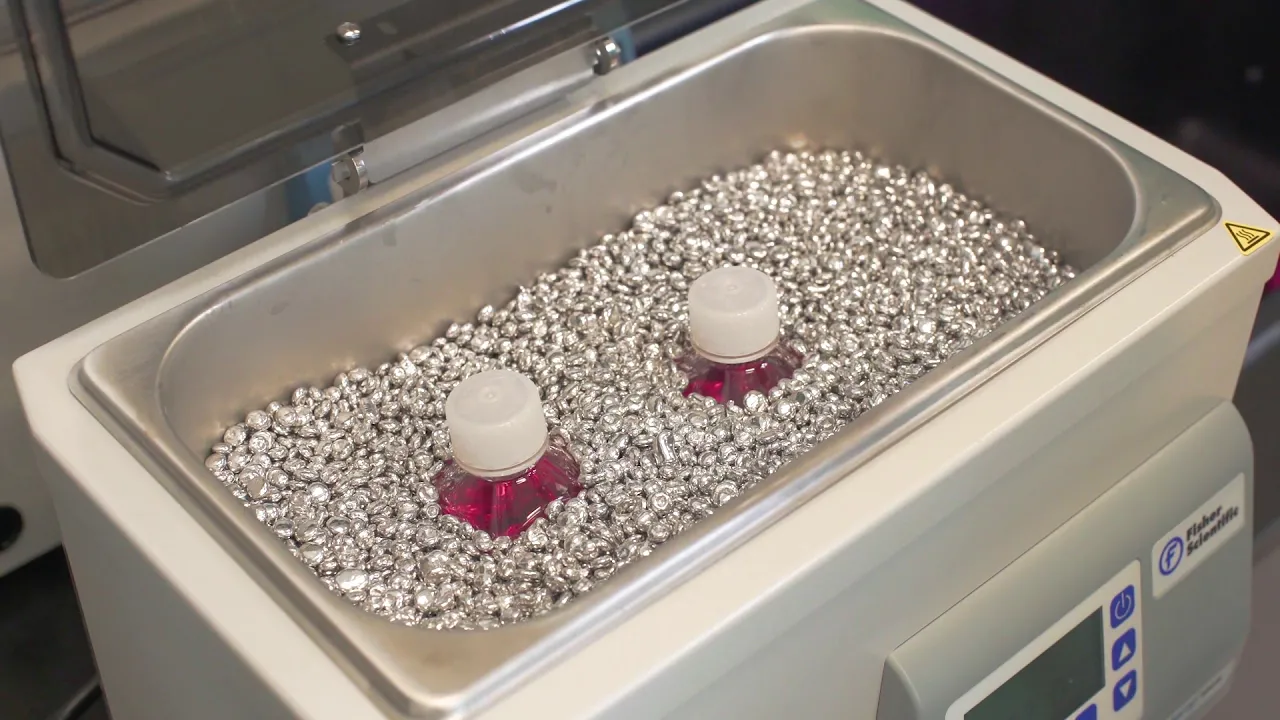
Aseptic Technique Training – Essential Cell Culture Video
457.2K
Successful cell culture requires proper laboratory setup with essential equipment including a cell culture hood, incubator, water bath, centrifuge, refrigerator/freezer, hemocytometer, and microscope; maintaining a sterile environment involves opening all materials within the hood, wiping surfaces with 70% ethanol, organizing items systematically (pipettes in front, reagents at back, waste containers in corners), and avoiding breathing or talking into the hood; mammalian cells should be cultured at 37°C with 5-7% CO2 and high humidity, with flasks evenly spaced for proper gas exchange; reagents should be warmed to 37°C for 10-20 minutes but not left excessive time to prevent degradation; daily microscopic examination is essential for monitoring cell health and detecting contamination.
Click to watch →

BIO 225: LAB EXAM 1 EXERCISE
699
Microorganisms are ubiquitous and omnipresent in nearly every environment. Contamination refers to the introduction of unwanted microorganisms into cultures or laboratory environments. Aseptic technique minimizes contamination of original and newly inoculated cultures while protecting laboratory workers. This technique ensures only intended microorganisms grow in cultures and prevents infection risks to personnel.
Click to watch →
Prerequisite Knowledge
- Concept 01Understanding of epithelial and endothelial cell biology, specifically the structure and function of tight junctions in forming cellular barriers.
- Concept 02Basic principles of electricity, particularly Ohm's Law (V = I * R) and how electrical resistance is measured in conductive solutions.
- Concept 03Familiarity with cell culture techniques, specifically growing cells on semi-permeable membrane inserts (such as Transwell systems).
- Concept 04Knowledge of aseptic technique and contamination prevention when interacting with live cell cultures and instruments.
Subsequent Learning
- Step 01How to calculate and normalize unit area resistance (expressed in ohms times square centimeters) by subtracting blank insert resistance and multiplying by membrane area.
- Step 02Troubleshooting variables that affect TEER measurements, including temperature fluctuations, cell passage number, media formulation, and physical electrode positioning.
- Step 03Exploring advanced, automated, or continuous TEER monitoring technologies (such as multi-well automated systems or microfluidic organ-on-a-chip platforms).
- Step 04Applying TEER data in functional assays, such as evaluating drug permeability, macromolecule transport, or pathogen disruption of cellular barriers.
Setup
Measure
Setup
0:01Playing Section
- 1
Power on the device and insert the USB drive for data storage.
- 2
Configure file naming with auto-indexing or a custom unique name.
- 3
Select plate size, measurement units, and resistance range.
Automated Continuous Impedance Spectroscopy vs. Manual 'Chopstick' TEER
While the EVOM Manual utilizing 'chopstick' electrodes is a widely used, budget-friendly method for measuring transepithelial electrical resistance (TEER), it faces criticism for lack of reproducibility and precision. Manual electrode positioning introduces significant operator-dependent variability, and removing cells from the incubator for measurement causes temperature and pH fluctuations that alter barrier integrity. Furthermore, static, single-frequency measurements fail to capture the complex capacitive properties of the cell monolayer. Critics advocate for automated, continuous impedance spectroscopy systems (such as cellZscope or integrated microfluidic systems) as a superior alternative. These automated platforms monitor barrier function continuously inside the incubator across a range of frequencies, eliminating human error, preventing environmental disruptions, and providing detailed kinetic data on cell barrier dynamics that static manual readings cannot offer.
Deep Dive
How to calculate and normalize unit area resistance (expressed in ohms times square centimeters) by subtracting blank insert resistance and multiplying by membrane area.

2 8 8 Membrane resistance Rm 1027
2.8K
Membrane resistance (R) differs fundamentally from resistivity (RM). Resistance depends on physical dimensions - larger membranes have lower resistance because current has more pathways. Resistivity characterizes the material property independently by measuring resistance per unit area (ohms/cm²). To calculate total resistance, divide resistivity by surface area: R = RM/A. This framework separates material characteristics from geometric factors, enabling standardized comparison across different membrane samples.
Click to watch →

1381 How To Make A Conductive Ink
40.9K
To properly compare conductive ink performance, normalize resistance measurements by dividing by the ink film thickness. The standard unit is ohms per square per mil (Ω/□/mil). Since ink films have varying thicknesses depending on application technique, normalization allows fair comparison between different samples. For example, a 121 ohm reading from a 10-micrometer thick film converts to approximately 48.4 ohms per square per mil when normalized.
Click to watch →

Clase 07 (Tarde): Conducción Nerviosa (22/09/2020)
132
This section explains membrane resistance (Rm) and its relationship to specific membrane resistance (Rm'). Unlike axial resistance which depends on length, membrane resistance depends on surface area. The formula Rm = Rm' / (2πrL) shows how membrane resistance decreases with increasing length and radius. The instructor addresses the counterintuitive unit of ohm·m² for specific membrane resistance, explaining that resistance decreases with increasing area (more pathways for current), so the relationship involves multiplication rather than division. This distinction is crucial for correctly calculating membrane resistance from tabulated values.
Click to watch →
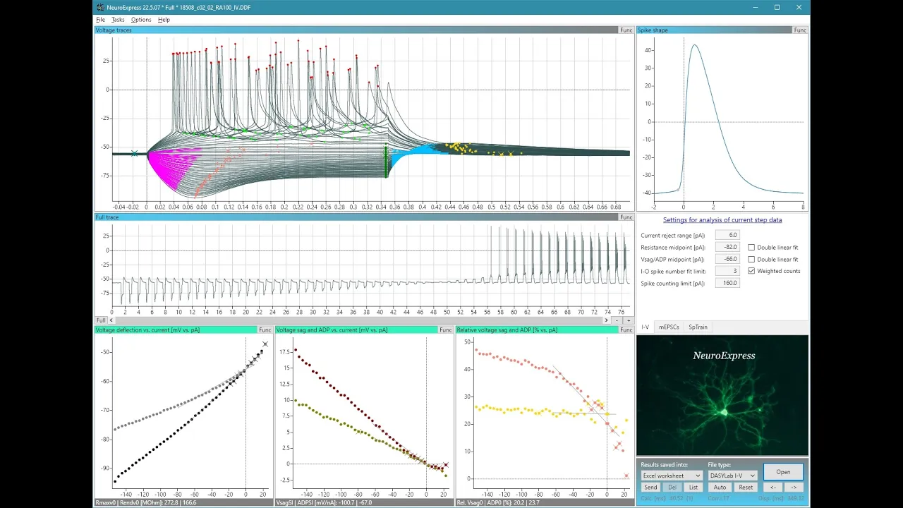
NeuroExpress Tutorial #1 - Physiological Properties and Excitability
424
The conventional method for calculating membrane resistance uses linear regression on the voltage versus current relationship. The program identifies the most negative voltage deflection under negative current steps and the steady-state voltage at the end of each current injection. By plotting these values against current and fitting a line, the slope of this relationship estimates membrane resistance at I=0 (resting membrane potential). For example, this method might yield a membrane resistance of approximately 272 MOhm.
Click to watch →

Episode #65: Tafel plot with multiple slopes - what does it mean?
466
Impedance is commonly reported in ohm·cm² (ohms times centimeters squared) rather than just ohms because it normalizes for electrode surface area. This normalization allows comparison between electrodes of different sizes. The units arise from using current density (A/cm²) instead of current (A) in the impedance calculation: Z = V/J, where J is current density. When impedance is reported in ohm·cm², it can be converted to absolute impedance by multiplying by the actual electrode area. This practice is particularly useful in electrocatalysis research where catalytic activity is compared across different electrode geometries. However, some researchers prefer reporting in ohms for simplicity, especially when comparing systems with similar electrode sizes.
Click to watch →
Deep Dive
Troubleshooting variables that affect TEER measurements, including temperature fluctuations, cell passage number, media formulation, and physical electrode positioning.

Primary Airway Cells in 3-D Models: Optimizing Culture at the Air-liquid Interface
593
Several factors can significantly affect TEER measurement accuracy in airway models: (1) Temperature variations - plates should cool to room temperature (~30 minutes) after removal from incubator, as interior areas tend to be warmer than edges; (2) Instrument preparation - ohmmeters need 30 minutes warm-up time and should be unplugged to avoid electrical noise interference; (3) Probe positioning - consistent angle and position between wells is critical, as probe angle greatly affects readings; (4) Well size - larger plates (6-well) produce non-uniform current distribution, so 12-well or 24-well plates are preferred; (5) Blank subtraction - raw TEER values must be subtracted by blank measurements (inserts without cells) and multiplied by insert area for accurate comparison across different insert sizes.
Click to watch →

Introducción a la Potenciometría: Medición Precisa de Potenciales
399
Proper electrode care extends lifespan and maintains accuracy. Avoid extreme temperatures and pH conditions that accelerate electrochemical reactions. Always rinse thoroughly between measurements; for organic contamination, use non-abrasive detergent in warm water. Never allow the membrane to dry out. Temperature affects electrode resistance (inversely proportional) and response time—lower temperatures require longer stabilization. Alkaline error occurs in high-alkaline solutions when metal cations compete with hydrogen ions at membrane sites. Lifespan decreases dramatically with temperature: 1-3 years at 25°C, less than 4 months at 90°C, less than 1 month at 120°C. Troubleshooting: replace defective electrodes, clean obstructed diaphragms, use fresh buffer solutions.
Click to watch →

Troubleshooting in Culture Media & How to overcome poor Embryo Development by Dr Robbie Kerr
2.8K
Temperature validation requires external probes rather than relying on device displays. Different dish designs (air gap vs direct contact) produce different temperature profiles. Direct contact dishes maintain better thermodynamic profiles than air-gap designs. Probe insertion causes thermal conductivity issues creating temperature gradients—T-type probes exert significant cooling effects on small volumes. K-type probes are more robust, while platinum resistance electrodes (PT100) provide most reliable measurements. Five-well dishes with 0.5ml media take ~13-15 minutes to reach optimal temperature on heated stages. Oil overlay affects temperature equilibration—direct contact dishes reach 36.3°C while air-gap dishes reach only 34.0°C. Higher temperatures are more damaging than lower temperatures.
Click to watch →
Does all Disease Begin in the Gut? Monitoring the Barrier Function of an In Vitro Gut Mimic
157
Temperature influences but does not significantly change TEER results. In control experiments, differences between time points were observed due to handling (taking plates out of incubator, putting back in), which changes temperature. However, these temperature-induced changes are not statistically significant compared to actual treatment effects.
Click to watch →

Real-Time TEER Monitoring for Organ-on-Chip Barrier Integrity
126
The organ-on-chip TEER platform offers PDMS-free design avoiding molecule absorption issues, simple scalable fabrication, wireless contactless Bluetooth readout, incubator compatibility, and intuitive operation. High biological reproducibility shows minimal TEER variation across experiments. Cells can be harvested as single-cell suspensions or whole tissue for downstream applications including flow cytometry and histological staining. Achieving stable TEER measurements requires fixed planar electrodes eliminating positioning variability and maintaining controlled incubator environments since TEER is temperature-sensitive. Gaps or holes in cell layers cause current leakage, producing artificially low TEER values that misrepresent true barrier function—requiring fully confluent monolayers before measurement. Perfusion initially increases TEER due to shear-induced tightening before stabilizing. The platform supports 6+ million cells in intestinal models and 700K-1 million cells in other organ models, sufficient for qPCR, flow cytometry, and biomarker analysis. TEER applications include assessing cell junction tightness, evaluating drug effects on barrier function, and real-time monitoring of barrier responses under disease-like conditions including inflammatory responses, infection models, and tissue damage scenarios.
Click to watch →
Deep Dive
Exploring advanced, automated, or continuous TEER monitoring technologies (such as multi-well automated systems or microfluidic organ-on-a-chip platforms).
Real-Time TEER Monitoring for Organ-on-Chip Barrier Integrity
126
Organ-on-chip technology provides microfluidic-based devices with chambers and channels for living organ substructures, creating controlled microenvironments that mimic in vivo conditions through perfusion systems and biomechanical stimulation. Standard readouts include supernatant sampling, cytotoxicity assessment, tissue recovery, live-cell imaging, and enzyme/protein transporter functionality. Tissue barriers are specialized cellular layers (epithelia/endothelia) acting as selective filters that permit essential nutrients while blocking harmful agents, maintained by tight junctions, adherens junctions, and desmosomes. TEER (Trans-Epithelial/Trans-Endothelial Electrical Resistance) is a non-invasive method measuring electrical resistance across cell monolayers to assess barrier integrity. Compared to permeability assays using labeled tracers (which take 1-24 hours), TEER provides faster results in seconds with less cellular interference, though accuracy depends on electrode type, membrane area, and temperature. Integrated organ-on-chip electrodes offer superior stability compared to movable chopstick electrodes.
Click to watch →

What is TEER? - Trans-Epithelial Electrical Resistance Assay
24.1K
Epithelial tissues form protective barriers in organs like skin, lungs, liver, kidney, and digestive tract, with endothelium lining blood vessels. Cells attach via junction proteins creating selectively permeable barriers. Disruption leads to adverse effects. TEER (trans-epithelial electrical resistance) measures barrier integrity by applying low-frequency current; easier current flow indicates lower resistance. Traditional methods use manual electrodes but suffer from low throughput and variability. Advanced systems integrate electrodes for consistent placement, enabling simultaneous measurement of up to 384 wells. Low frequencies detect barrier properties while high frequencies measure cell confluence, allowing researchers to distinguish between actual barrier changes versus cell coverage differences. This enables disease modeling and drug development studies, such as investigating CFTR protein function where ion channel activation causes rapid TEER drops.
Click to watch →

UCF Researchers Develop “Organ-on-a-Chip” Technology
318
Organ-on-a-chip technology uses microfabricated devices to measure electrical resistance (TEER) of cell layers, enabling non-invasive, real-time monitoring of cell health and drug responses in a controlled laboratory environment, which can replace animal testing for personalized medicine applications.
Click to watch →

Microbiome in Human Gut-On-A-Chip
1.9K
Three experimental groups are compared: static transwell culture, simple microfluidic device with 30 μL/hour flow, and microfluidic device with cyclic mechanical strain. All groups show confluent cells, but microfluidic devices show taller cells with different nuclear positioning—a six-fold increase in cell height compared to static cultures. Analysis reveals that increased cell height is caused by shear stress, not cyclic mechanical strain. Testing flow rates shows 30 μL/hour is optimal, falling within physiological ranges. Transepithelial electrical resistance (TEER) measures barrier integrity, with microfluidic groups achieving higher values. Permeability measurements show that only when cyclic mechanical strain is combined with microfluidic flow does permeability match in vivo conditions.
Click to watch →

Versatile & Automated Organ On A Chip Platform - Webinar - Fluigent
876
Automated microfluidic platforms like Omi address the critical need for precise, long-term fluid handling in organ-on-a-chip experiments by providing unidirectional recirculation, programmable protocols, and remote monitoring capabilities that maintain physiological relevance through controlled shear stress and continuous nutrient supply, thereby improving the reproducibility and predictive power of in vitro organ models for drug discovery and biomedical research.
Click to watch →
Deep Dive
Applying TEER data in functional assays, such as evaluating drug permeability, macromolecule transport, or pathogen disruption of cellular barriers.

Plenary lecture by Prof Nicholas Peppas (University of Texas at Austin)
503
Transepithelial electrical resistance (TEER) measurements provide a method to assess drug transport across epithelial barriers. In this assay, cells are grown on polycarbonate membranes, and particles are applied to the apical surface. TEER measures the resistance across the cell monolayer—high resistance indicates intact tight junctions and minimal transport, while decreased resistance indicates particle penetration and disruption of the epithelial barrier. This technique allows researchers to evaluate whether mucoadhesive particles can penetrate through epithelial layers and reach underlying tissues, providing valuable information about absorption potential.
Click to watch →

Intestinal Toxicity Assessment with Patient-Derived Organoids
342
Two primary assays measure intestinal barrier integrity: Transepithelial Electrical Resistance (TEER) measures electrical current flow across the epithelium, reflecting tight junction quality and thickness—higher TEER indicates better barrier function. FITC-dextran permeability assay tracks large carbohydrate molecules (too big to cross intact epithelium) labeled with fluorescent dye; increased leakage indicates barrier disruption. Compared to Caco-2 cells, organoid-derived ALI cultures maintain much higher TEER readings and show less baseline permeability, demonstrating superior barrier function representation.
Click to watch →

Organ on a chip MeetUp - Intestine & Intestine Microbiome 2024
183
TEER measurements are more sensitive than fluorescent dextran assays for detecting barrier disruption. Effects that are not visible in dextran permeability measurements can already be detected by TEER. However, adding dextran to each well is more labor-intensive than simply placing the entire plate in the OrganoTier for TEER measurements. This trade-off between sensitivity and practicality should be considered when choosing readout methods.
Click to watch →

3RsC Microphysiological Systems Workshop: Blood Brain Barrier
20.8K
Transendothelial electrical resistance (TEER) measurements provide quantitative assessment of BBB integrity in microphysiological systems. By placing organoplates in specialized measurement devices connected to computers, researchers can simultaneously monitor TEER values across multiple chips. Exposure to inflammatory agents such as LPS, TNF-alpha, NMDA, thrombin, and VEGF produces dose-dependent decreases in TEER values, demonstrating barrier disruption. Thrombin shows transient effects with partial recovery over time, while other compounds cause more sustained disruption. These assays enable screening of compounds for their potential to disrupt or protect BBB integrity.
Click to watch →
Product Evaluation Part 2: In vitro testing
281
Trans Epithelial Electrical Resistance (TEER) is a measurement of transepithelial electrical resistance that characterizes the integrity of the cell layer. Typically, a higher TEER value represents a more compact cell layer while a decrease in TEER indicates greater cell layer permeability. TEER is an important parameter characterizing the integrity of the cell layer on the tissue. However, a disadvantage of Caco-2 monolayer is its permeability to hydrophilic or paracellular transport, making it a better model for colon tissue rather than the small intestine. This limitation restricts the reliability of the technique as a model for the small intestine.
Click to watch →
Setup
Measure
Setup
0:01- 1
Power on the device and insert the USB drive for data storage.
- 2
Configure file naming with auto-indexing or a custom unique name.
- 3
Select plate size, measurement units, and resistance range.
Automated Continuous Impedance Spectroscopy vs. Manual 'Chopstick' TEER
While the EVOM Manual utilizing 'chopstick' electrodes is a widely used, budget-friendly method for measuring transepithelial electrical resistance (TEER), it faces criticism for lack of reproducibility and precision. Manual electrode positioning introduces significant operator-dependent variability, and removing cells from the incubator for measurement causes temperature and pH fluctuations that alter barrier integrity. Furthermore, static, single-frequency measurements fail to capture the complex capacitive properties of the cell monolayer. Critics advocate for automated, continuous impedance spectroscopy systems (such as cellZscope or integrated microfluidic systems) as a superior alternative. These automated platforms monitor barrier function continuously inside the incubator across a range of frequencies, eliminating human error, preventing environmental disruptions, and providing detailed kinetic data on cell barrier dynamics that static manual readings cannot offer.
Understanding of epithelial and endothelial cell biology, specifically the structure and function of tight junctions in forming cellular barriers.
Basic principles of electricity, particularly Ohm's Law (V = I * R) and how electrical resistance is measured in conductive solutions.
Familiarity with cell culture techniques, specifically growing cells on semi-permeable membrane inserts (such as Transwell systems).
Knowledge of aseptic technique and contamination prevention when interacting with live cell cultures and instruments.
How to calculate and normalize unit area resistance (expressed in ohms times square centimeters) by subtracting blank insert resistance and multiplying by membrane area.
Troubleshooting variables that affect TEER measurements, including temperature fluctuations, cell passage number, media formulation, and physical electrode positioning.
Exploring advanced, automated, or continuous TEER monitoring technologies (such as multi-well automated systems or microfluidic organ-on-a-chip platforms).
Applying TEER data in functional assays, such as evaluating drug permeability, macromolecule transport, or pathogen disruption of cellular barriers.
2.4K views8likes3:54@WPIIncOriginal Release: 2022-07-12
[Music] evon manuel is wpi's newest instrument to measure trans epithelial electrical resistance let's look at how to set it up and take resistance measurements power on the evon manual using the power switch on the rear panel then insert the usb thumb drive provided with the system into the usb port on the side of the meter by default the file name is set to plate 1 but you may choose a prefix that auto-indexes or unique file name press and hold the store button on the main screen for 2 seconds or press store screen on the setup menu to configure your stored data files to use a prefix and auto indexing activate the auto index radio button auto indexing sets the name to a numeric sequence with a name a prefix and a sequential number then you can press the store prefix to enter the prefix [Music] or you can use a unique file name to do that deactivate the auto index radio button and then select file name to enter the name of your file to do that you press clear use the keypad to enter a new name and press enter [Music] next press setup to access the setup menu select plate lets you choose your plate size select mode units lets you choose ohms or kiloohms for taking resistance readings select the resistance range either auto or 10 000 ohms you may press calibrate to calibrate the meter since the meter uses an internal resistance to adjust the internal measurement electronics the blank is the resistance value of a trans well with no cells only media subtracting the value of the blank gives you the value of the resistance of your cell culture to subtract a blank transwell including the fluid and the electrode resistances first press the blank handling button then place the electrode in a blank well and press reading press the blank enable radio button to automatically subtract that blank value from your future readings press return to navigate back to the main menu now you can begin measuring by pressing the foot switch or simply touch the store button on the screen repeat the sampling process for each well once the last well has been recorded the file storing selection notification appears press store the evon manual should respond with file saved within 15 seconds if saving file is still seen for two minutes press the return key and try a different usb drive once a file has been saved open the file on a computer to verify the contents once you get our system set up taking measurements is easy if you have any questions just give us a call [Music] you
Up Next

3:25
Choosing the Right EVOM Electrode for TEER Measurements
@WPIInc
148 views•2025-03-14

12:54
Triumph of Orthodoxy Icon: Byzantine Art & History Explained
@BenCallan
2.1K views•2024-08-06

9:49
FastAPI vs Flask vs Django: Choosing the Right Python Web Framework
@TechWithTim
302.5K views•2024-05-26

1:42
Game of Thrones Opening Credits: A Cinematic Analysis
@gameofthrones
46.3M views•2011-04-18
Related Study Plans & Knowledge Roadmaps
Structured learning paths in General & Interdisciplinary Studies
general-studies
Elevator Engineering: History, Physics & Cities
01From Hoists to Safety Brakes: The Dawn of Vertical Lift
02Hydraulic vs. Traction: The Engineering Physics of Elevators
03Modern Speed, Safety, and Algorithms
+ 2 more learning modules
12h5 modules
Explore Roadmap
general-studies
Rubber: Material Science, History & Geopolitics
01Origins of Natural Rubber & Mesoamerican History
02The Chemistry of Natural Rubber & Vulcanization
03The Geopolitics of the Rubber Boom & Colonization
+ 2 more learning modules
12h5 modules
Explore Roadmap
general-studies
Containerization: Evolution, Logistics & Trade
01Ancient Trade & Bulk Cargo: Amphorae to Barrels
02The Industrial Revolution & Cardboard Packaging
03The Containerization Revolution: Malcom McLean
+ 2 more learning modules
12h5 modules
Explore Roadmap
general-studies
Textile Tech: Evolution, Labor & Global Impact
01Pre-Industrial Textiles: Handloom & Cottage Industry
02Mechanization: The Spinning Jenny to the Power Loom
03The Factory System & Global Labor Transformation
+ 2 more learning modules
10h5 modules
Explore Roadmap