The EVOM™ Manual TEER meter offers three electrode options for different experimental needs: the STX 4 is a general-purpose electrode with replaceable blades for hands-free operation in 24-well plates; the STX HTS electrodes are designed for high-throughput screening with smaller tips that reduce contamination risk in 24 and 96-well plates; and the Endome cell culture cup chambers provide 1% tolerance precision with symmetrical electrode patterns for accurate readings in 6, 12, and 24-well plates.
Choosing the Right EVOM Electrode for TEER Measurements
Added:Deep Dive
Understanding the biological significance of Transepithelial Electrical Resistance (TEER) as a quantitative measure of cell monolayer integrity and tight junction barrier function.

What is TEER
1.3K
Transepithelial/Endothelial Electrical Resistance (TEER) is a quantitative technique that measures the electrical resistance across a cell monolayer grown on a porous membrane, reflecting the tightness of cell junctions and barrier function; it qualitatively assesses cell monolayer health and quantitatively evaluates cellular confluence by detecting changes in ion flow through transcellular and paracellular pathways, with values typically increasing from low levels immediately after cell seeding to higher values once a confluent monolayer forms, and varying significantly between cell types based on their junctional characteristics and ion channel expression.
Click to watch →
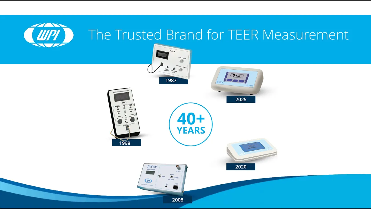
EVOM™ The Trusted Brand for TEER Measurement
229
Transepithelial Electrical Resistance (TEER) is a well-established technique used to assess the barrier function and integrity of epithelial and endothelial cell layers by applying a small AC current to the cell monolayer and measuring the resistance to ion flow, providing valuable insights into tight junction integrity and cell-cell interactions; WPI's EVOM family of meters, including manual and automated systems for 24-well and 96-well plates, serves as the gold standard in TEER technology, having been trusted for nearly 40 years and cited in thousands of peer-reviewed publications for applications in drug absorption studies, tissue engineering, disease modeling, and quality control.
Click to watch →

What is TEER? - Trans-Epithelial Electrical Resistance Assay
24.1K
TEER (Trans-Epithelial Electrical Resistance) is a widely accepted method for quantifying epithelial and endothelial barrier integrity by measuring electrical resistance across a confluent cell monolayer; impedance technology detects how much current is blocked by tight junctions and cell membranes, with low frequencies sensitive to barrier properties and high frequencies detecting cell confluence, enabling real-time, non-invasive monitoring of barrier function for applications in disease modeling and drug development.
Click to watch →
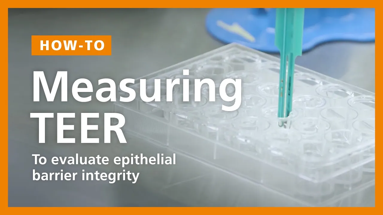
How to Perform a TEER Measurement to Evaluate Epithelial Barrier Integrity in ALI Cultures
34.5K
Transepithelial electrical resistance (TEER) measures epithelial barrier integrity by assessing the electrical resistance across an epithelial monolayer; the protocol involves sterilizing the probe in 70% isopropanol, preparing ALI cultures with calcium/magnesium-free PBS in the apical compartment and regular PBS in the basal compartment, inserting the probe at a 90-degree angle into the transwell hanger, recording stable ohm values until they level off, and calculating the corrected TEER value by subtracting the blank transwell control value and multiplying by the culture area in cm².
Click to watch →

The Literal Worst Food for Human Gut Health We’ve Found in 10 Years
283.3K
Research using transepithelial electrical resistance measurements has demonstrated that even at low concentrations (0.1%), polysorbates cause cellular death across all tested models. This cellular destruction leads to increased gut permeability and decreased expression of tight junction proteins, creating a cascade of negative effects on gut barrier function.
Click to watch →
Deep Dive
Familiarity with the setup and use of membrane-based cell culture inserts (such as Transwell systems) for growing polarized cell layers.

LCC of Airway Epithelial Calu-3 Cells to Study Respiratory Pathogens | Protocol Preview
242
This video demonstrates how to establish a liquid-covered culture of polarized human airway epithelial Calu-3 cells using a transwell culture system, where cells are seeded into the apical compartments and cultured for 2-3 weeks with media changes on a 3-4 day cycle, followed by verification of polarization through trans-epithelial electrical resistance and passive sodium fluorescein equilibration assays; Calu-3 cells are preferred over normal human bronchial epithelial cells because they are more accessible and develop more rapidly into depolarized cultures ready for use.
Click to watch →
A coculture system of polarized human endometrial epithelium and subject-paired stroma
1.1K
A hanging insert system (such as the Millicell system from EMD Millipore) allows researchers to grow primary endometrial epithelial cells in a polarized configuration. The epithelial cells are grown on inserts coated with Matrigel to replicate basal lamina components. Once confluent, these inserts are placed in standard 24-well plates containing patient-paired fibroblasts in the lower chamber. This setup enables measurement of basolateral secreted products from epithelial cells affecting underlying stromal fibroblasts, while also allowing assessment of stromal factors on epithelium.
Click to watch →
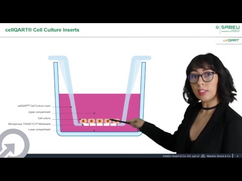
Introduction to Cell Culture Inserts – cellQART® at ASCCT 10th annual meeting
1.5K
Cell culture inserts are membrane-based devices containing semi-permeable membranes that split the culture chamber into apical and basolateral compartments, enabling cell polarization, co-culturing, and permeability assays. These inserts feature track-etched microporous membranes produced by breaking polymer molecular chains with accelerated noble gas ions to create defined pore channels, which are then chemically etched and treated with air plasma for optimal cell attachment. The quality of these membranes is critical for reproducible cell culture experiments, requiring uniform pore distribution, proper coating, and even welding to the plastic body. Membrane pore size selection depends on the application: 0.4-1 micron for epithelial and endothelial layers, while 3-8 micron sizes are suitable for migration and invasion studies. Clear membranes are preferred for phase contrast microscopy monitoring, while translucent membranes support faster cell growth. These membrane-based systems are widely used to model biological barriers including the blood-brain barrier, intestinal and respiratory epithelia, skin, hepatic and renal epithelia, and placental barrier, as well as for permeability and toxicity testing of drugs, chemicals, and cosmetics.
Click to watch →
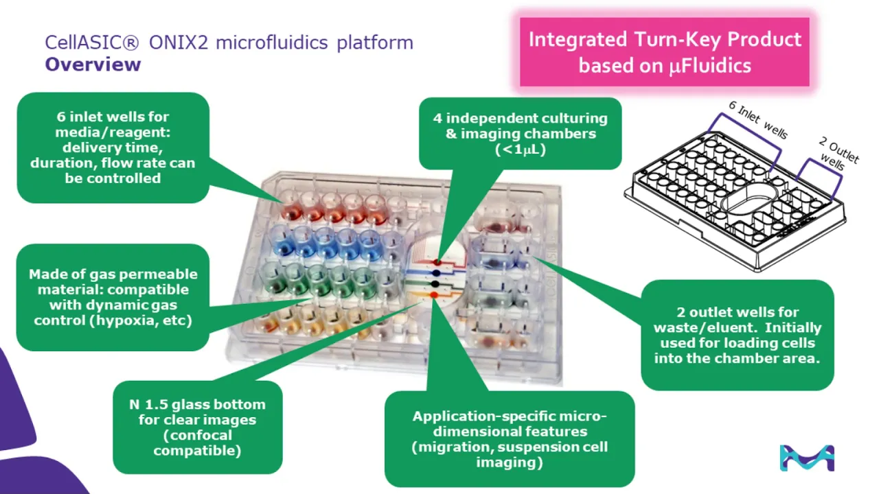
Beyond the Culture Flask: How Membrane- and Microfluidic-based Platforms
190
Membrane platforms enable long-term polarized cell culture that more closely resembles in vivo conditions compared to conventional plastic surfaces. For example, 21-day CaCo-2 cell cultures on 0.4-micron polycarbonate membranes produce cells with dramatic differences from those grown on plastic surfaces, exhibiting polarized morphologies characteristic of intestinal epithelium. Confocal imaging reveals the three-dimensional organization and functional polarization achieved through membrane-based culture systems, demonstrating their value for modeling epithelial physiology and barrier function.
Click to watch →

Millicell® 24 and 96 Well Plates: How to Access Various Ports
339.3K
Millicell® cell culture insert plates are membrane-based devices with 10-micron thickness and defined pore sizes, used to create in vitro models for air-liquid interface, blood-brain barrier, intestinal barrier (Caco-2), and cellular migration/invasion assays; they feature apical assist technology that creates a membrane-free area for safe apical media exchange without disturbing cells or puncturing the membrane, along with basolateral access ports for media exchange, available in 24-well and 96-well formats with either single-well feeder trays for initial cell culturing or multi-role receiver plates for individualized assay treatments.
Click to watch →
Deep Dive
Basic knowledge of electrical resistance, Ohm's law, and how electrical currents flow through conductive aqueous media (cell culture reagents).

CTET SEP 2026 SCIENCE दंगल by Sachin Academy live 8pm | 30/30 CTET PAPER 2 SCIENCE
72.5K
Ohm's Law states that current is directly proportional to potential difference: V = IR, where V is voltage, I is current, and R is resistance. Resistance is the opposition to current flow. Higher resistance means less current flows. The unit of charge is Coulomb (C), voltage is Volt (V), and current is Ampere (A). 1 Volt = 1 Joule per Coulomb, and 1 Ampere = 1 Coulomb per second.
Click to watch →

विद्युत धारा | Class 12 Physics Chapter 3 One Shot🎯 | Current Electricity | Bihar Board 12th Physics
20.8K
Ohm's Law states that if the physical state of a conductor remains unchanged, the current flowing through it is directly proportional to the potential difference applied across its ends (V ∝ I). This leads to V = IR, where R is the constant of proportionality called resistance. Resistance is defined as the opposition offered by a conductor to the flow of electric current, given by R = V/I. Resistance is a scalar quantity (अदिश राशि) with SI unit Ohm (Ω). The dimensional formula is [M^1 L^2 T^-3 A^-2].
Click to watch →

🎯 MP TET VARG 3 SCIENCE MARATHON 🔥 | Unit-16 Full Theory in 1 Science Class by Harish Sir
49.2K
Ohm's Law states that the current flowing through a conductor is directly proportional to the potential difference across its ends, provided physical conditions remain constant: V = IR, where V is potential difference, I is current, and R is resistance. Resistance is the opposition to current flow. The SI unit of resistance is the ohm (Ω). Resistance depends on: (1) Length - directly proportional; (2) Cross-sectional area - inversely proportional; (3) Material - different materials have different resistivities; (4) Temperature - resistance increases with temperature for most conductors. Formula: R = ρL/A.
Click to watch →

فيزياء الخامس العلمي | الفصل التاسع | محاضرة 1 منهج 2027
92K
Electrical resistance (R) is the opposition to current flow, measured in Ohms (Ω). Ohm's Law states that current is directly proportional to voltage at constant temperature: V = IR. The Ohm's Law triangle (V at top, I and R at bottom) helps solve for any variable: V = IR, I = V/R, or R = V/I. This fundamental law governs the behavior of most electrical circuits and is essential for circuit analysis.
Click to watch →

المراجعة النهائية شرح المنهج بالكامل علوم تالتة اعدادي | عام و ازهر | الترم التاني 2026
34.1K
Ohm's Law states that current (I) equals voltage (V) divided by resistance (R): I = V/R. Resistance opposes current flow and depends on material, length, and cross-sectional area. Fixed resistors have constant resistance, while variable resistors (rheostats) allow adjustment. The Ohm's Law triangle helps remember relationships: V = IR, I = V/R, R = V/I. Direct current (DC) flows in one direction with constant magnitude, produced by batteries. Alternating current (AC) periodically reverses direction and changes magnitude, produced by generators.
Click to watch →
Deep Dive
Awareness of the general operational purpose of an Epithelial Volt/Ohm Meter (EVOM) in cellular physiology laboratories.

How to Take Voltage Measurements with the EVOM Manual
849
The EVOM Manual is WPI's instrument for measuring Trans Epithelial Electrical Resistance (TEER); to use it, first power on the device and insert the USB flash drive, then configure file naming by choosing between auto-indexed prefixes or unique filenames, select your plate size and measurement units (volts or millivolts), equilibrate electrodes in liquid for at least three hours, perform a null offset by pressing the probe null button until the solution value reads 0.0, enable the offset function, and begin measurements by pressing the foot switch or store button, storing data files on the USB drive for later analysis on a computer.
Click to watch →
EVOM™ The Trusted Brand for TEER Measurement
229
Transepithelial Electrical Resistance (TEER) is a well-established technique used to assess the barrier function and integrity of epithelial and endothelial cell layers by applying a small AC current to the cell monolayer and measuring the resistance to ion flow, providing valuable insights into tight junction integrity and cell-cell interactions; WPI's EVOM family of meters, including manual and automated systems for 24-well and 96-well plates, serves as the gold standard in TEER technology, having been trusted for nearly 40 years and cited in thousands of peer-reviewed publications for applications in drug absorption studies, tissue engineering, disease modeling, and quality control.
Click to watch →
How to Perform a TEER Measurement to Evaluate Epithelial Barrier Integrity in ALI Cultures
34.5K
This comprehensive workflow covers the entire process of measuring trans-epithelial electrical resistance (TEER) in air-liquid interface (ALI) cultures using an epithelial volt ohm meter (EVOM). The procedure begins with probe sterilization in 70% isopropanol for 15 minutes, followed by washing in sterile PBS. ALI cultures are prepared by removing medium and adding calcium/magnesium-free PBS to the apical compartment and regular PBS to the basal compartment. The probe is inserted at a 90-degree angle into the transwell hanger, with the long tongue touching the bottom and the short prong resting above the culture surface. Ohm values are recorded after stabilizing, then the probe is washed and moved to the next culture. After completing measurements, the probe is returned to IPA, PBS is removed, and culture medium is restored. The blank transwell control is measured last, and the probe undergoes final cleaning before storage.
Click to watch →

Why I Like the EVOM Line of Epithelial Volt/Ohm Meters for TEER Measurement
421
Modern epithelial volt/ohm meters for TEER (Transepithelial Electrical Resistance) measurement offer significant advantages over older models, including faster stabilization times, higher resolution readings (down to 0.1 ohm), reduced electrical noise through battery power, intuitive touchscreen interfaces for setup and data navigation, and automatic data storage capabilities that eliminate manual data transfer requirements.
Click to watch →

EVOM Manual Electrode Options
1.1K
WPI's EVOM Manual provides stable and repeatable trans epithelial electrical resistance (TEER) measurements for assessing cell monolayer integrity; the system offers multiple electrode options including the STX4 general-purpose electrode with replaceable blades for 24-well plates, and STX HTS electrodes designed for high-throughput screening in 24 and 96-well plates, along with end dome cell culture cup chambers available in 6mm, 12mm, and 24mm sizes for different well plate formats.
Click to watch →
Prerequisite Knowledge
- Concept 01Understanding the biological significance of Transepithelial Electrical Resistance (TEER) as a quantitative measure of cell monolayer integrity and tight junction barrier function.
- Concept 02Familiarity with the setup and use of membrane-based cell culture inserts (such as Transwell systems) for growing polarized cell layers.
- Concept 03Basic knowledge of electrical resistance, Ohm's law, and how electrical currents flow through conductive aqueous media (cell culture reagents).
- Concept 04Awareness of the general operational purpose of an Epithelial Volt/Ohm Meter (EVOM) in cellular physiology laboratories.
Subsequent Learning
- Step 01Protocols for sterilizing, cleaning, and re-chloridizing silver/silver-chloride (Ag/AgCl) EVOM electrodes to maintain measurement accuracy.
- Step 02Methodologies to minimize environmental variables during measurement, such as controlling temperature and precise electrode positioning.
- Step 03Applying TEER measurements to specific in vitro models, such as the Blood-Brain Barrier (BBB), gastrointestinal tract (Caco-2), or pulmonary epithelial models.
- Step 04Transitioning from manual measurements to automated, high-throughput TEER systems for drug permeability and toxicity screening assays.
Electrode Options
Endome Chambers
Electrode Options
0:00Playing Section
- 1
Compares STX4 and STX HTS electrodes for TEER measurement.
- 2
STX4 suits 24-well plates, with replaceable blades for longevity.
- 3
STX HTS is designed for high-throughput screening on 24/96 wells.
Beyond Static EVOM: The Case for Automated, Continuous Impedance Spectroscopy
While selecting the right EVOM electrode (such as chopstick or Endohm chambers) is a standard laboratory consideration, a significant counterpoint advocates for moving away from manual EVOM measurements altogether. Critics argue that manual EVOM systems, which typically measure resistance at a single frequency, suffer from high operator variability, temperature fluctuations during handling, and a lack of real-time temporal resolution. Instead, the alternative perspective favors automated, continuous, multi-frequency impedance spectroscopy (such as ECIS or integrated microfluidic sensors). These advanced systems non-invasively monitor barrier integrity inside the incubator over time. Furthermore, by measuring across a spectrum of frequencies, they can mathematically differentiate between cell membrane capacitance and junctional resistance, providing far deeper biological insights than the static, single-point resistance values obtained via traditional EVOM electrodes.
Deep Dive
Protocols for sterilizing, cleaning, and re-chloridizing silver/silver-chloride (Ag/AgCl) EVOM electrodes to maintain measurement accuracy.

Tutorial 28-How to maintain Ag/AgCl reference electrode
18.1K
This video tutorial demonstrates the proper maintenance procedures for Ag/AgCl reference electrodes, including storing them vertically immersed in saturated KCl solution, removing air bubbles by gently tapping the electrode, polishing the silver wire with fine sandpaper or concentrated ammonium hydroxide when the surface becomes rough or discolored, and recoating the silver wire with silver chloride using either chemical methods or electrochemical methods (applying approximately 10 microamperes overnight with a platinum counter electrode). The video emphasizes avoiding basic solutions, ammonia buffers, and sulfide-containing solutions that can damage the electrode, as well as the importance of regular maintenance to ensure accurate and reproducible measurements.
Click to watch →
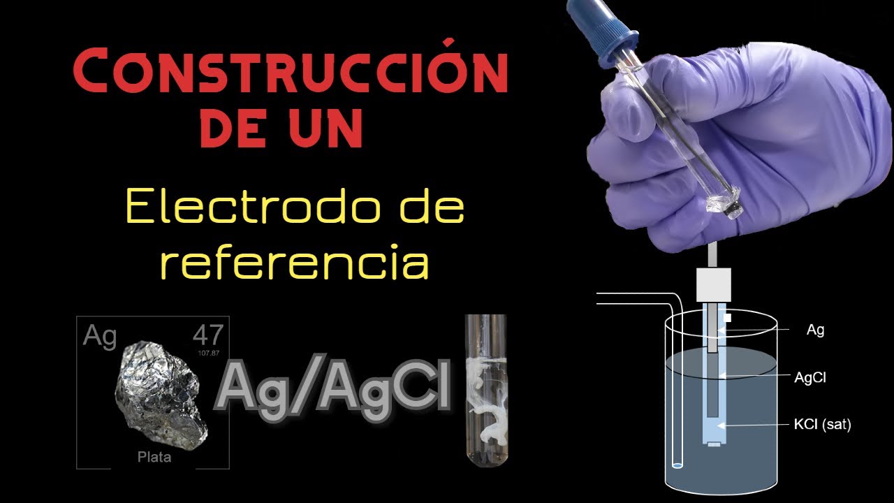
Construcción de un electrodo de referencia Ag/AgCl
10K
A silver/silver chloride reference electrode is constructed by first polishing a high-purity silver wire, then cleaning it with ammonium hydroxide and acetic acid, followed by electrochemical cathodic cleaning (hydrogen evolution) and anodic deposition of silver chloride in hydrochloric acid solution; the electrode is assembled with a cellophane membrane filled with saturated potassium chloride solution and must mature in darkness for about one week to achieve stable readings.
Click to watch →

Silver Chloride Electrode
64.7K
The Silver/Silver Chloride (Ag/AgCl) electrode is a metal-metal salt electrode consisting of a silver wire coated with silver chloride, dipped in a chloride ion solution. It functions as both anode and cathode depending on coupling, with oxidation occurring at the anode (Ag + Cl⁻ → AgCl + e⁻) and reduction at the cathode (AgCl + e⁻ → Ag + Cl⁻). Its electrochemical potential follows the Nernst equation E = E° - 0.0591 × log[Cl⁻], making it concentration-dependent—showing 0.223 V in 1M KCl and 0.199 V in saturated KCl. Key applications include serving as an internal reference electrode in glass electrodes, monitoring potential distribution in pipelines and ship hulls, and corrosion prevention systems.
Click to watch →
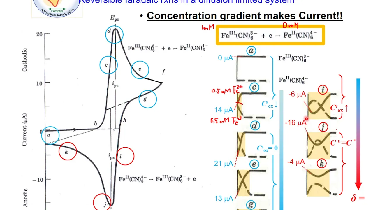
EC@2b-2. The Overview of Electroanalytical Methods (Part 2)
6.1K
Reference electrode performance is tested by comparing average peak potential against a benchmark electrode. Differences exceeding 40 mV indicate problems requiring maintenance: fill with fresh electrolyte, replace membrane, or replace the electrode entirely. Alternatively, compare against a known good reference using a voltmeter. Homemade silver/silver chloride electrodes can be prepared by coating polished silver wire with silver chloride via chronoamperometry at 0.75 V for 300 seconds, then inserting into saturated KCl electrolyte and sealing with paraffin wax.
Click to watch →
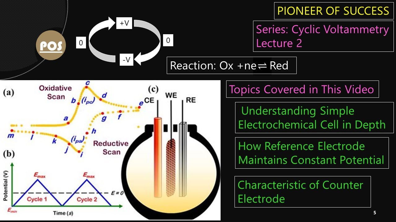
Cyclic Voltammetry Lecture 2 #CyclicVoltammetry #CV #Cell #Electrochemistry #research #pos
13.6K
A reference electrode maintains constant potential for accurate measurements. The Ag/AgCl reference electrode consists of three components: an AgCl-coated silver wire, a chamber containing saturated KCl solution, and a frit (porous plug) at the bottom. The frit allows connectivity between internal KCl solution and external electrolyte while preventing excessive leakage. The potential is constant because the electrode reaction AgCl + e⁻ ⇌ Ag + Cl⁻ depends only on chloride ion activity. Since the KCl solution is saturated, chloride concentration remains nearly constant, ensuring stable electrode potential for reference measurements.
Click to watch →
Deep Dive
Methodologies to minimize environmental variables during measurement, such as controlling temperature and precise electrode positioning.

Thermal On-Wafer S-Parameter Measurement Best Practices - FormFactor
5.1K
Environmental factors critically impact measurement stability—air conditioning vents and sunlight near measurement systems cause marked drift effects. Temperature logging correlates environmental changes with measurement anomalies, with real examples showing dramatic improvement in error set comparisons after addressing disturbances. Alignment markers directly above each standard on ISS substrates enable precise probe positioning even on manual stations, allowing wide translational movements with minimal risk of unintended Z-offset causing over-travel. Probe geometry changes significantly with temperature (probes expanding/contracting based on thermal expansion coefficients), with one probe warming while another cools during thermal cycling. Effective strategies include minimizing time away from thermal chucks, using instruments with good low-frequency bandwidth, and pre-positioning probes on separate ISS substrates before moving to main wafers.
Click to watch →
Dicas para soldar metalom galvanizado chapa finíssima.
107.3K
The electrode must be positioned at approximately 45 degrees, with the tip pointing slightly downward and forward to create a 'chapeuzinho' (little hat) shape. This positioning directs heat properly to melt the base metal without burning through it. The electrode should not be held parallel to the workpiece or at extreme angles. For thin materials, using only half of the electrode's coating helps prevent burning through. The coating on one side melts the base metal while the other side directs heat away from the joint, creating a controlled heat flow pattern.
Click to watch →

BTEUP 3rd Semester Measurement & Metrology | Chapter-1 Basics of Metrology Lec-10 #racevaengineering
1.5K
This section covers environmental factors (temperature, humidity, pressure, vibration, electromagnetic fluctuations, dust) that can reduce measurement accuracy. It also addresses methodological errors, where the measurement method itself is flawed (e.g., placing a thermometer in the ear instead of the mouth). The section explains that higher levels of environmental factors generally reduce accuracy, and that the method used must be appropriate for the quantity being measured.
Click to watch →

Подарили подписчику инвертор, маску, краги и электроды и научили варить!
24K
This segment teaches critical electrode positioning and weld pool management. The presenter explains that electrodes must be held at 15-45 degrees relative to the joint, not at a straight 90-degree angle, to allow molten metal and slag to flow properly. For T-joints, the electrode should be positioned at approximately 45 degrees. The presenter demonstrates how to adjust the electrode angle based on material thickness—closer for thinner materials, further for thicker ones. The segment also covers controlling slag by adjusting arc length and electrode angle to ensure slag moves away from the weld pool.
Click to watch →
Правильная методология регистрации ЭКГ. Сравнение с результатом ошибочной установки электродов.
520
Accurate electrode placement is critical for reliable ECG interpretation. The limb lead placement follows the 'traffic light' system: red on right arm, yellow on left arm, green on left leg, black on right leg. Chest lead placement requires precise anatomical landmarks: sternal line, midclavicular line, parasternal line, anterior axillary line, and midaxillary line. V1 and V2 are placed in the fourth intercostal space, 1.5 cm lateral to the sternum. V3 is placed between V2 and V4. V4 is at the fifth intercostal space at the midclavicular line. V5 is at the anterior axillary line, and V6 is at the midaxillary line. Electrode moisture significantly affects recording quality, and dry electrodes require re-moistening and re-recording.
Click to watch →
Deep Dive
Applying TEER measurements to specific in vitro models, such as the Blood-Brain Barrier (BBB), gastrointestinal tract (Caco-2), or pulmonary epithelial models.

Assessing The Blood Brain Barrier (BBB) Permeability of Waixenicin A
151
TEER measures the electrical resistance across the cell monolayer and is a standard method to confirm barrier integrity. A clinically relevant in vitro blood-brain barrier should have TEER values of at least 45 ohms per centimeter squared. This value was determined based on previous literature establishing what constitutes a functional barrier. The measurement uses electrodes placed in both compartments to detect resistance between them.
Click to watch →
Does all Disease Begin in the Gut? Monitoring the Barrier Function of an In Vitro Gut Mimic
157
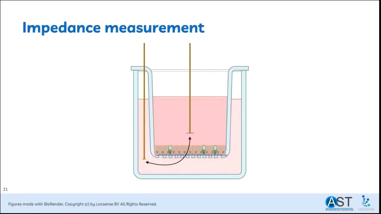
This webinar explains how to create and monitor an in vitro gut model using Caco-2 and HT-29 MTX cells in a 9:1 ratio on collagen-coated transwells, with transepithelial electrical resistance (TEER) measured via impedance spectroscopy to assess barrier function; the demonstration shows that EGTA disrupts tight junctions and reduces TEER to nearly zero, while Lactobacillus acidophilus does not significantly improve barrier function at tested concentrations, illustrating how this predictive model can evaluate compounds that modulate GI barrier integrity.
Click to watch →

Chemoinformatics and Computational Synthetic Biology: A Complete Course (Lecture 1)
588
In vitro blood-brain barrier models use transwell experimental protocols with cell culture membranes that mimic the basement membrane. Cells are grown on these membranes to create an artificial blood-brain barrier separating apical and basolateral compartments (representing blood and brain). The tightness of the barrier is measured using transendothelial electrical resistance (TEER), where increased resistance indicates a tight barrier and decreased resistance indicates barrier compromise. These models are useful for studying drug permeation and barrier function.
Click to watch →

3RsC Microphysiological Systems Workshop: Blood Brain Barrier
20.8K
Successful BBB modeling requires appropriate cell sources and rigorous validation. Primary human brain microvascular endothelial cells form tighter barriers than cell lines or iPSC-derived cells, which often exhibit epithelial characteristics. Barrier integrity is assessed using transendothelial electrical resistance (TEER) measurements and fluorescent dextran permeability assays. Inflammatory agents (LPS, TNF-alpha, thrombin) cause dose-dependent barrier disruption, with thrombin showing transient effects followed by partial recovery. These assays enable screening compounds for BBB-disrupting or protective properties, supporting drug discovery for neurological conditions.
Click to watch →
Real-Time TEER Monitoring for Organ-on-Chip Barrier Integrity
126
The intestinal chip model uses porous membranes to compartmentalize vascular and epithelial chambers. Endothelial cells seed first, followed by macrophages after 48 hours, then Caco-2 epithelial cells. Perfusion induces villous 3D structure formation. TEER monitoring reveals sequential barrier development: slight increases post-endothelial seeding, minor elevations post-macrophage addition, low values during endothelial monolayer formation, and dramatic increases upon Caco-2 seeding as tight junctions form. Perfusion initially raises TEER due to shear-induced tightening before stabilizing. Experimental validation using EDTA (a calcium/magnesium chelator) demonstrated immediate barrier disruption causing TEER collapse near zero within 30 minutes, with complete recovery within 24 hours upon EDTA removal. Multi-modal validation confirmed findings through brightfield imaging showing cell rounding/detachment followed by morphological restoration, and immunostaining demonstrating disruption and recovery of junction proteins (ZO-1, E-cadherin, CD31, PECAM-1).
Click to watch →
Deep Dive
Transitioning from manual measurements to automated, high-throughput TEER systems for drug permeability and toxicity screening assays.

REMS Robotic TEER Measurment for High Throughput Screening
2.2K
The REMS AutoSampler is a PC-controlled robotic system that automates electrical resistance measurements of transepithelial, transendothelial, or Caco-2 cell membranes grown on microporous filters in 24- or 96-well microplates, offering significant advantages including rapid measurement (96 wells in 5 minutes), elimination of human error from electrode placement, prevention of cross-contamination through optional rinse stations, and streamlined data organization with consolidated output formats for spreadsheet import.
Click to watch →

2017 REMS Robotic TEER Measurment for High Throughput Screening
1.5K
Automated robotic systems for measuring trans-epithelial electrical resistance (TEER) in cell monolayers on microporous filters significantly improve the efficiency and accuracy of high-throughput screening by enabling rapid, reproducible measurements across 24- or 96-well plates while minimizing human error, contamination risks, and operator fatigue compared to manual methods.
Click to watch →
EVOM™ The Trusted Brand for TEER Measurement
229
Transepithelial Electrical Resistance (TEER) is a well-established technique used to assess the barrier function and integrity of epithelial and endothelial cell layers by applying a small AC current to the cell monolayer and measuring the resistance to ion flow, providing valuable insights into tight junction integrity and cell-cell interactions; WPI's EVOM family of meters, including manual and automated systems for 24-well and 96-well plates, serves as the gold standard in TEER technology, having been trusted for nearly 40 years and cited in thousands of peer-reviewed publications for applications in drug absorption studies, tissue engineering, disease modeling, and quality control.
Click to watch →
Skin PAMPA Explorer™ Test System Demo
5.5K
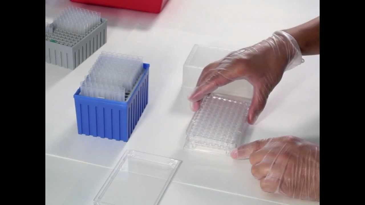
The Skin PAMPA Explorer™ Test System is a high-throughput, cost-effective alternative to Franz Cell testing for measuring drug permeability through artificial skin-mimetic membranes; the system uses pre-coated 96-well plates where topical formulations are applied to hydrated membranes, and permeability is measured by collecting and analyzing the acceptor phase at multiple time points, demonstrating strong correlation with human skin barrier permeability data.
Click to watch →
Introducing EVOM™ Auto
118
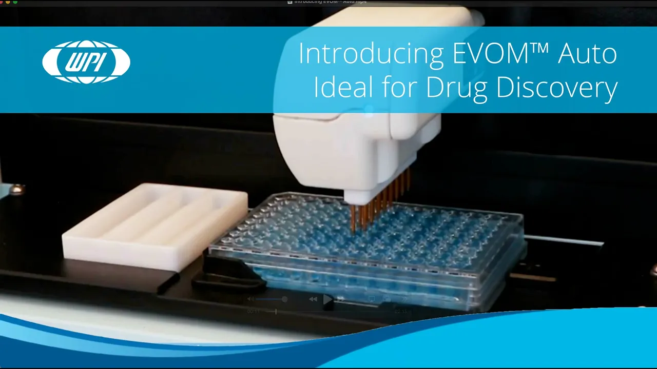
TEER (Transepithelial Electrical Resistance) measurements assess barrier function and cell integrity, which are critical data points in drug discovery research. Traditional TEER measurement methods are slow, error-prone, and labor-intensive, requiring manual operation and significant human intervention. Automated systems like EVOM Auto address these limitations by enabling rapid high-throughput screening, minimizing human error, maximizing precision, and increasing throughput. These systems can easily switch between 24-well and 96-well plates, maintain optimal cell growth conditions, prevent cross-contamination through automated disinfection, and provide wireless control options for researchers.
Click to watch →
Electrode Options
Endome Chambers
Electrode Options
0:00- 1
Compares STX4 and STX HTS electrodes for TEER measurement.
- 2
STX4 suits 24-well plates, with replaceable blades for longevity.
- 3
STX HTS is designed for high-throughput screening on 24/96 wells.
Beyond Static EVOM: The Case for Automated, Continuous Impedance Spectroscopy
While selecting the right EVOM electrode (such as chopstick or Endohm chambers) is a standard laboratory consideration, a significant counterpoint advocates for moving away from manual EVOM measurements altogether. Critics argue that manual EVOM systems, which typically measure resistance at a single frequency, suffer from high operator variability, temperature fluctuations during handling, and a lack of real-time temporal resolution. Instead, the alternative perspective favors automated, continuous, multi-frequency impedance spectroscopy (such as ECIS or integrated microfluidic sensors). These advanced systems non-invasively monitor barrier integrity inside the incubator over time. Furthermore, by measuring across a spectrum of frequencies, they can mathematically differentiate between cell membrane capacitance and junctional resistance, providing far deeper biological insights than the static, single-point resistance values obtained via traditional EVOM electrodes.
Understanding the biological significance of Transepithelial Electrical Resistance (TEER) as a quantitative measure of cell monolayer integrity and tight junction barrier function.
Familiarity with the setup and use of membrane-based cell culture inserts (such as Transwell systems) for growing polarized cell layers.
Basic knowledge of electrical resistance, Ohm's law, and how electrical currents flow through conductive aqueous media (cell culture reagents).
Awareness of the general operational purpose of an Epithelial Volt/Ohm Meter (EVOM) in cellular physiology laboratories.
Protocols for sterilizing, cleaning, and re-chloridizing silver/silver-chloride (Ag/AgCl) EVOM electrodes to maintain measurement accuracy.
Methodologies to minimize environmental variables during measurement, such as controlling temperature and precise electrode positioning.
Applying TEER measurements to specific in vitro models, such as the Blood-Brain Barrier (BBB), gastrointestinal tract (Caco-2), or pulmonary epithelial models.
Transitioning from manual measurements to automated, high-throughput TEER systems for drug permeability and toxicity screening assays.
148 views0likes3:25@WPIIncOriginal Release: 2025-03-14
[Music] wpi's evom manual is the gold standard for delivering stable and repeatable trans epithelial electrical resistance measurements the evom manual is the newest tier meter it qualitatively measures cell monol health and quantitatively measures cellular Confluence by determining an increase or a plateau in tissue resistance detected using our Innovative evom technology WPI State of-the-art ebom Technology provides you with realtime valuable feedback during experimental measurements electrod options are available for use with the new Evon manual here we will compare those options so you can choose the one that's right for your [Music] application the STX four electrode is the general purpose electrode for manually operated evom meters like the evom manual and the evom 3 with an adapter it may also be used with older versions of the evom and micel meters it is balanced and weighted for hands-free operation and the shallow electrode tips require less fluid than previous handheld electrodes its design offers greater Precision when compared with the stx2 and the STX 3 this electrode is the only one with replaceable blades this means you never need to Chloride the tips and you may replace the blades instead of the entire electrode this extends the functional life of your electrode the STX 4 is designed for use with the 24 well plates the STX HTS electrodes are designed for high throughput screening because the tips are much smaller and the electrode is keyed to fit neatly into the filter Wells the design improves accuracy over the stx2 and the STX 3 and it's more durable too the size of the electrodes and the design reduce the chances of sample contamination this is an excellent choice for 24 and 96 well plates the endome cell culture cup Chambers are built for stability and reproducibility to 1% tolerance these Chambers are ideal when Precision counts three sizes are available 6 mm 12 mm and 24 mm for 24 12 and six well plates the three-leg supports provide mechanical stability and holds the membrane parallel to the electrodes the symmetrical electrode pattern disperses the test current uniformally giving the most accurate reading of all the ivom electrodes however however each well must be individually positioned inside the endome chamber in order to make a reading the Eva manual requires an electrode for use choose an electrode suitable for your application if you have any questions just give us a call [Music]
Up Next

2:00
Organ-on-Chip Fabrication with Integrated Electrodes for TEER | Protocol Preview
@JoVEJournal
377 views•2022-05-27

12:54
Triumph of Orthodoxy Icon: Byzantine Art & History Explained
@BenCallan
2.1K views•2024-08-06

3:54
How to Measure TEER with EVOM Manual: A Step-by-Step Guide
@WPIInc
2.4K views•2022-07-12

1:42
Game of Thrones Opening Credits: A Cinematic Analysis
@gameofthrones
46.3M views•2011-04-18
Related Study Plans & Knowledge Roadmaps
Structured learning paths in General & Interdisciplinary Studies
general-studies
Elevator Engineering: History, Physics & Cities
01From Hoists to Safety Brakes: The Dawn of Vertical Lift
02Hydraulic vs. Traction: The Engineering Physics of Elevators
03Modern Speed, Safety, and Algorithms
+ 2 more learning modules
12h5 modules
Explore Roadmap
general-studies
Rubber: Material Science, History & Geopolitics
01Origins of Natural Rubber & Mesoamerican History
02The Chemistry of Natural Rubber & Vulcanization
03The Geopolitics of the Rubber Boom & Colonization
+ 2 more learning modules
12h5 modules
Explore Roadmap
general-studies
Containerization: Evolution, Logistics & Trade
01Ancient Trade & Bulk Cargo: Amphorae to Barrels
02The Industrial Revolution & Cardboard Packaging
03The Containerization Revolution: Malcom McLean
+ 2 more learning modules
12h5 modules
Explore Roadmap
general-studies
Textile Tech: Evolution, Labor & Global Impact
01Pre-Industrial Textiles: Handloom & Cottage Industry
02Mechanization: The Spinning Jenny to the Power Loom
03The Factory System & Global Labor Transformation
+ 2 more learning modules
10h5 modules
Explore Roadmap