















![X-ray Tube Physics, [Bremsstralung and Characteristic Illustrated for Rad Techs]](https://i.ytimg.com/vi_webp/K1S2Sz_T054/maxresdefault.webp)








This lecture covers the fundamental physics underlying medical imaging, including the four fundamental forces (gravity, electromagnetic, strong nuclear, and weak nuclear forces), energy units (electron volts and joules), atomic structure (Bohr model with electron shells and binding energies), and electromagnetic radiation properties. Key topics include x-ray production through bremsstrahlung and characteristic radiation, radiation-matter interactions (photoelectric effect, Compton scattering, coherent scatter, pair production, and photodisintegration), attenuation physics (linear and mass attenuation coefficients, half-value layer), and beam characteristics (quantity, quality, filtration, and the heel effect). The lecture emphasizes how these physical principles govern image formation, radiation dose delivery, and the design of diagnostic imaging systems.
Radiation Physics Basics for Radiology: X-Ray Imaging Principles
Added:thank you cynthia for inviting me and welcome everyone i want to tell everyone over the next um two evenings we're going to cover a lecture on the basics we're going to cover a couple lectures that are listed on x-ray imaging and we're going to do one on mammography and the times listed there are are not quite right so the basics is going to take us about an hour 10 minutes the x-ray imaging will take us about an hour 10 and mammography about 40. but we'll same three hours just a little bit different distribution and we're going to stop for the break here in a little bit but let's start off with a little bit of the basics so one thing um i i talk about sometimes is just some of the basic units that you should be familiar with and uh i've kind of skipped a little bit of that because you can look over some of those you have the slides but i just want to remind you of the fundamental forces right gravity is certainly important to us in radiology i mean we all know that this must be a left lateral decubitus film because the barium is layering there and this examination here must have been obtained prone rather than supine because the barium is laying anteriorly in the transverse colon rather than posterior at the hepatic and sigmoid flectures in addition another force that's going to be very important to us are the electromagnetic forces right we're going to talk a lot about what happens when we apply a voltage across an x-ray tube both in terms of between the cathode and the anode and across this little filament where we're going to boil off some electrons and those electrons are going to be accelerated across this very large voltage difference and strike that cathode material i'm sorry that anode material and um produce uh the x-rays that we're going to use to image you know there are a couple of other two other fundamental forces one is the strong force and that's really what binds the protons and neutrons together in the nucleus and you know it must exist right because two positively charged things should repel each other because of the electromagnetic force but yet they're bound together tightly in that nucleus and so for us i really just want you to realize that that must exist it must be stronger than the electromagnetic force in order to do that the weak force is is something more subtle and we actually need it to describe radioactive decay but really its purpose is kind of beyond the level of the lectures that we're going to be talking about but here they are in their relative magnitude okay the strong force stronger than the electromagnetic force both of these are stronger than the weak force and gravity the weakest of all of them we're going to talk a lot about energy we're going to talk about energy of x-rays that we create and normally outside of x-ray imaging if i mentioned energy to you you'd probably think of kinetic energy right the energy of motion you know how much energy does a car have and that's really determined by the mass times the velocity that it has squared the one-half term there and those units are are usually given in joules right a joule is a kilogram meter square per second square and that's much too large a unit to be useful to us in radiology we're going to work with something called the electron volt and it's the kinetic energy that an electron has an electron has mass remember when we accelerated across the voltage difference of one volt so here we have an electron we accelerated across that voltage difference of one volt when it strikes that positive terminal that catheter there that electron had a kinetic energy of one electron volt okay if we accelerated it across a thousand volts it could end up with it would end up with a kinetic energy of a thousand electron volts you know energy can exist without mass right that kinetic energy equation implied to you that well you've got to have mass to have kinetic energy and that's true but energy can exist without mass and we're going to see that a lot with electromagnetic radiation such as radio waves that we'll talk about you've already probably talked about when dr sirlen talked about mr imaging certainly visible light part of the electromagnetic spectrum very important to us and then the x-rays and gamma rays that we use to do our imaging there's also another important energy to us as we talk about x-ray imaging and especially nuclear medicine and that's the rest mass energy of a particle so if i could take all of the mass of an electron and convert it completely into energy okay that would give me 511 000 electron volts of energy you can calculate that by knowing the mass of the electron and just applying the simple equation e equals m c squared okay so it's mass times the speed of light squared and that number should be very uh recognizable to you because when we have a positron which has the same mass as an electron but the opposite charge right it's the antimatter equivalent of an electron and it annihilates with an electron we give rise to 200 to two 511 kev photons that we're going to use to image and pet imaging okay we're going to talk a lot about electricity and electricity right voltage equals current times resistance so here's a voltage think of it as a battery you apply it across a resistance perhaps that's a light bulb perhaps that's the filament in an x-ray tube perhaps that's the resistance between the anode and cathode in an x-ray too but once you apply that voltage the current that flows is determined by whatever that voltage was and whatever that resistance is so so in determin mentioning two of those right i have determined what the third one of those is and i'll make a point of that as we look at some x-ray tubes and talk about some of the fundamental little circuits that are there so here's one and really there are three electrical circuits that are going to be involved here one is we're going to apply a little voltage a small voltage across this filament this filament like in a toaster or like in a light bulb and when we do that it's going to heat that filament up and electrons are going to get boiled off the surface of that and with nothing else present they really wouldn't do a whole lot so we also apply a large voltage between this point on the filament negative terminal or the ground here and the positive terminal on this anode and that's on the order of right a hundred thousand volts right and that electron is now accelerated towards that cathode and strikes it with a lot of energy how much well a hundred thousand electron volts 100 kev when it strikes that that's how much kinetic energy it has that creates a lot of heat that ends up dissipated in this um anode here and so we really need to help dissipate that heat and one thing we do is then rotate the anode so there's another circuit right there's some a voltage that we apply across a motor here that rotates that anode so that the heat doesn't all get deposited at a single spot but all three of those are just examples of little electronic circuits in terms of electricity i also want to mention the concept of power and power if we know what the voltage is across an electric circuit and we know what the current is if we'll just multiply those two times each other we'll get an idea of the power the only thing i really want you to realize about this is a lot of our x-ray equipment consumes a tremendous amount of power typical voltage across an x-ray tube 100 kv typical amperage a thousand milliamps or one amps one amp that's a 100 kilowatts of power right think about a 60 watt light bulb in your in your house right this is a tremendous amount of energy it takes to to run an x-ray tube so just in general in terms of this first little introductory part i want you to know your units and their appropriate values for differentiate for different applications for example range of voltages on an x-ray tube right down in the 030 kv range from mammography up to the 100 20 140 kv range for ct maybe 80 100 for some x-ray imaging i want you to understand the range of mas for a ct study the energy of photons of technetium 99m if i say that to you all ought to be able to just tell me 141 kev effective radiation doses for some different studies speed of sound in tissues and realize that some of these are easily confused right kev which is a measure of energy right as a unit of energy is often confused with kv or kvp which is a measure of voltage the concept of ma which is a current and milliamp seconds mas which is is a charge right when we multiply by seconds are often confused and then some of the units of dose are also confused with each other we'll talk about those when we talk a little bit about radiation biology the next thing i want to talk about is uh the atom and just in terms of basics right we're gonna so long as we understand the bohr model of the atom we don't get need to get much more sophisticated than that to understand the physics we need to know so for us it's really going to be almost like planets orbiting a sun right we've got the nucleus the sun that consists of protons and neutrons and we can view these electrons as orbiting around that if you will it's a good idea for us to keep in mind some of the properties of those different elements that are going to come into play those that comprise the atom the electron we already talked about has a negative charge and it has a mass that's that's quite low a relative mass of one here i'm going to look at the mass of the other uh fundamental particles there relative to that so a proton has the same magnitude of charge but it's positive it's opposite in the sine and notice it's mass it's about 2 000 times more massive than an electron and so its rest mass energy is quite a bit larger right we already talked about the fact for an electron that's 511 000 electron volts and notice here this is in the mega electron volt range as opposed to the kilo electron volt range neutron very similar mass to that proton almost identical but remember has no charge so all atoms can be described using this notation we're all familiar with it right x is a unique letter o for oxygen c for carbon or sometimes a couple of letters for some of the elements uh a is the atomic mass number the number of protons plus the number of neutrons it also happens to be the number of grams per mole that that atom would weigh z is the atomic number it's the number of protons and of course in a neutral atom it's also the number of electrons and um you know this notation is a little bit redundant because o by definition is the atom that has eight protons right so we don't really need to put an eight subscript down here and so oftentimes oxygen is just listed as o16 if you will without that subscript just the superscript there and remember from back in your chemistry days how these orbital electrons line up we remember we've got this inner shell that contains two electrons we call the k shell and then just outside of that we've got our l shell that contains eight so this is our second shell the l shell so if we put a two in here for n two squared is four times 2 is 8.
so now if we go to the next shell 3 squared is 9 times 2 is 18 and that's how many electrons the m shell contains it's the electron shell structure of atoms that determines their chemical behavior and that's how mendeleev constructed the periodic table right these are all things that have full outer shells and they tend not to be very chemically reactive right these are things that have an extra electron just one electron in the outer shell and they'd really love to give it away and in fact they'd love to give it to these guys right here who are missing an electron from that shell so that's a chemical behavior it's not extremely important to what we're going to be discussing but i just wanted to mention it something that's more important to us is the notion of electron binding energy you know how tightly are these electrons held in place in the app well think about it a lot of that has to do with the electromagnetic forces that we talked about some of the coulombic forces so the closer the electron is to the nucleus the more tightly bound it's going to be right and and so the k shell electrons are more tightly bound uh than the l shell electrons and then the more positively charged the nucleus is the more tightly bound those electrons are going to be right the more positively charged the the nucleus is those negatively charged electrons are going to to be um more bound to it so removing an electron requires an energy that's greater than that binding energy if you don't have an energy greater than the binding energy you can't remove the electron from the atom and i put this list of binding energies here not because i want you to memorize anything from them but i want you to look at the number for hydrogen you know what if i want to take an uh electron from hydrogen well that's going to take about 13.5 electron volts of energy and then when we look at what is the level for ionizing radiation it's going to be right around in that number right that's the smallest number that occurs in this table so radiation that has energy below around that value in electron volts radiation that's less energetic than that isn't going to be able to ionize an atom okay and then the other thing that's important to us is are these binding energies on some of these materials barium uh iodine that we use for contrast agents those k shell binding energies are very important to us because part of the reason they work well as contrast agents is that these k shell binding energies determine uh the photoelectric effect when the photoelectric effect is likely to occur and that's really what makes these um these contrast agents have much appear much more uh highly attenuating at those energies so i wanted to show you that and then the other thing is we have some really interesting things in nuclear medicine like we we image with thallium and as thallium decays it does something really interesting and and i'll remind you of that on saturday when we talk a little bit about nuclear medicine so these binding energies become important to us for those reasons you know in chemistry we learned about the importance of removing or adding outer shells electrons from atoms you know creating ions right in physiology this can have fairly deleterious effects you know if you remove an outer shell electron you create some of these ions some of these free radicals uh the hydroxyl radical unfortunately we're about 70 percent water right so you can create a lot of o h minus ions there and they can result in in significant damage um to biologic tissues so so let's look a little bit about what happens if we start removing electrons from atoms so this is tungsten right it's got 74 protons and it's got you know it's 110 neutrons 184 minus the 74. this is the sum of the protons plus the neutrons i'm going to start to draw in its electron shell but i'm going to give up very quickly because i don't want to draw shells of 74 electrons and we'll make our point with a little subset of those so it turns out that the k shell binding energy for tungsten is about 69.5 kiloelectron volts and the l shell binding in a tree is about 12 kilo electron volt so now what if we had some electromagnetic wave or some particulate radiation that possessed enough energy to kick one of these inner shell electrons out of its shell what would happen next well what would happen is that one of these l shell electrons or perhaps one of the m shells electrons would come in and fill that space and after that came into fill we'd get a release of characteristic electromagnetic radiation here in the x-ray range 57.5 kev notice the difference between the two binding shell energies and this is called characteristic x-ray energy because it is characteristic of this particular atom if we looked at a different atom it would have different k and l shell binding energies and those differences would be unique to that particular atom so if you looked at some material out in space here on earth giving off radiation after you and shot radiation at it you could tell what the material was made of by looking at where the bands of characteristic radiation lie you know the that's not the only thing that can happen sometimes when we kick that out as we get this transition rather than getting that 57.5 or so 57 kev characteristic x-ray energy that energy gets transferred to one of these outer shell electrons and it heads off with a kinetic energy that's equal to the difference of these but also minus its binding shell energy and these are referred to as oj electrons or auger electrons depending upon guess whether you're from france or the united states right and the importance of those is now we've got a very energetic electron traveling around in these tissues and if this is biologic tissues it's going to wreak a lot of havoc there and so i want you to keep that in mind right we're talking about what happens not just in tungsten but this is also the same kind of thing that could happen if this were a biologic tissue so characteristic radiation uh we mentioned this they have those unique values and i mentioned that each element could be recognized by those you know we can't predict precisely which event will occur but we do know for a particular atom and imparting a particular energy on it we do know the probability that will each will occur heavier elements tend to be more likely to emit characteristic radiation while light elements are more likely to emit oj electrons again important to us because as biologic tissue we're predominantly made out of lighter right elements so what happens when we remove that inner shell electron we talked about when the difference in binding energy is large we can get those characteristic x-rays right when the difference is quite small and this isn't necessarily true for atoms but sometimes in molecules where some of these electrons are shared and the energy that binds them between the different atoms in the molecule the binding energy is even a little lower the binding energies might be in the few electron volt range and that's in the visible spectrum and these are tissues that either scintillate or phosphoresce right if you irradiate them they now give off electromagnetic radiation in the visible light range so when we talk about x-rays realize we're just talking about a phenomenon that's just much higher up in the electromagnetic spectrum in terms of energy than visible light but here this is also giving off some of the same kind of stuff i know we wanted to take a little break we're going to go a little bit longer here is that you set me to okay good all right so let's start start talking about uh electromagnetic radiation here so electromagnetic radiation is energy that travels through space or matter um i'm sorry radiation is energy that travels through space and matter there's two types particulate so that's stuff that has mass it may have charge remember the neutron doesn't have any charge but the positron the alpha particle those have charge uh the electron it fits in that class as well and then the electromagnetic radiation the pure energy has no mass or charge so let's just talk a little bit about electromagnetic radiation specifically now remember no mass it's unaffected by electrical or magnetic fields travels at a constant speed in a given medium and that maximum speed occurs in vacuum and is given it's about three times ten to the eighth meters per second in other media the speed is a function of the characteristics of that media and it can propagate through matter and as a matter of fact the behavior of electromagnetic radiation with matter is quite complex you know down at low at the radio wave end that radiation really tends to pass right through us when you get up to the microwave end it can interact with matter i mean that's how a microwave oven works but it it really works by rotating part of the the atom and you can vibrate or rotate that behind so you can get microwave energy to deposit in the tissues is heat if you will as you go higher up things change again at the x-ray energies things interact sometimes sometimes as it gets more energetic it's more likely that it may just pass through the interactions absorption scattering and we already mentioned radio waves going to be important to us in mr visible light x-rays as examples one of the first things i want to say is right notice this travels in straight lines right straddles in tr in straight lines if we're in a vacuum or in air for the most part it travels in straight lines and if you think about that that really explains to you the fir one of the first things you're probably told in radiology the inverse square law you know think of a little point source of radiation a little bit of technetium sitting in the front of the room right and it's emitting 141 kev gamma rays and for the most part in air in vacuum to be more precise those those gamma rays travel in a straight line away from that and so if you take a look at how many are passing through a particular area when you're a distance r from that source now if you move twice as far away notice those nine gamma rays are now spread out over four times as much area or if you will if you were the same size like this letter a in this picture only a fourth as many of them would pass through you right so as we double our distance from the source of radiation right the dose that we're exposed to decreases by the square of that distance okay but this is true for all electromagnetic radiation right i mean this is true for radio waves so if the source of radio waves is sitting at that point where do you want to put your mr coil you want to put it as close as possible to the source of those waves okay so true for all electromagnetic radiation electromagnetic radiation you know has both wave and particle-like behavior and sometimes as i talk i'm going to talk to you and i'm going to say a gamma ray and that sort of accentuates the wave-like properties when i say that and other times i'm going to say to you an x-ray photon and that sort of accentuates the particulate properties of electromagnetic radiation because the truth is at this subatomic level you know there's this duality where where things can behave both wave-like and particle life that's really unfamiliar to us in the macroscopic world where you know you kind of behave like one or the other um like any wave the velocity of electromagnetic radiation is equal to its frequency times its wavelength and but for electromagnetic radiation you know that velocity is constant it's the speed of light in a vacuum and it it propagates as a pair of magnetic fields and that's only important for us i want you guys to remember that if i have a time varying electrical current then i have created a magnetic field right or if i have a time varying magnetic field i can create a current but but the first way that i said it is more important to us because that's how we're going to make our gradient coils and things work in the mr scanner right we're gonna put a current through some wires and that's going to change the nature of the magnetic field the add to or subtract from the main magnetic field of the magnet the wave-like properties of electromagnetic radiation are probably best understood by this double split slit experiment i mean think about it if light were a particle right light is an electromagnetic wave if you shot particles through this slit then oftentimes they just hit this wall over here except occasionally they'd go through one of these other slits and they'd hit this screen and they'd have to hit this screen in this fairly small region right in here and the same thing they could pay depending on your angle sometimes make it through both slits with the bottom slit here and you'd see the distribution of those particles hitting in this area but that's not what we see we see something that looks much more like the behavior of a wave where that wave of light propagates out and then when it hits this wall with these two slits we get two individual wave fronts propagating through each of those slits and those constructively and destructively interfere with each other to create this pattern on that screen so just intuitively giving us the idea that light electromagnetic radiation has a wave-like property that particle-like property i want to show here and as soon as i finish we'll take a break with this next slide here's the best example i can give you that and that's the photoelectric effect which is going to be really um important to us you know potassium the m shell binding energy of potassium the lowest energy at which any of its electrons are bound is 18.3 electron volts and you can beat potassium all day long with multiple 18.2 ev photons okay multiple ones they never add up together like waves might and knock out one of these electrons from their shell but once you go above that threshold you immediately can kick out some of those electrons from the shells of potassium emphasizing that particulate nature of electromagnetic radiation okay so that's the photoelectric effect it'll be really important to us a little while as we talk about um uh some of the x-ray interactions with matter that happen when we we make a radiograph so i i show this image just to kind of remind you where the visible spectrum is you know i don't particularly like this image because it it lists x-rays and gamma rays as being separate entities and they really can have very very similar entities and for us we're really going to designate them based on how we produce them but but also to give you an idea of you know mr is really interesting right we're gonna we have electromagnetic phenomena with a wavelength the size of a kind of a skyscraper if you will and yet we're imaging um you know the protons in water predominant in fat predominantly and then up here where we are you know we're on about the level of the diameter of an atom i like this a little better right because it shows the overlap between x-rays and gamma rays our visible spectrum right here the ultraviolet infrared so let's talk a little bit about electromagnetic radiation gamma rays are really amount from the nucleus of radioisotopes and we use them to make images of the distribution of those radiopharmaceuticals within the patient so anything that's you know this electromagnetic phenomenon that's produced from the nucleus we're going to call a gamma ray and x-rays are produced outside the nucleus we're going to talk about how we get those in an x-ray tube but we use them in radiography and ct we've already mentioned the radio waves in visible light i want to mention ionizing radiation right electromagnetic radiation of a higher frequency than the near ultraviolet has enough energy per photon to remove electrons from atoms and molecules if you look at that right in water that requires about 12.3 electron volts right they're sharing some electrons there so it's a little bit lower than what we saw in hydrogen and if you look back at the chart that i had or on this one right here where we've got that ionizing radiation it falls right in that range that where we were talking about around 12 electron volts or so so below that level there's just not enough energy to remove electrons from the shells of atoms or or molecules so let's talk a little bit about how radiation interacts with matter and i want to start off by saying look this is all matter it some of this is going to be important to us when we talk about how it interacts with biologic tissue some of it's going to be important to us when we talk about how radiation interacts with materials like tungsten and x-ray tube and some of it's going to be important to us when we talk about how radiation interacts with scintillators in detectors like digital imaging detectors or a pet ct scanner so radiation interaction with matter so energetic charged particles right interact with matter by coulombic interactions and lose their kinetic energy via excitation ionization and radiative losses so these are things that have mass particulate radiation mass and charge here so here we have an energetic charged particle and as it passes close to an atom it may excite one of these um outside shell electrons and that may move that electron may move to a slightly higher shell temporarily and then move back and it may give off some emission of radiation as it returns to that lower level and this is usually in in the infrared range so this is heat this excitation results in heating and the vast majority of the interaction in human tissues results in this occurring frankly in most materials ionization of course a very similar thing happening but now the interaction is strong enough so that that electron breaks free creating an ion and we've already talked about what happens once this occurs right once this occurs we can get some of those um characteristic x-rays and oj electrons and approximately 70 percent of the interaction of charged particles uh is deposited via excitation when ion ionization occurs that ejected electron might possess sufficient energy to kick additional electrons out of their shells in the neighborhood in neighboring atoms and we refer to those as delta rays the importance of that to us is right if i kick an electron out of its shell and in part to it a lot of kinetic energy it can end up resulting in some fairly deleterious effects to the surrounding tissues and i mentioned the fact that with both these processes we can get those uh characteristic x-rays and those oj electrons talk when we talk about dose radio biology i'm going to talk about linear energy transfer because really the effect that radiation is going to have on biologic tissue is going to have to do with how much energy it transfers to that tissue per unit distance that it travels but i just want to mention that things like electrons that are have relatively low mass and they're charged they travel a very awkward path bouncing around and i usually say you know think of um the billiard ball bouncing around striking other billiard balls and you know here here think of a bowling ball hitting other billiard balls right it really just keeps going the direction it's going as it strikes everything in its path and those are things that are quite massive and have a lot of charge let's say like an alpha particle right the nucleus of um of helium two protons two neutrons um the proton falls in that that group as well by the way so what about radiative interactions because this is going to be important to us in terms of how we generate x-rays well charged particles can also undergo interaction with the nucleus of that atom right and so here we have this charged particle it's going to swing close to by the nucleus of this atom and its direction is going to be changed by the interaction of its charge with the charge in the nucleus and that's going to change the kinetic energy of that particle so it's reduced let's say to 40 kev in this example but conservation of energy says we're going to get the emission of this 100 kev photon so notice our initial charged particle has slowed down and we've gotten production of this radiation and that's the bremstro lung or breaking radiation that we see produced as we slow down and change the path of that particle it turns out that when the energy of that electron is low that brimster along radiation is emitted preferentially in a particular angle now this picture is a little bit misleading and i want to make sure to say right when this electron comes in here i want you to realize that we get radiation right if this is a material consisting of many of these atoms and we're bombarding it with particulate radiation we're going to get bremstrolung radiation that heads in all different directions but it doesn't quite hit in all different directions equally okay it has a slight preference for this direction and if you'll notice that's where we put the x-ray window on our tube right why is our tube have the x-ray window in the location it does it does here because that's the greatest number where the greatest number of those x-rays are produced what about other things well the positron undergoes annihilation with the electron there's another thing that has mass and charge same mass and the as an electron but opposite charge the antimatter equivalent that interaction is going to be very important to us in pet imaging the neutron we haven't talked about you know it has no charge but it has mass and so it can transfer energy via collisions or be captured by the nuclei and when it has collisions frankly what it does is it ends up launching out some of those other particles be they electrons which we've talked about here or protons or other things neutrinos i'm not going to talk about they have no charge extremely low mass they don't really interact with anything i mention them only because i want people to keep neutrons and neutrinos separate from each other okay so what about x-ray and gamma ray interactions right we just finished talking about particulate radiation interacting with matter now i want to talk about electromagnetic radiation and how it interacts with matter and there those interactions are complex they depend on the x-ray energy beam ener i'm sorry the energy of the x-ray beam the particular material that we're talking about it's atomic number and then also its electron density so how many electrons are located in a cubic centimeter of that tissue this is an equation for electron density remember this is just density so that's the amount of grams per cubic centimeter this is avogadro's number so that tells you how many atoms there are right per gram this is the atomic number that tells you how many grams there are per cubic centimeter so when you cancel these things the things out here you've got the number of atoms per um mole and so now multiplied by the uh grams per cubic centimeter i'm sorry you've cancelled out the grams there so you've got the number of atoms per mole when you make this multiplication and then if i multiply by z well that's the number of protons but it's also the number of electrons in the neutral atom so i have the number of electrons times the number of gra of atoms per per cubic centimeter so this is the density of the electrons it turns out that the molecular structure of material really is negligible in terms of its effect on the attenuation properties up at diagnostic x-ray imaging right any of you who wear glasses realize it's not negligible down at the visible light range right you can bend light refract light with a pair of glasses there but the molecular structure is really negligible where we're going to be talking about in diagnostic imaging x-ray photons predominantly transfer their energy to electrons of the atoms right which are going to lose their energy by interacting with other adjacent atoms in other words x-rays when they interact with matter are going to produce energetic electrons and how do energetic electrons interact with matter via the ways we just discussed when we talked about particulate radiation but i want you to keep that in mind right because when we image with x-rays one of the first things that's going to happen inside the patient is we're going to create a bunch of energetic electrons that's going to deposit the dose in the patient there when we take an image about a third of the x-ray energy is going to end up getting deposited in the patient as does i'm sorry about two-thirds of the energy about one-third of it's going to be scattered back out of the patient and only a small fraction of it is what we're going to use to actually make the image of the patient in air a kinetic energy an electron with kinetic energy 100 kv goes 10 15 centimeters before it is interacted enough that it stops if you will in soft tissue that distance is only one tenth of a millimeter about that's very helpful right i think most of you is a resident right you've had to do six i-131 doses to patient right and the thyroid accumulates i-131 very nicely right so those electrons those energetic electrons that are radiated off in the beta beta minus decay of i-131 they interact and give all their energy off within a tenth of a millimeter typically of where they're produced so it treats the thyroid very well locally for that reason all right so now remember we're talking about x-ray interactions i've just made the point that x-rays interact with matter and the first thing they do is produce energetic electrons and i don't want you to forget that because that's where the radiation dose to the patient comes from there but but let's talk about how those x-rays do that the first thing i want to talk about is coherent scatter so in coherent scatter this x-ray interacts with that atom kind of as a whole it kind of if you want to almost think of it you know it vibrates it to a slightly excited state and as that atom relaxes back to its normal state it gives that energy back off but in a slightly different direction okay so coherent scatter changes the direction of the energy but not by a whole lot we're lucky because coherent scatter really is a minority of the interactions that occur at diagnostic energies um much more at mammography kvs k k e v's energies but very little up at the diagnostic energies for let's say abdominal plane film ct those kind of things so we're going to ignore it for our purposes here it is here's the total interaction from some of these different events notice that um classic scatter coherent scatter rayleigh scatter really drops off pretty quickly and at a hundred kev it's really dominated by compton that we're going to talk about photoelectric dominates it fairly well lower down there so we're going to ignore it another thing that can happen when you have an energetic x-ray or gamma-ray photon is it can end up interacting with the nucleus and producing a pair of an electron ejecting an electron and a positron from the nucleus of the atom this pair production this doesn't happen you it should make sense that in order to have that occur you've got to have a an x-ray or a gamma ray that's at least two times 511 kev which is 1.022 mev that i have there and so for us at diagnostic energies this really is not an issue right if you were maybe doing uh um radiation therapy this would be more of an issue to you so here's pear production incident there we get that negatron and that positron or that electron and positron produced there another thing that can occur is photodisintegration and now here the x-ray is so energetic that it actually breaks the nucleus apart and it takes about 15 mega electron volts to even be able to do this so this is way above the energies that we're interested in and it just makes sense if you look at the binding energy per nucleon in the nucleus of an atom how much energy does it take to keep the nucleons bound together by the strong force you can see it's in the range of eight nine seven eight nine mega electron volts per nucleon so you've got to have be quite energetic to make that happen so that leaves us two things to talk about photoelectric interactions and compton interactions and before i talk about those two those are the two that are important to us i want to say something about the image formation process look in order to make an image right in order to make an image some of my x-rays have to go through the patient and some of them have to be stopped by the patient right if none of them made it through the patient we would just make an image that looks like a shadow of the patient right it would just be a black area if you will where none made it through with white in the rest of the area if all of them made it through the image would just be uniformly white neither of which is helpful we rely on the fact that there's differential absorption of those x-rays as they go through the patient okay it would be great if when the x-rays interacted with the patient they were just stopped and didn't continue on and there's one effect that actually is going to do that for us the photoelectric effect unfortunately there's another effect where they impart some energy and then change direction they scatter from there and they may scatter and still hit our image plane and that's going to result in a degradation in the quality of our image reduce our image contrast and that's the compton scatter that does that we wish we could really minimize that if at all possible we'd like to be unable to understand how can we select our kv or kilovolt or kilovolt peak on the x-ray tube or ma our type of detector our type of filtration our type of contrast all these things to really make those interactions happen the way we would like them to happen to get the best possible image that's our goal so let's talk about the photoelectric effect with the photoelectric effect when this energetic x-ray photon comes in it's going to interact with one of these inner shell electrons typically and it and it kicks that out of its shell that's the photoelectron right so there's our 65.96 kev photoelectron and what's that going to do it's going to deposit all its dose there in the patient very locally like we already talked about that's how particulate radiation interacts with matter okay that l shell electrons then going to come in and fill that l shell and we'll get rise to some characteristic radiation you know if we're talking about this interaction occurring in high z materials that characteristic ener x-ray energy might be fairly high but in human beings where calcium is about the highest z material you can have notice that energy is quite low so most of these that dose gets deposited in the patient as well the photoelectric fa the probability that the photoelectric effect is going to occur is roughly proportional to the z cubed of the material divided by the energy cubed okay divided by the energy of the x-ray cubed so as the energy of the x-ray goes up this drops off very quickly right as we go from 10 kev to 100 kev 10 times 10 times 10 is a thousand a hundred times a hundred is ten thousand times another hundred is um what ten million right the the the denominator gets small very quickly the probability of this interaction drops precipitously it turns out though what's interesting is there's an abrupt increase in the probability of the photoelectric effect just above the k shell binding energy remember i showed you those binding energies and i told you they'd be important to you and that's why the probability that the photoelectric effect is going to occur it's not a continuous function that just gradually goes down one over e cubed it has some discontinuities in it that correspond to the binding shell energies and we can take advantage of those by using a a particular x-ray energy and using a particular contrast material that have k shell binding energies matched to the x-ray energies to really take advantage of that okay so here are some important um k shell binding energies you know very low for human tissues like oxygen but i put them up here for iodine notice about 33 kev barium kev lead 88 kev here's a picture this is cesium iodide so this is relatively high right iodine we use as a contrast agent cesium's fairly close to it on the periodic table so the k shell binding energies are fairly high much higher than for biologic tissue and notice here the photoelectric effect and the compton effect they're about the same contribution to the total attenuation of the x-ray beam up at around 200 kev we're going to see a biologic tissue that occurs much lower down than that because biologic tissue has a much lower average z so let's talk a little bit about compton now so in compton the x-rays tend to interact with those outer shell electrons the the x-ray photon gives some of its energy to one of those outer shell electrons typically breaking it from its shell and giving it some kinetic energy and that scattered electron loses its energy by interacting with those adjacent atoms through those mechanisms that we talked about excitation radiation um interactions the x-ray photon then has less energy and unfortunately heads off in a new direction right so here's this case right now we come in we interact with this outer shell electron it heads off with some kinetic energy let's say we gave it five kev of energy well now if we want to know the energy of the photon after that this x-ray photon after that we've got to subtract the binding shell energy plus the kinetic energy that we gave this electron and if we do that we get about 94.56 kev that that heads off with and unfortunately it now may strike our detector but you know the focus of our image has to do with our we're using this point source of radiation and so now this scattered x-ray hits our image plane and it looks like it came from a completely different direction than the original x-ray actually did so that scatter degrading our image they maintain a large percentage of their energy even at large scattering angles so you know we it doesn't help us to kind of think about well could we get rid of them at the detector if we could somehow do some energy discrimination we'd have to be really good to make that happen a hundred kev compton scattered uh x-ray at 90 degrees so if we started off at 100 kv it compton interacted and scattered off at 90 degrees it still has 83.6 kev of energy these will escape the patient right and that's bad for us when we're doing fluoroscopy procedures right when we're doing ir because these highly energetic scattered x-rays are going to hit us they're going to scatter 180 degrees 90 degrees from the patient and have enough energy to escape their body and potentially interact with us these account for the majority of scatter at diagnostic x-ray energies the probability is proportional to the electron density of the atoms and it drops off as one over e approximately one over e not one over e cubed like the photoelectric effect so as energy increases photoelectric effect is dropping one over e cubed compton is dropping one over e so very quickly compton dominates the effects they're both equal to each other remember i showed you cesium iodide at cesium iodide they're both equal to each other at about 200 kev because those materials have much higher z than human tissues in human tissues soft tissues they're equal to each other at about 25 so here's photoelectric effect here's compton here's where they're equal to each other the percentages right this is a log scale so this is right down around 25 kev that's great for us in mammography right but that unfortunately when we get up to ct energies or so we really are not able to take as much of the advantage of the photoelectric effect as we want to so i've mentioned attenuation and linear attenuation a couple times as i've said them and i haven't really told you what we mean right the linear attenuation coefficient is the rate at which x-rays are removed from an x-ray beam a monochromatic single energy beam let's think an ideal case as i pass it through tissue if i put a thousand in how many do i see on the other side that are completely unaffected we've mentioned the fact that some comptons may still get through but they've been affected their energy is less how many make it through completely unaffected it turns out that that's just a kind of a decaying exponential the same kind of thing that we see in radioactive decay the number of x-rays that make it out is a decaying exponential that has to do with the attenuation the linear attenuation of the material and the thickness of the material that you've passed through how could we figure out what that is for particular materials for a particular uh energy of x-rays well here's our monochromatic x-ray source of a particular energy just take yourself some layers of that material let's say one centimeter thick and first put down one layer and see how many x-rays you detect know how many you transmitted and then put down two layers how many of the transmitted now do you tack put down the third layer and if you drew that for different numbers of layers centimeters thickness layers you'd make a curve that looks like that a decaying exponential like we talked about and if you just plotted that on a semi-log plot right that the slope of that line right there is the linear attenuation coefficient that we're talking about the rate at which we remove those things from the beam now that linear attenuation coefficient really is the sum of the behavior of those five different things that we talked about what's the linear attenuation coefficient due to rayleigh scatter what's the linear attenuation of due to the photoelectric effect to compton to pair production to photodisintegration but i told you we're going to ignore three of those and so i can get this to simplify quite a bit remember we did describe the fact that linear attenuation is dependent on the energy of the x-ray beam so at 30 kev right notice that the decay constant the decay constant here is bigger than it is at 60 right we you're gonna take more out of this beam through soft tissue than we are at 60. higher energy x-rays pass through soft tissue or any material frankly more easily and notice that of course it's dependent on the material so here's the linear attenuation coefficient for soft tissue at 30 and here it is for bone right bone is going to stop many more of the x-rays per unit thickness than soft tissue is so just a rough idea what those numbers mean i mentioned the fact that for us we're really going to think of linear attenuation really only having to do with photoelectric effect and compton scatter right we're going to simplify that equation for that and so really this linear attenuation is really just the function of the average z of the material right the z has to do with the the how tightly bound those inner shell electrons are where the k edge is so the photoelectric effect and also that electron density that's going to be the compton effect that we're going to see now before when i showed you those plots those plots actually were mass attenuation coefficients not linear attenuation coefficients and the main reason for that is if you think about water you know if you think about this the gaseous form the liquid form and the solid form steam water and ice they all have different linear attenuation coefficients and so the mass attenuation coefficient says look just divide by the density of the material and then all states of water will have the same mass attenuation coefficient so when people make graphs of them they tend to make graphs of mass attenuation coefficients just so that the particular density of that molecular structure doesn't come into play so here's a mass attenuation coefficient before i was showing you cesium iodide and soft tissue i just wanted to show you these this is gold right this is iodine this is uh calcium this is i'm sorry this is calcium this is gadolinium this is calcium here and this is soft tissue so someone tell me sorry what is this discontinuity due to right this is due to the photoelectric effect right this is the k shell binding energy for gold and so just above that energy gold appears more highly attenuating to x-rays just above that energy than it does to x-rays just below that energy right that's really kind of odd look at look at here look at iodine and gadolinium do you notice that iodine is below gadolinium and then all of a sudden iodine is more attenuating than gadolinium right after we go over the kh and it stays there for a little while right so if we're going to image with something like iodine what we would really love to do is make sure that our kvp that we put on the x-ray tube is such that it generates x-rays that have energy that are just above the k shell binding energy of iodine right that's how we'll utilize iodine as a really good contrast agent make sense now what's going on down here in soft tissue and calcium why don't we see those discontinuities there well do you notice that this starts at 20 kev so those exist right for those but those are much lower z tissues and so therefore those little k those little discontinuities would be much lower down on the graph okay good you know i wanted to make sure i talked to you about linear attenuation coefficient but frankly this rate this decay rate which is what uh linear attenuation coefficient is it's a hard concept to put your your mind around sometimes we're much more familiar with something like the half value layer right how much thickness of material does it take to reduce the number of x-rays in one half right that's a much easier thing for us to understand conceptually and that's a measure of the quality of the x-ray beam and i want to introduce that concept quality right it really tells us how energetic the x-ray beam is right because the more energetic an x-ray beam is the greater the thickness the greater the half value layer is going to be have to be in order to decrease the number of x-rays in that beam by 50 percent there's a concept called the tenth value layer that's just the thickness that reduces it to 10 but usually we work with half value layer so so here we are we've got a i want to start off with kind of a medium energy monochromatic x-ray beam so a single energy x-ray beam and it's kind of medium energy we put 2.5 millimeter aluminum sheet in front of it and notice now we've reduced its number of x-rays by one-half so the half value layer of this x-ray beam is 2.5 millimeters of aluminum if we wanted to we could put gold in front of it and and we could figure out what its half value layer was for gold and that would be a different thickness of gold a slightly lower thickness because gold is more highly attenuating has a higher z than aluminum and then if we put another 2.5 millimeters here it should again reduce the number of x-rays in the beam by one-half that's what we mean by the concept of the half-value layer here's a low-energy monochromatic x-ray beam we put 2.5 millimeters of aluminum in it and look only one makes it through so so this is too thick to be the half value layer for this lower energy x-ray beam right so we have to go to a thinner piece of aluminum to find the half value layer so right away it should say to you right okay this thickness that they're reporting as a half-life value layer it tells me something about the energy the average energy of that x-ray beam right and so you'll notice that in most states there's some sort of legislation that says that if you've got an x-ray machine that the half value layer from the x-rays produced by that machine has to be at least 2.5 millimeters of aluminum and what they're saying is the average energy has to be above a certain level right otherwise you're just imparting a lot of dose in the patient without getting good information for your image okay so a lot of times that's expressed that way the half value layer has to be above a certain level but you know we have polychromatic x-ray beams and things get a little more complex right our x-ray beam that we create from an x-ray tube isn't a single energy but it's a bunch of energies and so here's that polychromatic beam we've got the different energies here we put 2.5 millimeters of aluminum and now notice we've got half as many x-rays notice a couple things though we've got much fewer of the lower energy x-rays and kept quite a bit more of the higher energy x-rays the average energy of the x-ray beam is now quite a bit higher so now we put another 2.5 millimeters of aluminum in front of that and notice that now we don't drop the intensity of this x-ray beam by half not right it's not quite dropped in half anymore because of this preponderance of the higher energy x-rays when you have a polychromatic x-ray beam the second half value layer is always greater than the first because with a polychromatic x-ray beam we preferentially take out lower energy x-rays the mean energy of the beam gets higher the quality of the beam gets higher the quantity always gets lower with filtration but the quality the average energy of the x-rays that you have gets higher okay all right we already mentioned this fact here that most states there's this legal minimum we talked about the fact that monochromatic beams second half value layer exactly the same as the first in polychromatic second is always greater than the first here's the half value layer for some tissues we've got monochromatic x-rays here half value layer of soft tissue bone and lead i just want to make a couple of points right think about it about 360 kev which is kind of the average energy of the x-ray beam for an abdominal radiograph or so right the half value later is 30 millimeters of tissue right i'm just going to assume i'm only made out of soft tissue right but from from my left side to my right side you know maybe 30 centimeters right so 300 millimeters so 10 half value layers so we start off with our x-ray beam the first half value later gets reduced in half second a quarter third an eighth fourth a sixteenth fifth a thirty-second sixth a sixty-fourth 128 256 right 512 10 24.
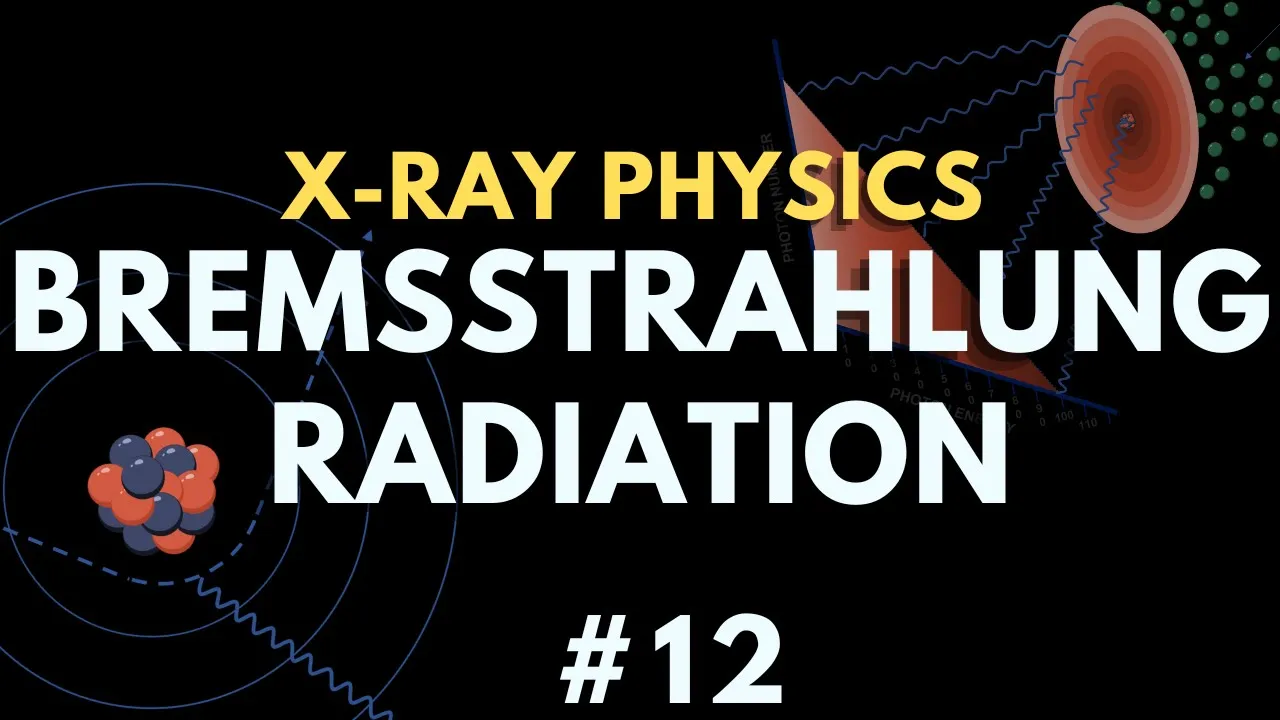
when you get to the other side only one in a thousand x-rays are making it through unaffected okay remember i told you that most of the x-ray dose right two-thirds of it is going to be absorbed by the patient a third of it is going to be scattered out he probably said well what are we making the image with well we're making the image with that one in a thousand x-ray that strikes the film plus unfortunately the scatters that strike the film as well okay so just keep that in mind in terms of that so let's talk a little bit about x-ray production here so the x-ray generator has this power supply on the x-ray tube it has some timing and exposure circuits you know here's some typical values that we've i've shown you already for an x-ray tube we've talked about how big a sink of power it is here's an old control panel right this has been replaced with you know computerized units that select these things automatically but back in the old days right you hit your voltage by picking a button here and your current here and your time here a similar thing is happening now it's just more under kind of computer control rather than human control and you can just see how large one of these generators are to power these x-ray units so we place that large voltage across that x-ray tube between that cathode and that anode and we put a small voltage across that filament which is part of the cathode and we boil those electrons off the filament and because of that large voltage between the anode and the cathode they get accelerated towards that cathode and strike it with a tremendous amount of power so that kinetic energy of those electrons is transformed into heat and x-rays as those electrons strike the anode right we've talked about how with that bremstrolung radiation we produce x-rays when an electron interacts with matter so here's an old x-ray tube very simple right you applied uh you screwed this into a socket here which would give your voltage across your little filament as well as your common terminal for connecting your high voltage between here and here positively charged here so now when those electrons boiled off the filament they accelerated towards this struck it and x-rays were produced modern tube is much more complicated with this rotating anode i apologize i've misspoken a couple times when it said cathode when i went to say anode here's that filament when the x-rays are just are boiling off the surface of it and accelerating to there here's our motor right rotating as those strike so here's that brem strolling we talked about right that kinetic electron gets its path changed by the nucleus of that atom and we give rise to that uh energetic x-ray photon bremstrolung produces a continuous spectrum sometimes we get bremstrom along x-rays that are down very close to zero energy and we can get them as high as the kvp applied across the tube after all what is one electron volt it's the kinetic energy that electron has when we accelerate it across one volt so if all of its kinetic energy is converted to x-ray energy then the most that that could be would be the kinetic energy that the electron had when it struck it which is determined by what the voltage was across those two the closer the electron passes to that nucleus the higher the resulting x-ray energy the efficiency of production if you take the kvp you've applied across the tube and you multiply it by the z of the anode material that you're using and multiply by 10 to the minus six it'll give you a rough idea of the efficiency of production of x-rays remember we talked about when those electrons interact producing bremstro lung radiation is only one of the things that they do they do some uh excitations and some ionizations and those kind of things and they produce some bremester-long radiation less than one percent of the energy is used to produce the x-rays this is basically a big heater that produces an occasional x-ray for us right so here's that unfiltered bremsstrahlung right if you applied 90 kvp across the tube this is what you would get there it is often in books they just show you this filtered okay i want you to realize that's what the spectrum looks like if you could somehow measure it before it got outside of the tube because even the glass of the x-ray tube is going to filter out some of the really low energy x-rays right so that's the unfiltered here's the filter i want to mention characteristic radiation we've also talked about the fact that these electrons may kick out some inner shell electrons of the tungsten and then those k shell electrons are going to be replaced by their l shell electrons coming into place and when that transition from l to k occurs we're going to get the admission of k to l characteristic radiation so we're going to get those bands those peaks and here it is for k shell so these are the transitions from l to k m to k we also have some low energy ones down at the l shell right and here's where an l shell gets kicked out and gets replaced by an m shell or a shell further out so those are the characteristic x-rays of tungsten by the way our electron better have at least kinetic energy 69 well 60 some uh 69.525 k vp right if we didn't put that across the x-ray tube um then our electrons are going to not have an uh enough energy when they strike the anode to kick out any k shell electrons and we won't see those peaks right so if we applied 60 kvp across the tube we wouldn't get the k shell peaks only the l shell peaks here's molybdenum right that we use sometimes in mammography imaging here's the k shell characteristic x-rays for molybdenum so we've got to combine those together right because the characteristic and the bremstro lung are happening together so here's that unfiltered spectrum the l shell characteristics with the k shell characteristics but most books again show you this picture right where it's already been filtered a little bit all right so beam quantity i've already mentioned that once it's the intensity it refers to the number of x-rays in the beam just the number of x-rays beam intensity can be affected by beam filtration if we put a filter in front of the beam we're going to decrease the intensity of the beam if you put anything in front of the beam the patient in front of the beam the intensity of the x-ray beam on the other side of the patient is decreased the further we get from the x-ray source the less the quantity is going to be we talked about the r-squared effects time the less amount of time you're in front of the beam the deep the quantity is going to be decreased and the current if you change the current that you apply to the x-ray tube which is the number of electrons that are being boiled off the filament right so if we increase the small voltage across the filament so that more electrons it heats up more and more electrons get boiled off of it keeping the voltage across the really high voltage across it the same now more electrons are flying across when they hit the anode they're hitting them with the same kinetic energy but there's just more of them we've increased the quantity of the beam doubling that current doubles the number of x-rays produced doubling the amount of time that we image for doubles the number of x-rays that were used and so we usually say that that product the current times the time the milliamp seconds is the important quantity certainly if you want to image something that's moving you'd like to get that time small and maybe bump that current up right because they're not quite equivalent in living humans who may be moving right time may induce some blur so here's that beam quantity tube is doubled right and notice the output at every energy is doubled as well the quality of the beam refers to the average energy of the beam right we've got this polychromatic beam and its average energy and it's just it's not just the kv across the tube that determines that it's what the target material is tungsten and then the amount of filtration that we use before that so what determines this is a little bit complex but in average in most of our radiographic imaging it's about one-third to one-half the k-v-p that we put across the tube if you'll divide that number by one-third or one-half it'll give you an idea of what the average kev the average energy of your x-ray beam is okay we talked about increasing the voltage increases beam quality but if you increase voltage you also increase the quantity of x-rays that were produced you remember our efficiency equation our kv times our z times 10 to the minus six if you increase kv you make more x-rays of every particular energy okay so here's that picture notice if we increase to 75 to 90 yes we can make some more highly energetic x-rays but we also increase the number of x-rays at every single energy so increasing kv both increases the quantity and the quality of the x-ray beam okay here's our tube filament notice in a lot of tubes you've got two the bigger this is the larger the focal spot is and so the bet the poorer the resolution properties of our our imaging system here's a nice modern tube where we've got this cathode we're boiling electrons off it has this focusing cup this positively charged focusing cup because after those electrons are produced they're all negatively charged they want to spread away from each other as they travel over towards the anode so we kind of force them towards each other with that focusing cup here's that focal spot we've got our x-ray beam coming in right from the cathode it strikes the x-ray beam and the bigger this filament is the broader this electron beam is and so the larger area it ends up striking on the anode and the larger area it strikes on the anode you know our sharpest image would be if all those x-rays look like they came from a single point source right and so the broader the area that it looks they come from the poorer resolution properties of our imaging system but i want you to notice something else right notice that when this comes in x-rays are produced via bremstrolung the ones that head this direction have to pass through more tungsten than the ones that had this direction so the x-rays here are lower in intensity the number of x-rays here than they are here this talks about some of the techniques that we use kind of different values that we use for some of those we deposit an awful lot of energy in that x-ray tube right we deposit a tremendous amount of energy here which is why we rotate an anode we make it out of something like tungsten we we try to coat it in some oil to help dissipate that heat and prevent it from being destroyed filters preferentially absorb those low x-ray energies thus removing x-ray photons that would add to patient dose without adding to image quality so filtration reduces x-ray output at all energies it reduces the quality the quantity of the x-ray beam but it increases the average energy it increases the the the the quality so here we go right filter decrease number of x-rays decreased quantity but higher average energy higher quality of the x-ray beam that's that filtration the average energy is higher but the number of x-rays is smaller than the unfiltered beam hardening refers to the preferential loss of those lower energy photons from a polychromatic x-ray beam what what does that mean filtration hardens the x-ray beam right filtration hardens the x-ray beam it makes the average energy higher so if we want hard beams beams that are going to penetrate things very nicely like if we're imaging through this pelvis with the bones there we want a higher cave b with some heavier filtration beam hardening i i just showed you right that's the same picture as for filtration because that's what filtration does harden the beam the heel effect i want to mention one more time remember we mentioned that the intensity of the beam is lower here higher towards this side with respect to the central ray quite a bit of drop off how can we eliminate that or minimize that effect so that our image isn't brighter towards one side and and dense and lighter i'm sorry denser towards the other side well we've got a couple things you can change your anode angle so that the variation across the x-ray beam is smaller there's less variation across it by making this anode angle bigger you could move further away from the x-ray beam instead of here where the variation is 75 to 120 if we'll move to this distance away we're 90 to 105.
remember the in the intensity of the beam is decreased so we might have to image for longer or increase the ma on the tube there right but this is what we do for chest imaging we image from six feet away so that we get less of that kind of effect or in mammography where we take advantage of the heal effect and we put the cathode side of the tube where the intensity of the beam is higher towards the thicker part of the soft tissues okay you
Up Next

MRI Physics Made Easy: A Beginner's Guide to T1 and T2 Relaxation
@theneuroradiologist
23.2K views•2024-10-01

21cm Hyperfine Transition in Neutral Hydrogen: Radio Astronomy Basics
@AaronRobertParsons
12.4K views•2011-10-13

NMR Spin Physics I: Zeeman Effect, Resonance Condition & Larmor Frequency
@nptel-indianinstituteofsci8064
2.3K views•2024-01-17

Entropy and the Second Law of Thermodynamics Explained
@veritasium
27.5M views•2023-07-01
Related Study Plans & Knowledge Roadmaps
Structured learning paths in Physics