
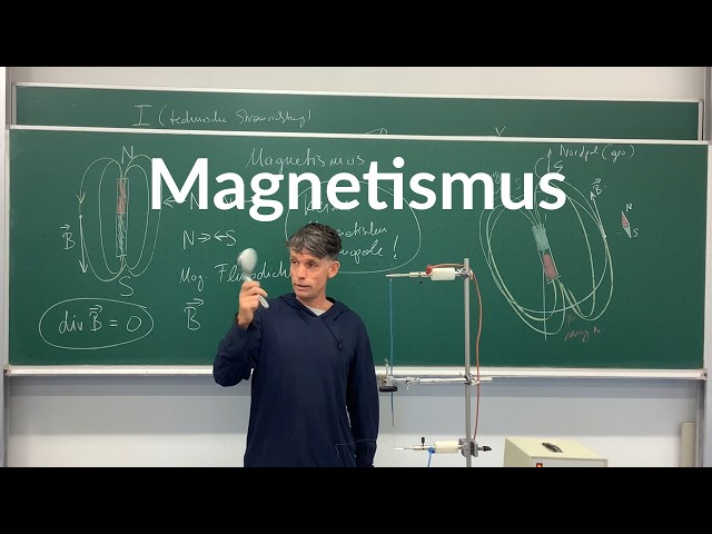


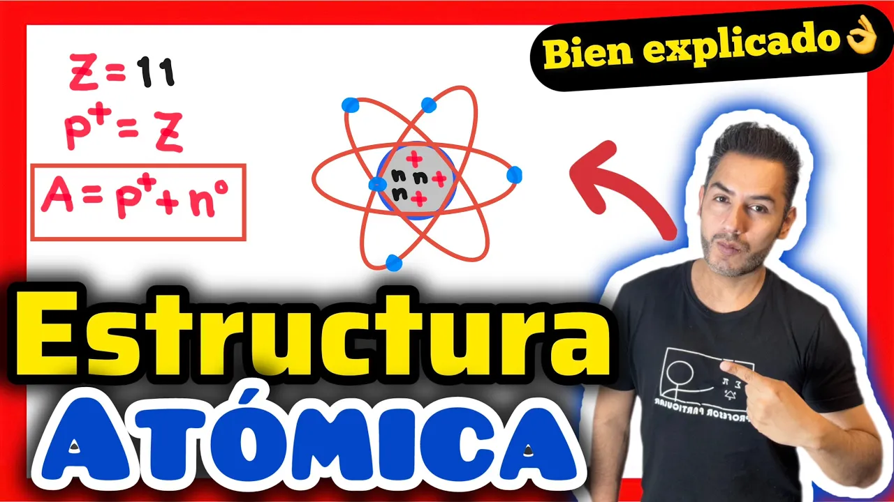


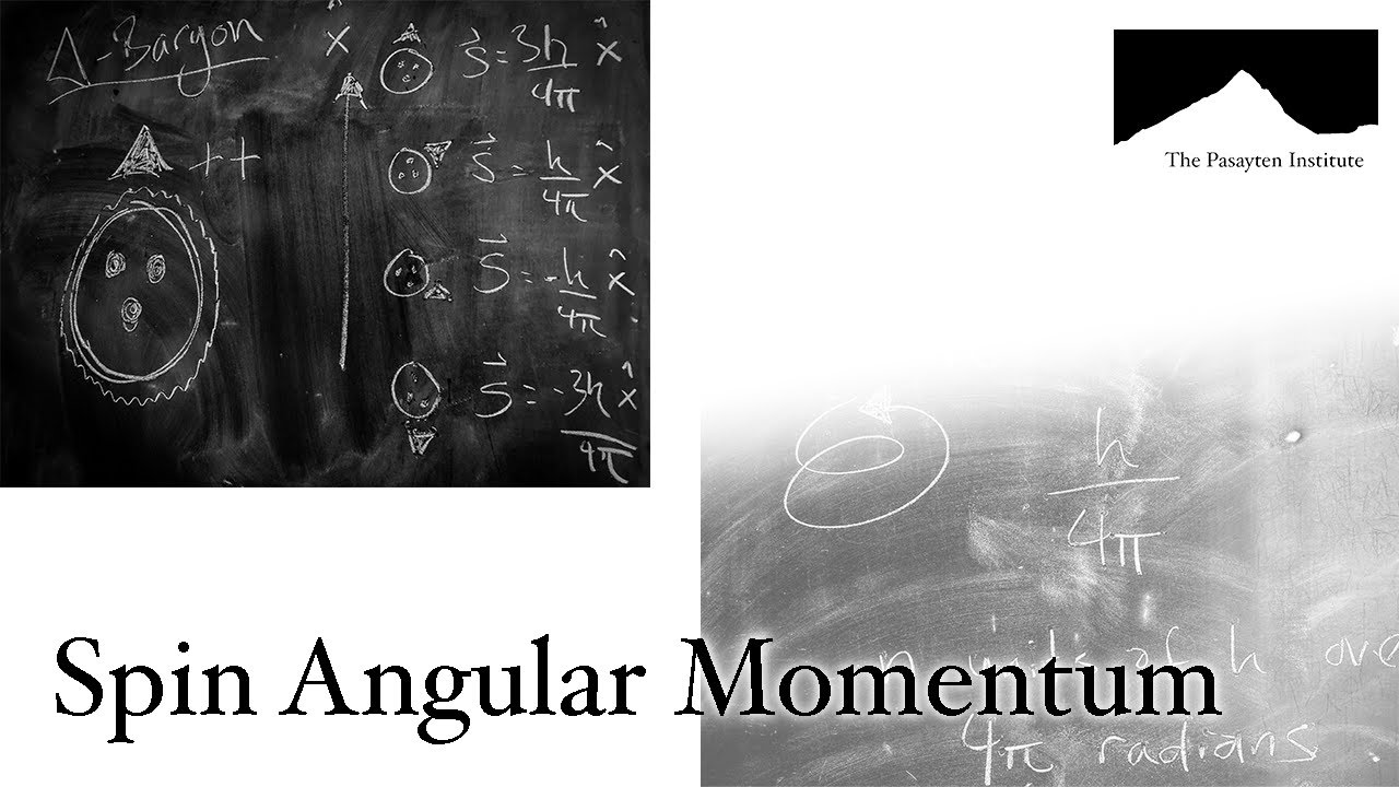







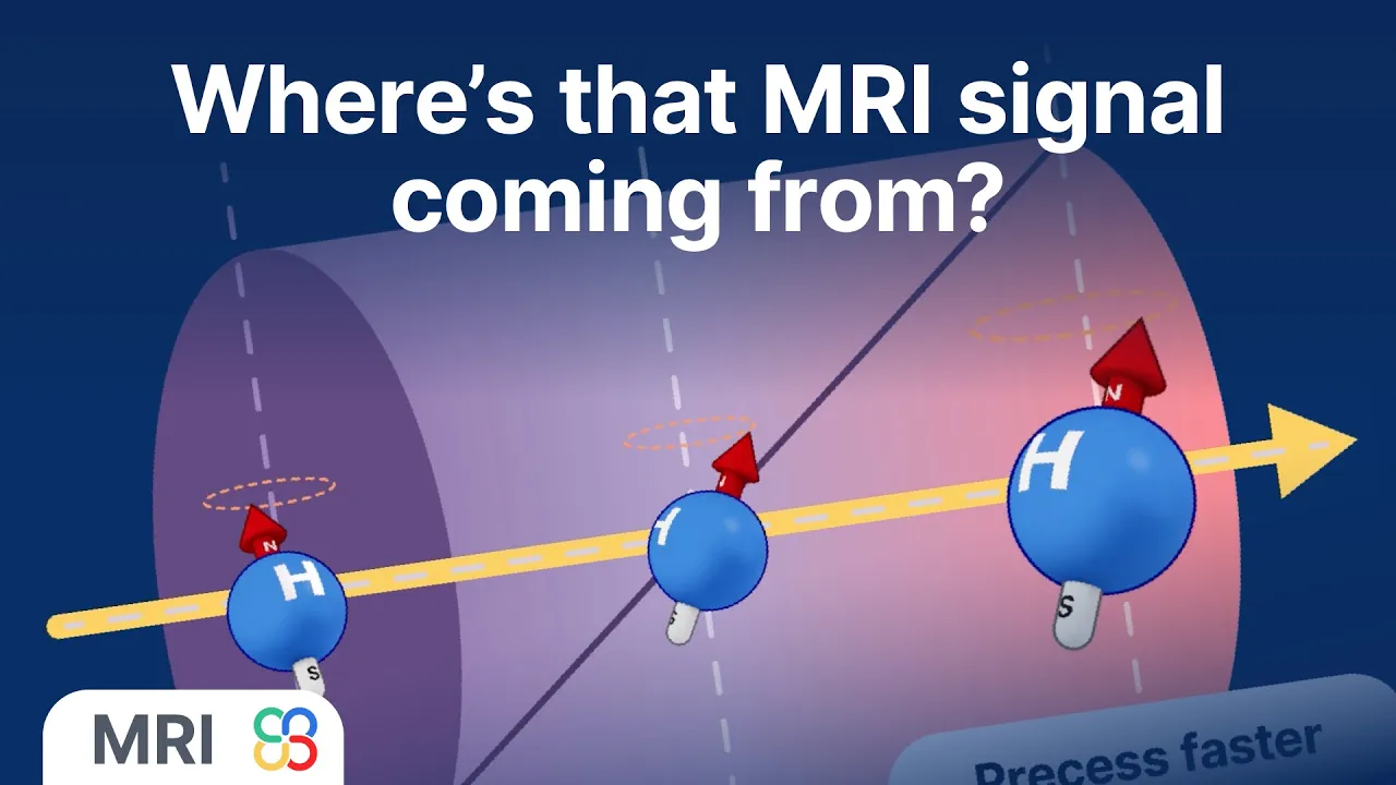






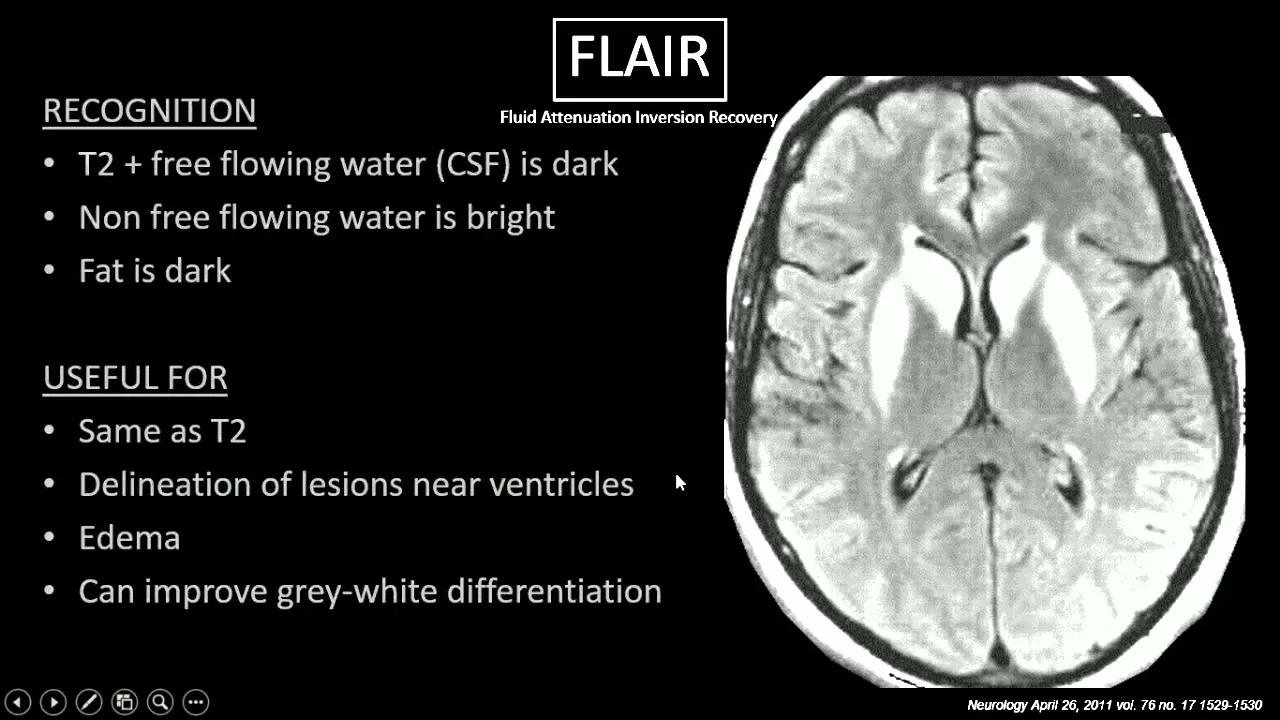

MRI imaging relies on the quantum mechanical property of atomic nuclei with odd proton or neutron counts possessing spin, which generates magnetic fields; when placed in a strong external magnetic field (B0), these spinning protons align and precess at the Larmor frequency (γ × B0), and by applying a radiofrequency pulse at this resonance frequency, the body magnetization is flipped into the transverse plane where it can be detected; the subsequent relaxation processes—T1 relaxation (returning magnetization to the longitudinal plane) and T2 relaxation (loss of phase coherence)—determine tissue contrast, with T1-weighted images emphasizing differences in longitudinal recovery times and T2-weighted images emphasizing differences in transverse signal decay times, controlled by adjusting repetition time (TR) and echo time (TE) parameters.
MRI Physics Made Easy: A Beginner's Guide to T1 and T2 Relaxation
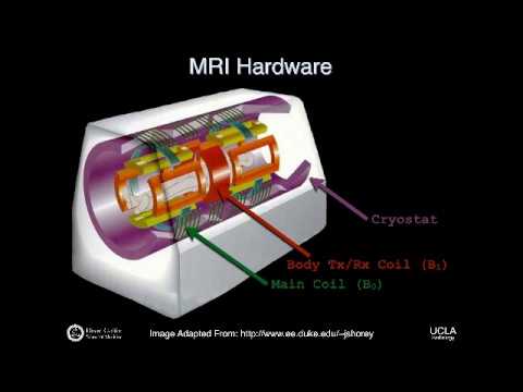
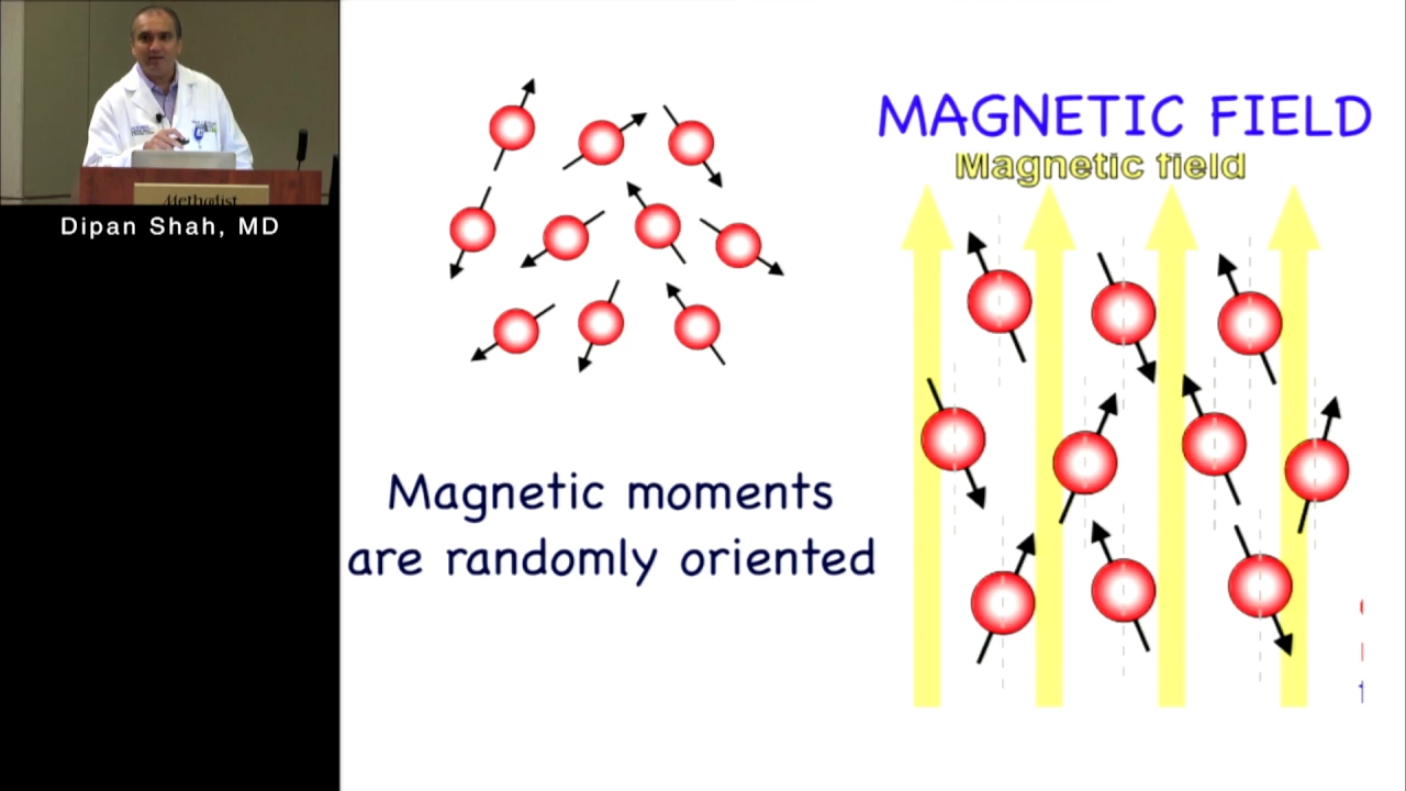
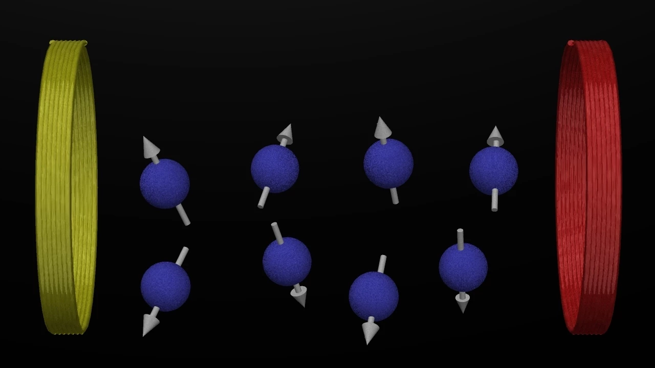
Added:hello my name is fand gazer I a neur radiologist working in Gent University Hospital in Belgium and welcome to this presentation on the basics of MRI physics not an easy topic often a daunting topic it's something a lot of Radiologists struggle with it's difficult to master so I try to explain this as easy as possible not just for you but also for myself and let me know at the end of this presentation I succeeded or not what I'm what I'm what am I going to talk about I'm going to start with the basic principles of MRI physics we're going to apply that to how we can use MRI to generate those MRI images what determines the Imaging characteristics and the image contrast we see in for instance T1 weighted images and T2 AED images I'm going to talk about some of the basic MRI sequences what things look like on them and how we generate them some more specific MRI sequences like 3D sequences for instance and some Advanced MRI techniques like fmri spectroscopy MRI profusion and diffusion tensor Imaging let's start with basic MRI physics now what is the absolute basis on which everything is built two principles first of all atoms with an odd number of proton or neutrons have Spin and spin is basically a quantum mechanical um o my apologies is a quantum mechanical uh concept but we can try to visualize it as uh an atom spinning around its own axis that's not what really happens but it helps to understand what's happening there and the second thing we have to know is that a moving electric charge produces a magnetic field and as law of Faraday and vice versa a moving magnetic field or an osculating magnetic field will induce an electric current in a conductor and that's basically what MRI is based on so on this image on the previous image we see a spinning atom normally there was but it's not happening this was supposed to spin but it's not doing that apparently and this illustration shows us uh moving electrons and these moving electrons generate an atom a magnetic field around them and here we see electrons um being induced by a magnetic field around the magnetic field and this is an illustration of the magnetic of an MRI bar uh which is basically a gigantic magnet with a North Pole and a South Pole so to summarize we start with the fact that atoms with an odd number of protons or neutrons have a spin spinning atoms have a magnetic field because well basically they're moving electrical charge so they have a magnetic field oruse a magnetic field and we can consider those spinning atoms like tiny magnets and luckily for us our body is filled with these tiny little magnets we can basically use any kind of atom with an odd number of protons or neutrons uh as the basis of our MRI signal but we have chosen protons protons just consist of one proton so we have an odd number of uh protons there um and our body is filled with them uh we have about four uh multiplied by 10 to the 27 I can't even visualize that number I don't even know I have to call it a billion billion billion or something like that uh probably more protons so that's a lot and these are tiny little magnets but they are completely randomly aligned in the body and that explains why why our body in a normal situation has no uh magnetic characteristics that's because all these tiny little magnets are randomly aligned and they kind of neutralize each other's magnetic effects now what is the basics of MRI basically we have five steps the first one is we place the page in a very strong external magnetic field and that external magnetic field is generated by the MRI machine secondly and I'm going to explain what these things mean later we sent a radio frequency pulse which is basically an ulating magnetic field uh we send that in the patient and it is transmitted by a device called a transmitter coil and this is basically sending energy under the form of an an ating magnetic field in the patient then we turn that radio frequency pulse off and as a consquence the patient will emit an electromagnetic signal more specifically a magnetic signal and that magnetic signal is once again captured by the coil I talked about that coil can transmit things it can also receive things in this case it receives the magnetic signal coming from the patient and it translates that into by the laws of electromagnet uh electromagnetic uh induction it translates that in an electrical signal and an electrical signal is analyzed by a computer and the end result is our uh image or MRI image so that's basically what involves or entails an MRI examination these are five steps let's go over these five steps one by one the first one being the patient being placed in an extern magnetic field so here we see the MRI machine again which is basically a very large magnet and which creates a very strong external magnetic field with a strength of in most uh clinical machines 1.5 Tesla or tree Tesla and this is the magnetic field denoted by b0 and it points uh in a specific Direction so it's a magnet so it has a North Pole and a South Pole and what happens if we put a body in there if you put a human being in there well that human being is composed of 10 uh 4 * 10 to the 27 number of protons so a lot of tiny little magnets completely randomly aligned and the net magnetization of the body in a normal situation is zero if we put that body in a very strong external magnetic field these magnets will align along the axis of that strong magnetic field and the result is that our body will become magnetized mind you it's still a very tiny magnetization the body is very weakly magnetized but it suffices we're going to use that magnetization to create the MRI image and that magnetization is small because well we have a lot of protons in the body but the protons can align themselves along the axis of the main magnetic field in two directions either parallel to the axis which is a low energy state or anti- parallel which is a high energy State there are a limited number of protons uh more that align themselves parallel than antiparallel so the result is we get a very weak net magnetization Vector given by MZ uh in uh the human body so we have generated some magnet ization there that's fine what's happening next well first thing is the protons will align themselves along the axis of the external magnetic field but they will also do something else they will start precessing and what is that that's not the same as spin so spin is basically the axis uh spinning around uh the uh proton spinning around its own axis it's an intrinsic property of nuclei giving the magnetic momentum of the nucleus proc Precision on the other hand is something else is the circular movement of that magnetic momentum when placed in an external magnetic field and we can compare it a bit with a spinning toll so that um tolls around its own um tolls around its own um and is almost falling but isn't so it's a bit similar so possession is not the same as spin so these protons perform that Precision movement with a very specific frequency and we call that the lore frequency and I'm going to avoid mathematical or physical formula as much as possible but this one is I believe basic for most radiologist the lore frequency is given by the gyromagnetic ratio given by the simple symbol gamma over here multiplied by the external magnetic field and for protons the gyromagnetic ratio is 24 85 mahz per Tesla so that means that if we place a human body inside a main an external magnetic field or an MRI machine uh of 1.5 Tesla the frequency or the L more frequency of the protons and the body will be uh 6387 so so this is for a 1.5 MRI if you want to know what it's going to be in a tree Tesla machine just multiply uh 43 with three and that will be about like uh 29 if I'm not mistaken if I'm mistaken let me know in the comment section so final result protons have aligned themselves along the axis of the main magnetic field and they are also processing along along the main magnetic axis with a specific frequency called the lore frequency okay onto the next step basically we've magnetized the body now but we can't really measure that magnetization why it's very very tiny and it's located along a very strong external magnetic field if we were going to use some kind of uh detector there's no way we can distinguish that a very tiny amount of magnetization coming from the human body amidst that huge or massive external magnetic field but we can do it if you were to be able to flip that body magnetization in another plane so that it's no longer along the plane of the main magnetic field so the direction of the main magnetic field as along the Z axis if we were able to flip the magnetization and the body give by this orange arrow over here into the X Y AIS we would be able to measure it so that is what we want to do we want to flip the body magnetization in another plane in this case a plane completely perpendicular to the external magnetic field how do we achieve that well we're going to use a radio frequency post for that sent in by a so-called transmitter coil this probably require Rees a little explanation so what is an MRI machine composed of this is extremely basic but we have main magnetic coils gradient coils and radio frequency coils and coils are basically able to generate magnetic fields what do they do the main magnetic coil is responsible for the generation of the main magnetic field which in clinical MRI machines is generally 1.5 or three Tesla then we have the magnetic gradients which are created or induced by the gradient coils what is that it's also a magnetic field but it's not uniform it doesn't have the same strength the strength varies L varies linearly with the location of the gradient and it is superimposed on the main magnetic field so what are we doing we are basically adding some magnetization and that can be along the axis of the m of the main magnetic field but it can also be in another plane and we going to change the magnetic magnetization the main magnetization a tiny little bit and that change will vary depending on the exact location so we have a very weak additional magnetization over here and a stronger additional magnetization over here so we can perform that in any plane so generally we have three sets of coils one in the Z plane so along the the main magnetic field and also in the X and Y planes perpendicular to the main magnetic field and these are important for the spatial encoding of the MRI signal I'm not going to talk about that because I consider it consider it already a bit more advanced but just remember it so this is important for the spatial encoding so basically the body emits a magnetic signal but if we detect that magnetic signal we want to know where it is coming from that's why we need spatial encoding but I'm not going to talk about that as such and we also use the gradients for the creation of specific MRI sequences but more about that later and then what I wanted to uh show you is the radio frequency coil what is the radio frequency coil it's a coil that transmits energy in the body under the form of an oscillating magnetic field that's important oscillating so it's like moving while the main magnetic field and the gradiance are basically uh static magnetic fields so they don't change um so okay we transmit energy but we can also detect magnetic signal emitted by the body so those R radio frequency coils can do two things they can transmit a magnetic field or an oscillating magnetic field or they can detect an MRI signal a magnetic signal and they will then translate that into an electrical signal um in this part of the examination also notice that I have drawn two coils one is built into the MRI machine another one is basically a coil that can be put over the head and we have other types of coils that can be used for body Imaging or muscular skeletal Imaging so we can use coils that are located in the machine or we can use coils that are put over specific body parts like a head coil in neuro radiology and we have uh coils that are just transmitting coils that are just receiving and coils that can do both things they can transmit a radio frequency pulse so this oscillating magnetic field is also called a radio frequency pulse they can emit it or they can detect it so okay in this step we use a radio frequency coil we put it on and it will um emit an oscillating magnetic field a so-called radio frequency pulse and it will do that in a direction perpendicular to the main magnetic field and what happens if we do that so this is the body inside the main magnetic field we see a coil we see a head coil over here and we're going to assume that it can both receive and detect radio frequency pulses in this presentation and notice that uh at this moment the protons are aligned along the main magnetic field now what happens if we emit a radio frequency pulse perpendicular to the main magnetic field as shown over here this will change the direction of the proton alignment how does it do that or how does it work well basically we send in a lot of energy so in a normal situation as shown over here the body magnetization given by MZ will be located along the Z plane along the axis of the external magnetic field then we send in a radio frequency po so basically we send in a lot of energy and that energy will push the body magnetization out of the longitudinal plane and into the transverse plane into the XY plane I told you that the radio frequency pulse is an oscillating magnetic field um which means that it also has a specific frequency um the angle under which the body magnetization is pushed uh out of the longitudinal plane is called the flip angle and basically that can be any flip angle but for spin Echo sequences generally it's a flip angle of 90° and if we use a radio frequency pulse that flips the body magnetization over uh 90° we call it a 90° radio frequency pulse so this radio frequency pulse is given with a specific frequency namely the lore frequency oh wait a minute the lore frequency is the frequency which which the protons are pressing in the body is this a coincidence no why do we use the lore frequency because we want to use a physical concept called resonance and what is resonance well it's something every parent knows so you have a lovely boy old girl who likes to uh swing on a how do you call that in English a swing set I don't know but you see the picture so you know what I mean so to do this efficiently we just need to give a push each time our darling boy or girl reaches us if we were to push too soon for instance if you were to push here or here that won't do anything if you were to push here but not here well nothing is going to happen we are not transmitting energy from ourselves to our darling child and we won't push him higher resonance means that I give energy to an object with a frequency that is exactly the same as the frequency which which that object or in this case a person is performing a specific movement and if I do that I will transmit energy and I will push my child higher and higher with with each swing this is resonance and we use that to push the protons which are precessing with a lot more frequency out of the longitudinal plane by applying an oscillating magnetic field a radio frequency pulse with the exact same frequency which protons are resonating we use uh resonance and we have a very efficient way of giving energy to these pressing protons for pushing them into the transverse plane so I hope this is clear because it's a very important concept so just compare the radio frequency pulse with a parent and the recessing protons in the body with a child on a swing and you know why the radio frequency PSE needs to be given with the L more frequency so in immediately after the 90° radio frequency POS something else happens so we then see that protons are spinning in Phase what does that means what what does that mean it means that their magnetic moments are synchronized and they are process processing so they started with a procession movement along the Z AIS we flip them in the XY AIS and the transverse plane and they keep doing that procession movement with a lot more frequency and initially they all press in Phase they all uh preset at exactly the same rate and all nice together and now we have um a movement of magnetic structures in a plane perpendicular to the external magnetic field and this is a magnetic signal that we can detect it's a moving magnetic signal so it will induce an electric current in the radio frequency coil which can also be used as a detector as said and that electrical signal is the basis of our MRI signal so we're done right we managed to do it we managed to flip the body magnetization in the transverse plane and now we have created a moving magnetic field uh that can be detected and translated an electrical signal and images we're done well no we're not oh this is another illustration uh showing you the concepts of moving in phas and out of phas in case that wasn't clear so in Phase means all moving uh in Pace out of phase is what you will see at a marathon it's not um the intention of marathon runners to run together in Phase no you want to be the first but some people have better stamina or better trained just have better phys physique and you will always have people who run faster who will run slower so you don't see people running in PH at a marathon for instance so okay to summarize we give a 90° radio frequency pulse this is adding energy or giving energy to the protons and the body pushing them out of the longitudinal plane and this is what happens so the longitudinal magnetization of the body flips in the X uh XY plane and protons are now processing in Phase with the L more frequency and then we turn off the radio frequency pulse what happens then uh now those protons can relax again so we were giving them energy and energy to push them out of uh the longitudinal plane out of the Z axis uh now we remove the energy and it's like if we stop pushing our child on the swing eventually if he doesn't try to keep going higher himself and the protons don't do that they're lazy uh they will start to relax again and they will return to their initial situation and what is that well two things will happen the uh the body magnetization will return into the zip plane and this is determined by a phenomenon called T1 relaxation and what's happening also the protons will lose their phase coherence so initially they were uh pressing in Phase they will lose that precession in Phase so these are two independent well not completely but let's consider them completely independent for the sake of this presentation to keep it easy these are two independent mechanisms that happen simultaneously which complicates a bit to understand it but we're going to address them one by one they happen simultaneously and T2 relaxation so the def phasing of the protons uh is something that happens faster than the T1 relaxation so let's talk about T1 and T2 relaxation uh separately so T1 relaxation once again let's summarize I'm going to repeat myself a lot but I think that's important to really understand it when the radio frequency pulse has turned off protons will return uh to the axis of the main magnetic field um so let's look at what happens basically so initially all magnetization after the 90° radio frequency pulse is flipped in the transverse plane if we now um turn off the radio frequency pulse we will see so the magnetization along the Z AIS in this picture where is my mouse yes in this picture is zero if you uh put out radio frequency pulse gradually the magnetization will return not at one once but if you just wait long enough we'll see it gradually increase until it reaches its normal value again the value it had before the radio frequency PSE and if we look at these um diagrams or how should I call them uh and if we try to connect them so this is what happens over time we see that the longitudinal magnetization gradually increases and it follows an exponential curve we call that the T1 recovery curve until it reaches its normal equilibrium value this is the T1 relaxation curve and the T1 relaxation time which is an important Concept in MRI physics is defined as the time it takes for longitudinal magnetization to reach 63% of its original magnetization for those of you who like mathematics here is the formula the only thing that's important that you know that is an exponential curve as given by the formula and what causes T1 relaxation well basically protons want to be in a lower energy State they were pushed into a higher energy State they hate it they don't want it so what do they do as soon as the radio frequency pulse is turned off they release all the energy they received from the radio frequency pulse into the surrounding environment which is also called the lce under the form of heat and that is why we slightly uh become warmer discretely but it happens during an MRI examination and this also explains why synonyms for T1 relaxation or longitudinal relaxation because we get relaxation towards the longitudinal plane thermal relaxation because we release heat or spin elat relaxation because we give energy to our um to the surrounding environment or the lce and also important the T1 relaxation time will defer between tissues and that is something we can use to create MRI images uh because it will depend on the interactions between protons and the surrounding environment and that surrounding physical or chemical environment will be different in different tissues so let's once again uh let's now move on to T2 relaxation so in immediately after 90° radio frequency poles protons were pressing in phase in the transverse plane with a lot more frequency um but that doesn't last very long um nevertheless so this is if we were to look at the XY plane from above we see the protons moving in Phase but only for a very short amount of time now these are moving protons so uh they are charged particles uh so this induces a magnetic field and each time those moving protons pass here for instance where a detector is located or a coil is located this will create a signal that can be picked up so it is basically this sweep of magnetization in the transverse plane that induces a current in the receiver coil but oh this is another slide protons however start recessing out of phas very quickly so if we now look at another way to visualize it so this is nice I'm going to show it again just because it took me an awful lot of time to create this PowerPoint slide so let's admire it a bit we see these P turns moving out of face is beautiful okay and let's now look at this other depiction of the2 relaxation time just to make it really really clear so this is basically um the net vector magnetization and all protons are spinning in Phase immediately after the 90° radio frequency pulse but then we put it off and they start spinning out of phase some of them start pressing a little bit faster some a little bit slower and if we just wait long enough and basically it doesn't really have to be that long because the2 relaxation happens pretty quickly NO phase coherence is left and it also means that the magnetic signal and the transverse plane will have become very very weak and for some reason I found it necessary to illustrate the same concept with yet another image but this time take a careful look at this here this is the magnetization in the XY plane this is wrong and we see that of course as protons start uh def phasing this will become weaker and weaker and weaker and this is basically simp SAR to T1 relaxation in which the longitudinal relaxation increases and T2 relaxation the transverse magnetization becomes weaker and decreases so and because this is the basis of the measurable signal that will also become very weak so this depiction of the magnetic signal once again this is wrong this should be XY so here we have once again seen from above protons precessing uh and phase initially but normally this vector or this Arrow which basically denotes the strength of the magnetic field and the XY plane it should become smaller with every spin due to Def phasing so basically it should form this shape so not a circle but a spiral gradually becoming weaker and weaker and weaker and this here shows us the magnetic signal that is detected by a receiver coil and with each sweep so here it reaches let's say this point where it is located then it goes back to the other side so the signal becomes weaker inverts then it comes back again but we see that it has become smaller the peak is smaller due to phase uh loss of phase coherence and this happens with every sweep and then we get a curve looking like this and this also follows this is also an exponential curve but a decreasing exponential curve this is the t2 relaxation curve so let's look at this curve over here how do we Define D2 relaxation it is the time it takes for transverse magnetization to be reduced to 37% of its initial maximal value so it's a decreasing exponential curve um and2 relaxation is also known as spin spin relaxation or transverse relaxation transverse relaxation because it is relaxation happening in the transverse plane spin spin refers to one of the possible mechanisms responsible for this uh decrease in transverse magnetization um now T2 relaxation and T1 relaxation happens simultaneously and any process resulting in T1 relaxation will also result in T2 relaxation so as T1 relaxation increases T2 relaxation will also increase that is also T2 relaxation independent of T1 relaxation and that has two components or two reasons one is intrinsic the so-called spin spin aspect and one is due to extrinsic factors so let's explain those a little bit further and I hope you are still with me what is causing protons originally processing in Phase to lose that phase coherence we have true to Decay which is due to Intrinsic factors and we have extrinsic factors now the intrinsic factors is a result of interactions between spins socalled spin spin interactions and we we also call that true T2 relaxation which is irreversible it occurs because these spins have tiny magnetic fields and they will influence the tiny magnetic fields of other neighboring spins and basically simply by being in each other's vicinity spins will cause each other to lose phase coherence in the transverse plane so this is true the2 D2 dek spin spin interactions and then we have extrinsic factors which have nothing to do with uh the fact that spins are located next to each other and will influence each other no this is caused by factors located at the level of the the uh magnet at the level of the MRI machine or at the level of the body and what exactly are those well first of all we also we always assume that the external magnetic field generated by the MRI is like this perfect uniform magnetic field but that's simply not true uh a perfect magnetic field uh exists somewhere in the universe or in physicist's mind but in reality a main magnetic field is not perfect there are very small variations in the main magnetic field and these very tiny uh imperfections uh also called in homogene inhomogeneities inhomogeneities uh will of course cause protons to have slight variations in their in their lore frequency and this will induce uh def phasing of the spins there are also Vari ations in the magnetic properties of tissues or materials which create local disturbances in the magnetic field for instance if you have a hemorrhage in the brain even if it's small you will find a lot a lot of iron there because blood hemoglobin contains iron and iron is paramagnetic it's it's very magnetic so that will change the magnetic properties of the tissue and have an influence on the magnetic field it will cause Pro protons to defas more rapidly so the combination of these factors so imperfections and the Magnetic machine and small variations in the magnetic properties of tissues um will also contribute to T2 relaxation so let's now also introduce the concept of T2 star relaxation what is T2 star relaxation it's basically a combination of what I just called the true T2 Decay caused by spin spin interactions combined or with the t2 Prime effects so basically the t2 relaxation due to extrinsic factors like imperfections in the main magnetic field or susceptibility um differences between the body tissues and this I already explained this is true T2 relaxation and because T2 star relaxation as a combination of true T2 Decay and these extrinsic factors T2 star relaxation will always be faster than true T2 relaxation and T2 star relaxation is basically what we see after we have given that first 90° radio frequency pose It's a combination we see loss of phase coherence due to true2 Decay and due to extrinsic factors the T2 Prime effects now T1 and T2 relaxation as said happen simultaneously but T1 relaxation will generally always be faster than T2 relaxation theoretically they can be the same and practice T2 relaxation will always be faster and T2 relaxation will never be uh faster than T1 relaxation so T2 relaxation happens faster than T1 relaxation okay I think this is clear or I I hope this is clear so we can move on to the next step what happens now the patient will emit an electromagnetic signal we have flipped um the magnetization of the body and the transverse plane so now it can be detected we've already seen this figure this is looking from above uh on the X uh y plane and we see proton spinning in Phase from for a very short amount of time in the XY plane and as already said the sweep of magnetization can be detected by a receiver coil and will induce an electric current in that coil but as also said due to phase coherence the signal and the XY plane becomes weaker with each sweep so we have a receiver coil here it picks up the signal uh best when the signal is over here and if the the signal moves away again it will become weaker and it will also become weaker with each sweep so in the end we get this as a signal and this signal is called the free induction Decay signal free because we have removed the 90° radio frequency pulse so the protons are free again to lose their phase coherence induction because it's a moving magnetic field and it will induce an electrical current in the receiver coil and Decay because it comes it becomes weaker with each sweep and it's basically so uh it becomes so weak that we can't really use it to generate or create the MRI image no we want a stronger signal for that how do we do that so I think everything on this slide I've already told you here we see a receiver coil which is located perpendicular to the moving magnetic field notice that it is a spiral as well the result will be the free induction Decay uh curve and this is due to T2 star relaxation um so what do we do to create another signal also in the transfers plane that's more suitable that's stronger uh that lasts a bit longer and that can be used for to create MRI images so this we have seen this is Phase coherence which occurs after we return of the 90° radio frequency pulse um how do we get a new signal that's stronger we apply another radio frequency pulse but this time not a 90° radio frequency pulse no we apply a 180° radio frequency po so what do we do we basically uh push the transverse magnetization into the transverse magnetization but towards the other side we make it flip 180° why do we do that well as said the signal the free induction Decay signal is to weak we want a new signal a better signal we can compare this with our marathon runners I already told you about the marathon runners they are running out of phase this guy is running really fast this guy is running a lot slower this guy will the more time pass be farther ahead of this guy so the phase uh this coherence will only increase with time but now imagine that somebody were to say stop turn around everybody still keeps running at the same speed this guy is still running quite slowly this guy is running very fast now if they were to turn around they will eventually meet each other again because this guy is slowly going back and this guy very quickly so he will catch up with that guy and that's what the spins do after we applied a 180° radio frequency PS they catch up with each other they were defacing they were losing their phase coherence but after the application of a 180° radio frequency po they will regain their phases again they will become coherent again so this is just a lot of text but I think I've set everything that's on here so we flip the spins in the transfers plane transverse plane by 180° and Spins that were pressing faster or now behind spins that were lagging or no ahead but they are able to catch up with one another and eventually we will get reasing and a new strong signal so this is the same concept but Illustrated in a different way so here on top we see what happens after Return of the 90° radio frequency pulse so the magnetization and the XY plane becomes weaker but then we turn on the 180° radio frequency pulse and what do we see we start with a weak signal but the protons start catching up with each other and they gradually come together again and as a result the magnetization and the transverse plane will increase until we get a moment of perfect phase coherence again and that signal that we create this way is called the spin Echo and this spin Echo is the basis of our MRI image so the same concept once again Illustrated in a different way because I think these kind of visuals really help you understand this quite difficult topic this is the 90° radio frequency pulse which abuses the free induction Decay signal too weak we can't use it so we apply a 180° radio frequency pulse and we generate the spin echo which lasts longer and can be detected uh by the radio frequency coil and is the basis of the MRI signal I'm going to introduce A New Concept here which will return later on the echo time and the echo time is the time between the start of the application of the 90° radio frequency pulse and the time the spin Echo was created the 180° radio frequency pulse is given at the echo time divided by two so this here is Echo time divided by two and this half is also Echo time divided by 2 so the spin Echo signal is used for the creation of the MRI image this is a conventional spin Echo uh sequence and well one spin Echo is not enough we're going to repeat it a number of times to create a lot of spin Echoes here we get the this um helps us introduce A New Concept the repetition time which is the time between the start of successive pulse sequences we call this a pulse sequence the N the 90° 180° radio frequency pulse there are a lot of variations on it but it's called a pulse sequence and repetition time is the time between the both sequences and this is the spin Echo signal and as already said the echo time is the time between the 90° radio frequency pulse and the center of the spin Echo okay this brings us to the last step the detection of the signal by a receiver coil and using it for the generation of an image um I am not going to talk about that uh why exactly because well for image generation I already touched upon that we need to to be able to detect what part of the body the signal is coming from spatial location I could do a presentation on spatial location of the MRI signal but it will definitely take me uh a lot of time and it makes the topic even more complicated and it already is I'm going to focus on the basics now and what are the basics let's repeat we start with spit protons in the body spinning randomly in all directions the nrom magnetization of the body is zero we place these spinning protons in a strong external magnetic field and they will align themselves along the axis of the main magnetic field and they will also start precessing along the external magnetic field with a frequency given by the L more frequency then we want to flip that body magnetization and the transverse plane and we do that by applying a radio frequency pose basically we give a lot of energy uh to these spinning protons and this pushes them in the XY plane now normally they shouldn't this is the wrong animation they should stay there so they're not coming back and then we turn off the radio frequency pose and because of T1 relaxation and T2 relaxation the protons will become autom magnetic field and the XY plane will become weaker T1 relaxation will make the protons return to the longitudinal plane and T2 relaxation will make them lose their phase coherence in the transverse plane now by applying a 180° radio frequency pulse we create another signal the spin Echo signal and this signal is finally used for the creation of our MRI image so this is the absolute basis of how we create MRI images so I'm not going to talk about spatial location I want to talk about tissue contrast because when I said we use this to create an MRI image well there are a lot of different types of MRI images we have T1 weighted sequences and T2 weighted sequences which are the work courses of MRI images and basically most of our sequences are derived from that uh to put it easily or to put it simply but what makes these sequences different why do these images look different the basic principles are the same what makes them different let's talk about that now so here we have T1 weighted images and T2 weighted images what determines the difference or how do we get these different images using the same physical principles but first we need to Define what tissue contrast is exactly what do we mean with tissue contrast if you want to examine uh a certain body part or if you want to detect pathology it's important that we can distinguish the various components of that organ for instance the brain contains white matter gray matter there is water in the ventricles it is surrounded by bone by a scalp we want to be able to distinguish all these parts so we need to see a difference in the signal intensity of these different tissues to be able to recognize them on an image that is what tissue contrast is um so tissues have that's uh what determines the tissue contrast and MRI basically three things tisues have differences in their number of protons and their T1 relaxation times and their T2 relaxation times and we can use these different es between tissues to generate different types of images each showing different contrast between the various part uh parts of an organ this is an example of T1 and T2 relaxation times for various tissues in the brain or the central nervous system so the number of protons is quite similar not 100% but I just assume it's quite similar we're going to focus on differences in T1 and T2 relaxation time look at T1 relaxation time of water and fat for instance there's a huge difference there so if you were to create an MR image emphasizing the difference in the T1 relaxation time we should be able to see a lot of difference between water and fat how how do we do that we're going to uh my apologies we're going to work further upon this example here we have the T1 relaxation time of water water and fat so if we were to look at the T1 relaxation curve for water that would look a bit like that so water has a long D1 relaxation time so it reaches 31% um 63% router of its equilibrium after a pretty long time and fat has a very short T1 relaxation time so the curve for fat would look a bit like this so we see that there's a huge difference at this time Point here here if you wait a very long time so um T1 relaxation happens after we flipped our protons and the transverse plane we turn off the radio frequency pulse they return so if we wait a long time the magnetization will have completely returned in the Z plane so if we were done to do another P well we would basically flip the same amount of magnetization in transverse plane and there will be very little differences between water and fat this is the point we have to use where there's a huge difference and the T1 relaxation of water and fat for the creation of our MRI image so how do we do that I already explained it a little bit but let's illustrate it with figures to make it more clear so this is 90° radio frequency pulse it flips the body magnetization and the transverse plane and of course the magnetization is a bit different um or tissues have different magnetic properties if we wait a short amount of time let's say 200 Mill 200 milliseconds we will see that a lot of fat magnetization will have already returned completely in the longitudinal plane it will have almost completely recovered however for water in the same time period only a little bit of magnetization has recovered so if we were to perform another po sequence now we would actually flip a lot of fat magnetization in the transverse plane but not a lot of water magnetization because this is the signal used for the creation of the MRI image fat will have a high signal because there's a lot of fat magnetization in the transverse plane water however will have a low signal because there is a very little water magnetization in the transverse plane the result is we will see a high contrast between fat and water so how do we do that how do we exploit the T1 relaxation differences between water and fat for the creation of high contrast between these two structures we have to perform a new sequence after a short amount of time why if we wait a long time both water and fat will have completely recovered the longitudinal magnetization we need to do it when f has compl has recovered a lot and water a little so where the difference is the biggest so we basically need a short repetition time if we choose a long repetition time these structures will look exactly the same by choosing a short repetition time the these structures will look very differently and fat will have a high signal water low signal this is a figure I did not make myself it's one I took from the internet but I found it very interesting this is basically what happens if we uh give uh or start a new pulse sequence after a certain amount of time so this is the recovery of the longitudinal magnetization for fat gray matter and water so if we were to start a new pulse sequence after th seconds we will see that most fat has recovered its longitudinal magnetization and if then flipped in the transverse plane will have a high signal it will be lower for gray water uh for gray uh matter and will be very low for water so let's maybe look at the video oh I can't okay let's look at that video again so this is the recovery of the longitudal magnetization then we give uh start a new pulse sequence and we get a lot of transverse magnetization for fat very little for water because water had recovered yet in the longitudinal plane and this slide is basically for my students for for study purposes because they can't remember everything I'm telling them so every now and then I spell it out on a slide but there's nothing new here okay let's now talk about using differences in T2 relaxation I'm going to illustrate differences in T2 relaxation between water and white matter water has a very high T2 relaxation time of 2,000 Mill uh milliseconds so it takes lot of time for the protons water molecules free water molecules uh to lose their phase coherence and for white matter that happens very rapidly after 80 milliseconds so if we were to draw the t2 relaxation curve for these two structures that's what it would look like for water and this is what it would look like for white matter uh we see that there's a lot of contrast between the two or a big difference and T2 relaxation times at this time Point um how can we use this information to create an image with a lot of contrast between water and white matter we do that by choosing a long Echo time and why is that imagine if you were to choose a short Echo time then basically both structures will have the same degree of magnetization in the main magnetic um and the transverse plane with very little difference between them if we wait a longer time for the generation of the echo time we will see a lot of difference um why this Echo time or um what's the relationship between the t2 relaxation and the echo time uh well remember that the signal is basically that the signal that is used for the generation of the Mr is the spin Echo and the spin Echo occurs after the echo time which is the time between the application of the first 90° radio frequency pose and the actual spin Echo so T2 weighted images are images in which the tissue contrast is mainly determined by differences in the t2 relaxation values of tissues and we need if we want to uh create create images that show the difference and T2 relaxation values of tissues we need to perform or sequence with a long Echo time uh because then the degree of Def phasing of water and white matter or the difference between the degree of the phasing will be greater than if we do a short Echo time so let's summarize we have talked about T1 and T2 relaxation and repetition time and Echo time um repetition time as the amount of time between successive pulse sequences and if we choose a short repetition time this will emphasize T1 differences between tissues the echo time is a time from the center of radio frequency pulse to the center of the echo or from the beginning I believe of the radio frequency I'm not sure uh maybe Center um anyways if we choose a long Echo time this will emphasize the2 differences and we basically can control that by choosing when we're going to apply the 180° radio frequency pulse uh because um that's basically the echo time divided by two this should be t e / 2 not T2 / 2 so I hope this is clear if we perform sequences and we use a long repe ition no a short repetition time we will accentuate the T1 relaxation differences between tissues so T1 weed images or images made with a short repetition time and what we see then is basically a vis visualization of the different T1 relaxation types of tissues and a T2 weighted image we choose a long Echo time uh and then we get images in which the image contrast is mainly determined by differences in the t2 relaxation times of course you also have T1 and T2 effect in uh both kinds you never have a pure T1 image the image contrast is determined by both T1 and T2 effects but if you want a T1 weed image we're going to minimize the impact of differences in T2 relaxation times and how do we do that by choosing a uh short Echo time so an1 weighted images we have a short repetition time and a short Echo time and two weighted images we use the long Echo time to make use of the difference and T2 relaxation values between structures but we also use a long repetition time to minimize the impact of uh differences in T1 relaxation times between tissues so this is the same Illustrated in yet another way T1 weighted images have a short Echo time uh a short repetition time and a short Echo time the2 weed images are made by using a long Echo time and also a long repetition time then there's a thing called proton density images what are those well if we use a long repetition time uh the longitudinal magnetization due to T1 relaxation will have almost completely recovered so the tissue contrast will not be determined by differences in T1 relaxation values if we use a short Echo time almost all magnetization of tissues any tissue will still be in the transverse plane and still be strong in a transverse plane so the difference in uh contrast between tissues will not be determined by the t2 relaxation values what then determines uh the image contrast in this image well basically the number of protons and that's why we call them proton density images and finally what if we were to do images with a short repetition time when there's barely any longitudinal magnetization and a long Echo time when basically um all magnetization in the transverse plane has disappeared or has become very small then we get very crappy images with very poor image contrast so and another slide to illustrate once again what I just said I'm not going to read it out loud this is more for my students so they can use that for their study purposes okay now we are ready for the next part recognizing MRI sequences I will upload this part in a separate video because this presentation is otherwise becoming very very long
Up Next
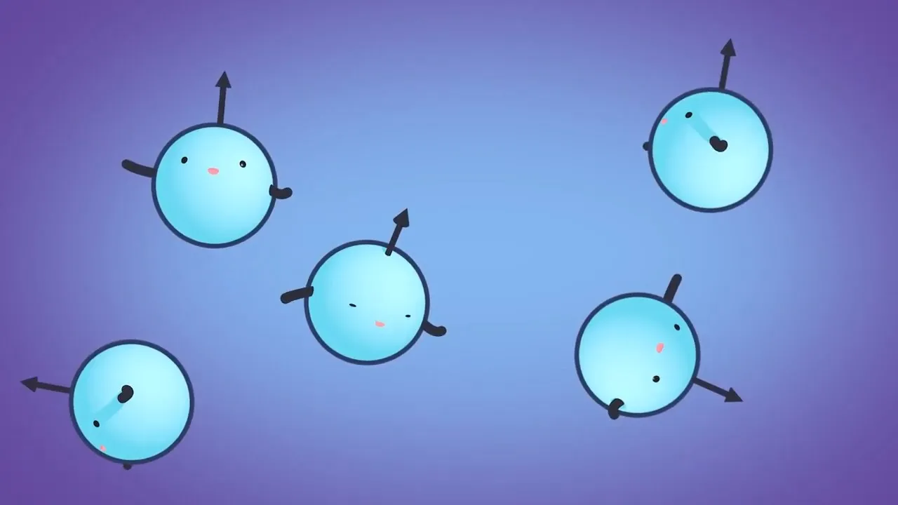
MRI Physics Explained: Magnetic Resonance and Spin Echo Sequences
@JohnsHopkinsMedicine
445.8K views•2022-07-05

21cm Hyperfine Transition in Neutral Hydrogen: Radio Astronomy Basics
@AaronRobertParsons
12.4K views•2011-10-13

NMR Spin Physics I: Zeeman Effect, Resonance Condition & Larmor Frequency
@nptel-indianinstituteofsci8064
2.3K views•2024-01-17

Entropy and the Second Law of Thermodynamics Explained
@veritasium
27.5M views•2023-07-01
Related Study Plans & Knowledge Roadmaps
Structured learning paths in Physics