




















The neural crest is a transient multipotent and migratory cell population that arises at the border between neural and non-neural ectoderm during late gastrulation and early neurulation, forming through a multi-step process involving BMP signaling gradients and contact-mediated tissue interactions; these cells undergo epithelial-mesenchymal transition and migrate to specific locations where they differentiate into diverse derivatives including craniofacial structures, heart tissues, peripheral nervous system components, melanocytes, and the enteric nervous system, making them the 'fourth germ layer' due to their extensive contributions across multiple organ systems.
Neural Crest Development | Embryology Tutorial Part 3
Added:Understanding of gastrulation and the establishment of the three primary germ layers (ectoderm, mesoderm, and endoderm).
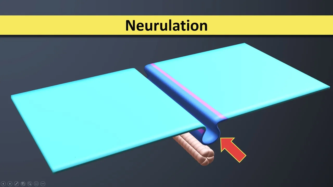
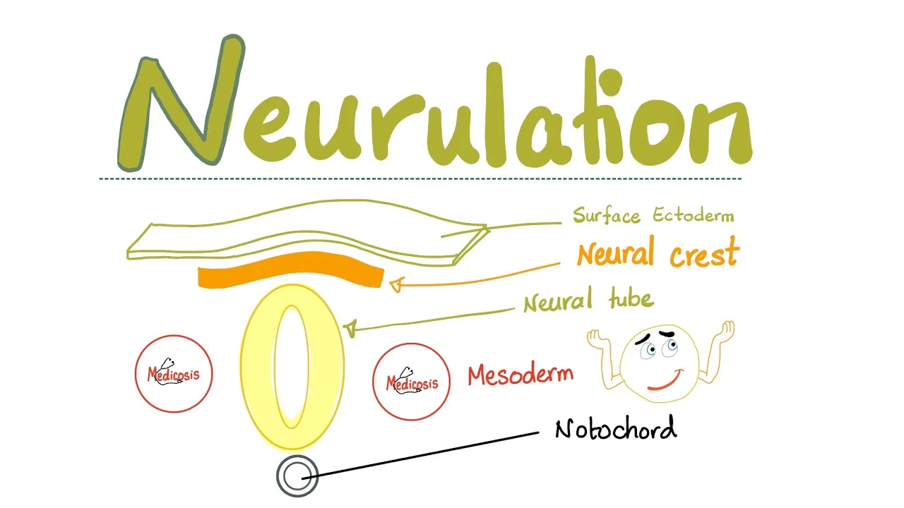
The process of primary neurulation, including the formation of the neural plate, neural folds, and neural tube.
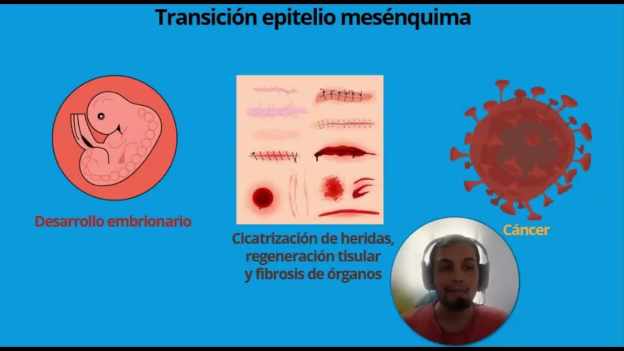
Basic concepts of cell biology, specifically the Epithelial-to-Mesenchymal Transition (EMT) mechanism.
Introduction to embryonic cell signaling pathways, such as BMP, Wnt, and FGF, which pattern the neural plate border.
In-depth clinical pathology of major neurocristopathies, such as DiGeorge syndrome, Hirschsprung's disease, and Treacher Collins syndrome.
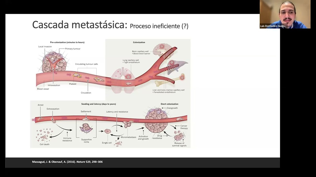
The molecular mechanisms of neural crest cell migration and how these pathways are hijacked during cancer metastasis (e.g., melanoma).
Detailed organogenesis of specific neural crest derivatives, including craniofacial development and the formation of the peripheral nervous system.
Evolutionary developmental biology (Evo-Devo) perspectives on the neural crest as the 'fourth germ layer' and its role in vertebrate evolution.
15.5K views201likes13:52Original Release: 2020-08-02
welcome to neural development part 3 the neural crest a key feature that separates vertebrates from other craniant organisms is the neural crest a transient multipotent and migratory cell population that generates an astonishingly diverse array of cell types during development previously we've talked about neuralation and how the early neural tube and cns are patterned this tutorial starts off around day 28 of development and focuses on what happens to the neural crest i'm dr catherine moore the histology wizard now by day 28 we've altered the fate of some of the surface ectoderm to make neuro ectoderm that neural ectoderm has formed a closed tube and that tube is now inside the body and the neural crest cells have formed this temporary cell population here in green consists of bilaterally paired strips of cells that arise at the border between the neural and non-neural ectoderm during late gastrulation and early neurulation these cells form along that whole cranial caudal axis the formation of neural crest cells is a multi-step process and it requires contact-mediated tissue interactions between the neural ectoderm seen here in pale blue the non-neural ectoderm seen in darker blue and the underlying mesoderm and this is controlled by an intricate series of molecular signals i've talked about some of these signals before including bone morphogenetic proteins or bmps wince fibroblast growth factors and retinoic acid you can recall that low levels of bmp are required to form this neural ectoderm high levels seem to drive skin ectodermal fates well it turns out that neural crest cells seem to form at the border with intermediate levels of bmp now after induction the newly formed neural crest cells will undergo epithelial to mesenchymal transition and they will delaminate from their tissue of origin and migrate out from the entire nerve axis of the embryo to specific locations these cells are multi-potent now remember that means they have the ability to give rise to many cell types within the embryo many different systems such as nervous system skin teeth face heart adrenal glands gastrointestinal tract will have contributions from neural crest i'll talk more specifically about those fates in just a minute but first i want to touch on what controls the fate of neural crest cells how do they know where they're going and how do they know what to become well here you see an electron micrograph of a chick embryo and cross section just showing the neural tube and those neural crests migrating out we know from a lot of experiments in model organisms that neural crest cells migrate along very well defined roots we also know that this migration is controlled by a large array of inhibitors permissive factors and positive guidance cues as well as a lot of cellular interactions and all together these signals are going to organize the crest into distinct subpopulations and then guide them into specific territories now interestingly this overall dynamic of neural crest migration is reminiscent of cancer cell metastasis which follows very similar steps of epithelial to mesenchymal transition and then this kind of guided cell migration so we can use crest to actually learn about cancer so as these cells are traveling along they actually lose their multi-potency and they become more lineage restricted so what do they actually become well neural crest cells have a diverse set of derivatives and what cells and tissues form depends somewhat on what part of the neural tube the crest cells came from in the first place this cartoon shows the major divisions of the crest cell populations the cranial the vagal the trunk and the sacral populations now cranial and caudal neural crest can actually give rise to some identical cell types so for example both can form neurons but there is some specificity for example only cranial neural crest can form cartilage in bone so let's take a look at some of the derivatives and we'll start with the cranial crest the neural crest of this region migrates into the pharyngeal arches then they form ecto mesenchyme and that's going to contribute tissues to the face which in the body region are normally derived from mesoderm so this is cartilage bone connective tissue so a great deal of the face is formed by neural crests they also form the middle ear bones and the odontoblasts which give rise to the dentin of the teeth and in addition they give rise to some cranial nerve ganglia and parasympathetic neurons moving caudally to the vagal neural crest there's actually a subpopulation called the cardiac neural crest which gives rise to some tissues of the heart including the kona truncal septum and the cardiac septa the entire nervous system the entire enteric nervous system is of crest origin and trunk neural crest also gives rise to neurons and glia of the peripheral nervous system the adrenal medulla and all of the melanocytes in the body and finally sacral neural crest contributes to the caudal part of the enteric nervous system and sympathetic ganglia now this seems like a long list it actually is you can see why neural crest is often called the fourth germ layer i'll tell my students if you don't know the embryonic origin of something in the body neural crest is a pretty good guess now i know that students have different ways to remember lists and although i'm not a great singer i find that songs can actually help me to remember so here's one way i remember some of the neural crest derivatives [Music] cells gave to me myelinating swan cells the adrenal medulla inner brain and ninjas [Music] [Music] that's just the way i remember it you can make up your own song now we're going to talk about cranial crest derivatives when we talk about the pharyngeal arches and head and neck development since they're so involved in there but i want to briefly highlight the contributions of neural crest to the cranial nerve sensory ganglia cranial nerves and their ganglia originate from the neural ectoderm of the brain and spinal cord the crest cells and epibranchial placodal ectoderm now placodyl ectoderm is just this thickened surface ectoderm patch that that actually forms in pairs rostral caudally in the head region and together these form a lot of our cranial sensory ganglia now here's another cartoon that shows the relative contributions of neural crest in green and the placard in blue to some of the cranial nerve ganglia i'm not going to go into all the details here and you don't have to memorize what ganglia comes from what tissue but what you do need to remember is that the cranial nerves listed here all have some contribution from neural crest and also you want to remember that cranial nerves one the olfactory nerve and two the optic nerve actually arise from neural ectoderm now as i mentioned previously in the body region neural crest cells also contribute to the peripheral nervous system both neurons and glia they give rise to sensory ganglia sympathetic and parasympathetic ganglia and neural plexis within specific tissues and organs so let's just briefly review the organization of the autonomic nervous system now recall that this system is a component of the peripheral nervous system that regulates involuntary processes including heart rate blood pressure respiration digestion sexual arousal it contains three anatomically distinct divisions the sympathetic the parasympathetic and the enteric which i'll talk about next so the sympathetic nervous system and the parasympathetic nervous system contain afferent and efferent fibers that provide sensory input and motor output respectively back to the cns activation of the sympathetic system leads to a state of overall activity or tension so this is that flight or fight response so in this case we would have blood pressure and heart rate increasing while for example peristalsis would cease conversely the parasympathetic nervous system promotes that rest and digest process heart rate and blood pressure are going to lower peristalsis and digestion will actually restart or increase sympathetic nervous system innervates nearly every tissue in the body because mostly because it includes all of the smooth muscle in all of the blood vessels now the parasympathetic nervous system innervates only the head and the viscera and the external genitalia so you'll see that it's notably vacant in much of the musculoskeletal system this actually makes it much smaller than the sympathetic nervous system if we look at spinal levels the crest gives rise to the sensory ganglion the dorsal brute ganglia sympathetic chain ganglia and the pre-vertebral pre-aortic ganglia and in addition as i said before the crest cells give rise to all parasympathetic and sympathetic motor neurons and all of their glia now let's look at the enteric nervous system so this system is composed of reflex pathways that control the digestive functions of muscle contraction and relaxation secretion absorption and blood flow now you'll see this system sometimes listed as part of the parasympathetic nervous system sometimes not but we do know that it can act independently of the central nervous system and the p s now in this cartoon i've put the neural tube on top and color coded it with the rough locations of the different neural crest cell populations and on the bottom we have the digestive tract with the different regions of the system forget mid-gut and hindgut beginning at the end of week 4 neural crest cells are going to migrate and form nerve plexi in the gastrointestinal tract so first cells from the vagal population are going to form the enteric system within the foregut and then the mid gut and then those will also migrate and a few of those will end up in the hindgut but mostly the lumbar sacral crest will populate the hindgut okay now that we know where neural crest cells go and what they become let's talk about what happens when there's problems with this important cell population abnormal specification migration differentiation or death of neural crest cells during development leads to a set of syndromes and diseases called neurocrystopathies which comprise a broad spectrum of congenital malformation one clue to neural crest involvement is that seemingly unrelated and multiple organ systems are often affected because they share the same embryonic origin now this cartoon shows the neural crest populations in the embryo color coded as before and neurocrystopathies can be classified according to the neural crest origin and also according to which developmental step of neural crest formation is actually disrupted so let's look at an example george syndrome which is now called 22q 11.2 deletion syndrome is actually a subset of conditions that includes cardiac defects a hypoplastic thymus mild craniofacial defects and parathyroid gland abnormalities structures affected in this syndrome are derived from cardiac and cranial neural crest which makes sense given the phenotype let's look at a few more examples treacher collins syndrome or first arch syndrome and craniosynostosis arise from problems with induction and specification of neural crest cells and that affects their final numbers albinism pheochromocytoma neurofibromatosis all arise due to alterations in neural crest cell differentiation while her sprung disease melanoma charge syndrome and pierre robin secrets all result from problems in neural crest cell migration now unfortunately only a few causative genes for neurocrystopathies have actually been identified and these only account for a minority of the patients now we do know that environmental factors play a role in neural crest cell migration and differentiation and exposure to medicinal drugs and teratogenic agents can also affect crest formation so for example ethanol exposure causes fetal alcohol spectrum disorder or sometimes known as fetal alcohol syndrome and this is characterized by reduction in cranial and cardiac neural crest migration and by reduction in cranial neural crest numbers so these patients have very characteristic facial abnormalities other factors that can affect neural crest include high glucose levels from uncontrolled diabetes in the mother retinoic acid many drugs of abuse some herbicides and a lot of drugs that are actually used to treat different disorders such as lithium tamoxifen thalidomide and valproic acid just to name a few i hope it's clear that the neurocrystopathies comprise a large number of congenital abnormalities again reflecting the wide contribution of neural crest cells to the body and the importance of this cell population thanks for stopping by
Up Next

37:23
Hydrothermal Vents Ecosystem Explained | Deep Sea Science
@OctopusLady
626.5K views•2023-10-31

50:12
Bacterial Communication: Quorum Sensing and Biofilm Formation Explained
@JHUAAP
12.6K views•2011-06-28

11:43
Neural Tube Patterning: Early Nervous System Development (Part 2)
@thehistologywizard5783
8.6K views•2020-08-02

7:08
Bacteriophages: Earth's Deadliest Killers and Future Antibiotics
@kurzgesagt
34.6M views•2018-05-13
Related Study Plans & Knowledge Roadmaps
Structured learning paths in Biology
biology
RNAi: Biogenesis, RISC & Gene Silencing
01Molecular Biology Foundations: DNA to Protein
02Introduction to RNA Interference (RNAi)
03Molecular Biogenesis of miRNA and siRNA
+ 2 more learning modules
10h5 modules
Explore Roadmap
biology
Mycorrhizae: Signaling, Biophysics & Ecology
01Foundations of Mycorrhizal Symbiosis
02Molecular Signaling and Root Colonization
03Biophysics and Bioenergetics of Nutrient Exchange
+ 1 more learning modules
11h4 modules
Explore Roadmap
biology
Circadian Clocks: TTFL, SCN & Periphery
01Foundations of Molecular Biology & Gene Expression
02Introduction to Chronobiology and Actograms
03The Core TTFL: Molecular Architecture of the Clock
+ 3 more learning modules
14h6 modules
Explore Roadmap
biology
Population Genetics: Math, Drift & Selection
01Genetic & Probability Foundations
02The Hardy-Weinberg Principle and Baseline Models
03Mathematical Modeling of Natural Selection
+ 3 more learning modules
18h6 modules
Explore Roadmap