










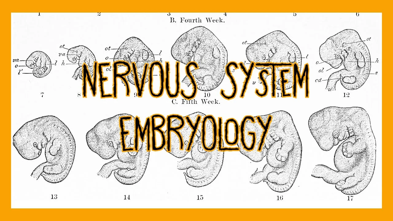





The neural tube is patterned along two axes—the cranial-caudal axis using morphogens like FGFs, Wnt, and Sonic Hedgehog, and the dorsal-ventral axis through signaling centers (roof plate secreting BMPs and Wnt, floor plate secreting Sonic Hedgehog)—with Hox genes providing positional codes that determine cell fates and establish the basic CNS architecture by the end of the first month of development.
Neural Tube Patterning: Early Nervous System Development (Part 2)
Added:The process of primary neurulation, including how the neural plate folds and fuses to form the early neural tube.
Basic embryonic axes and anatomical terminology, specifically anterior-posterior (cranial-caudal) and dorsal-ventral orientation.
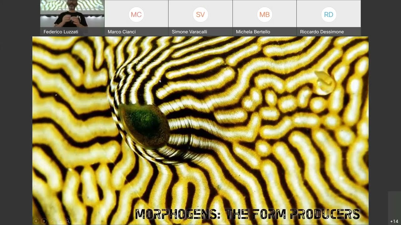
The fundamental mechanism of morphogens and concentration-dependent cell signaling (e.g., how a gradient of a signaling molecule instructs different cell fates).
An introductory understanding of transcription factors and gene expression regulation during embryonic development.
Neurogenesis and gliogenesis, exploring how patterned progenitor cells differentiate into functional motor neurons, sensory neurons, and glia.
The regionalization of the brain, specifically how the cranial neural tube develops into the primary and secondary brain vesicles (e.g., forebrain, midbrain, hindbrain).
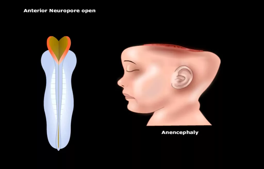
Clinical implications and developmental disorders resulting from patterning defects, such as holoprosencephaly (Shh pathway disruption) or neural tube defects.
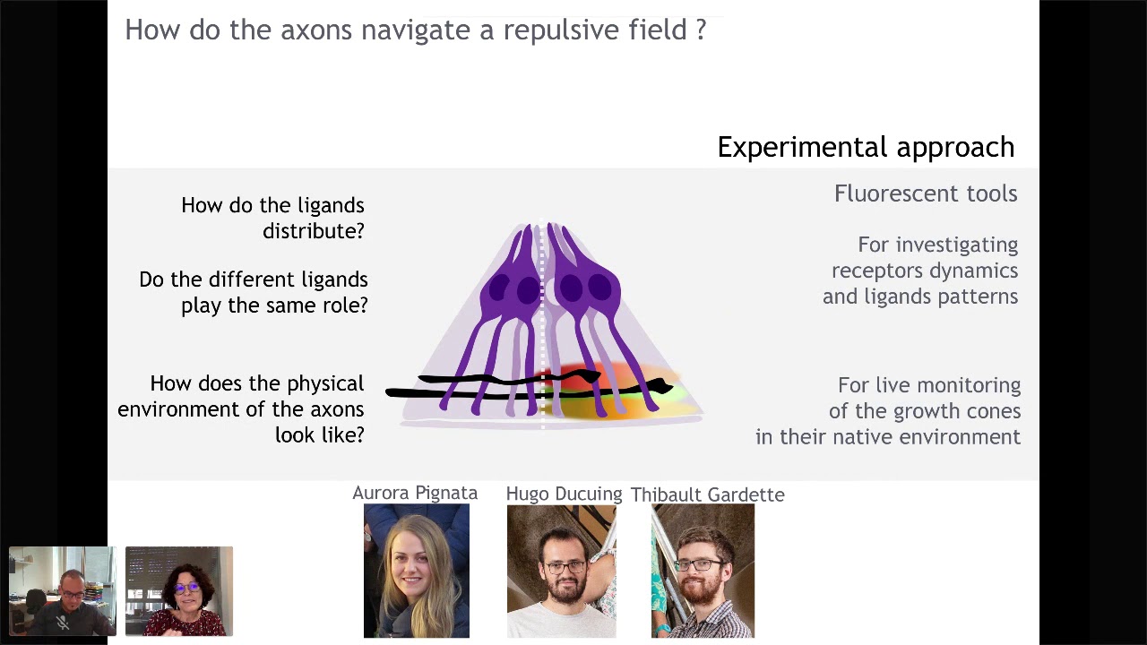
Axon guidance mechanisms, analyzing how newly formed neurons navigate and establish precise synaptic connections throughout the developing body.
8.6K views147likes11:43Original Release: 2020-08-02
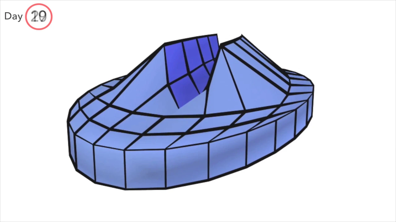
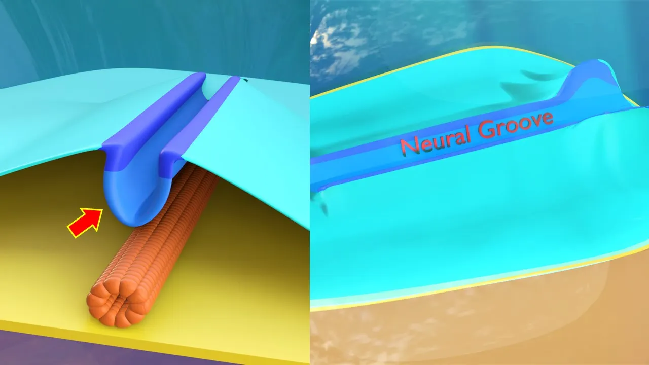
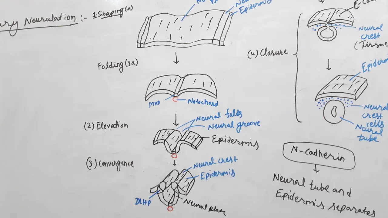
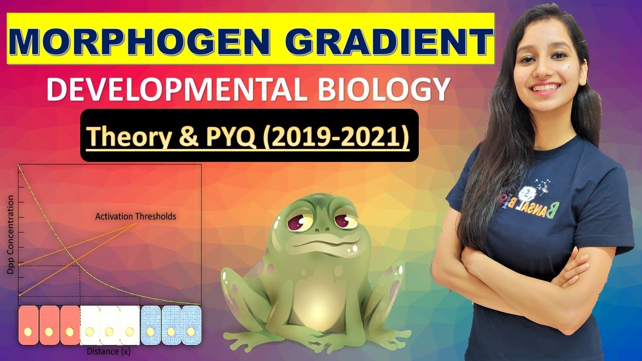
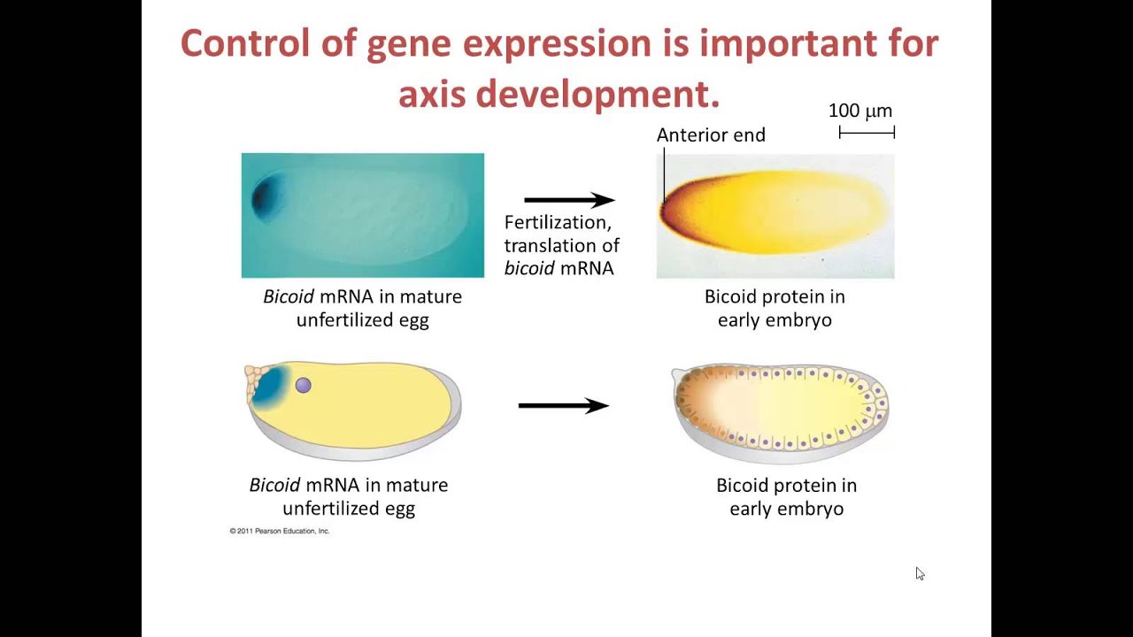
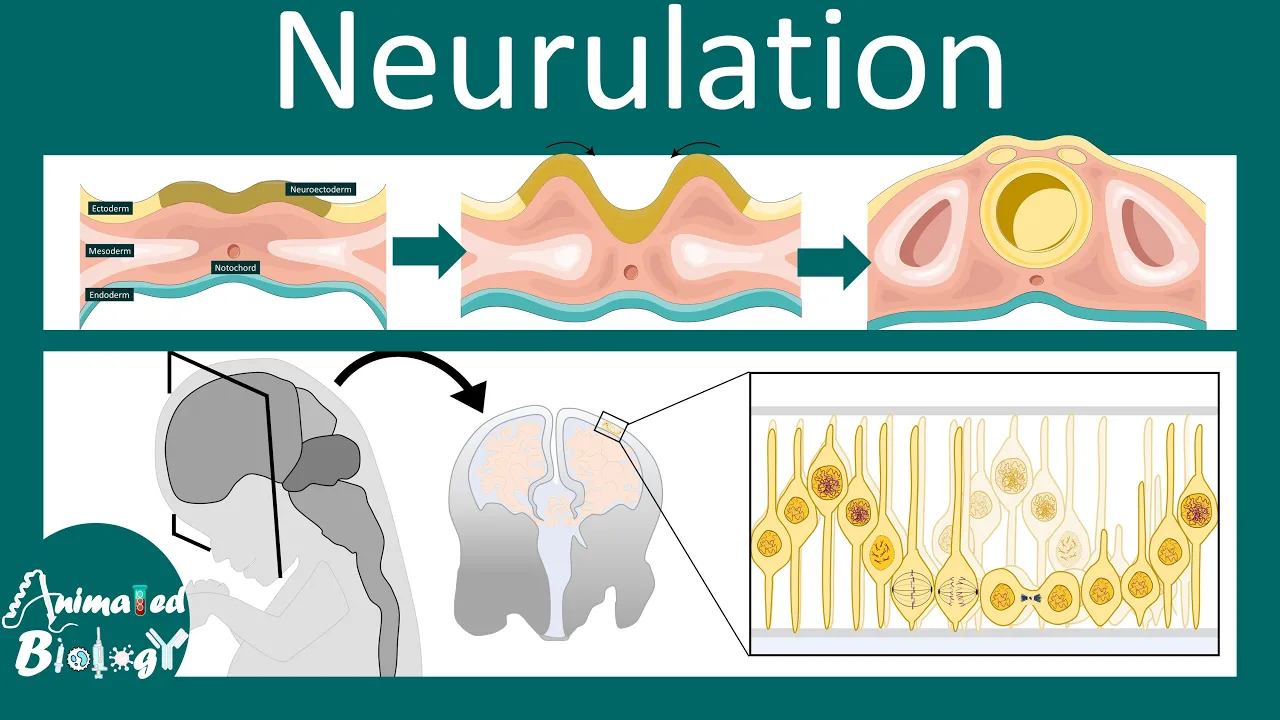
welcome to my second video on neural development this is part two early neural tube patterning in my first video of this series i talked about the process of neurallation and here i'll continue by describing some of the early patterning events of the nervous system i'm dr katherine moore the histology wizard at the end of my first video we were here at day 28 where we'd altered the fate of some of the surface ectoderm to make neuro ectoderm that neural ectoderm had formed a closed tube and the tube was now inside the body and the neural crest cells have formed but how do we get from this relatively simple design to this extraordinarily complex system how do we get different structures that are going to play different roles all in the right place and all wired together to work now that of course could be the subject of about 100 videos but here i'm going to skim across the surface and hopefully just give you a basic understanding of how early neural tube requires a basic plan the nervous system is patterned by a series of signaling events that turn on different genetic programs to pattern along two basic axes the cranial caudal axis and the dorsal ventral axis so now let's take a look here we have a simple cartoon of the neural tube and i've colored in the cranial and hot pink and the caudal end bright yellow these colors are going to represent morphogens and you can see that there's a kind of gradient of pink that decreases as we move caudally now what are morphogens morphogens are simply singling molecules that emanate from a restricted region of the tissue and then they spread away from the source that forms a concentration gradient and the gradient is going to then prefigure the pattern of development the fate of cells in the field depends upon the concentration of the morphogen signals that they receive so each cell will receive signals and then interpret those signals and that will essentially give them coordinates similar to getting a latitude and a longitude on a map different coordinates or combinations of these signals will activate or suppress various genetic pathways which will drive different fates so now let's look at an example cells in this cranial region are in the highest concentration of the pink morphogen and the lowest concentration of the yellow morphogen and they'll become cells in the forebrain while cells in this region have a high yellow morphogen concentration but low pink morphaging concentration and they'll become cells in the hindbrain now this is clearly a very simple example but i hope that it illustrates how multiple gradients can affect cell fate now importantly this very first set of morphogens is going to set up initial boundaries that will later be refined by more signals this is one form of patterning second form is dorsal ventral patterning and this cartoon shows the neural tube with two signaling centers at the dorsal and ventral poles again we have morphogens that are released forming two gradients now a cell will have to interpret the signals and determine its identity in this case this cell is detecting low amounts of both morphogens so it's going to take on the fate of a purple cell now we know that cells are going to have to interpret a combination of multiple signals from both cranioconnal and dorsal ventral axes to interpret their coordinates now i'd like to dive just a bit deeper into cranial caudal patterning here's our neural tube again and i've labeled the pre-chordal plate and the notochord the pre-chordal plate is an area of thickened endoderm that's directly in contact with ectoderm it's going to give rise to the endodermal layer of the oral pharyngeal membrane or the primitive mouth but it's important here because it participates in the patterning of the cranial neural tube now you can see that from very early on there are morphological differences between the cranial neural tube and the caudal neural tube now the early signals that pattern the neural tube come from multiple sources they come from pre-chordal plate they come from neural tube the notochord and other locations and some of these signals are fibroblast growth factors wince and sonic hedgehog as well as many other genes including the hawks gene family now for the most part although we know what the signals are the patterning process remains poorly understood but one of the gene families that we do know more about is the hawx gene family now hawks genes encode a family of highly conserved transcriptional regulators that elicit distinct developmental programs along the head to tail axis of animals as shown in this cartoon these genes have a conserved chromosomal order that's actually reflected in their expression pattern the specific regional functions of individual hox genes in development largely reflect their restricted expression patterns and sometimes we call this the hawks code now if the code is disrupted this can lead to developmental defects and disease invertebrates hox genes often in combination will help define somite identity directing them to develop differently depending on where they are in the body they also help specify the difference between an arm and a leg between a humerus and an ulna as well as between a pinky and thumb and in the nervous system their expression in segmented embryonic structures called rhombomirs directs the development of different brain regions this cartoon illustrates how some of these hox genes are expressed in the rhombomeres and in this example you can see that if you have expression of only the a2hawks gene this would give an identity for rhombomere 1 while expression of more of these genes would be associated with an identity of rhombomer 8.
now disruptions in hox genes can alter identity of a specific brain region now that's all i want to say right now on cranial caudal patterning because as i said it's not really well understood and i want to move to the dorsal ventral axis where we understand a lot more about how fates are established but before we get into specifics of that patterning i want to just back up a little bit and talk about the cell's neural tube now here's an electron micrograph of the neural tube with an inset showing the cells these are neuroepithelial cells and they are pseudostratified columnar cells that will give rise to all the neurons and macroglia of the cns these cells are going to divide and migrate to different locations in the different cns tissues now next let's look at a cartoon of a cross section of the neural tube where i've colored in the different layers of the tube so here we see our ventricular zone in orange and these are the cells that are proliferating and that will form the neurons in glia those cells are going to migrate from the ventricular zone to the mantle layer or zone as neuroblasts these are your primordial neurons and they'll form the gray matter of the cns so these are going to be the neuronal cell bodies the neuron and glial cell bodies the marginal zone is shown in yellow and this represents the axons of the cells in the mantle layer so this is the white matter of the cns and finally we have the ependymal cells in red these are the cells that line the brain ventricles and will produce cerebral spinal fluid now these cells differentiate in order after the neurons and glial progenitors differentiate let's look at some early patterning of the spinal cord the spinal cord itself develops from the caudal part of the neural plate and from the neural tube caudal to the fourth pair of somites here you can see again the ventricular mantle and marginal zones this early post neuralation tube is going to develop into dorsal and ventral halves the dorsal or alar and the ventral or basal plates and these plates are going to produce longitudinal bulges of cells that are in that will extend the length of the spinal cord and these are going to be separated in the embryo by the sulcus limitands and here you can also see the neural canal in the center so in the adult as seen in this example those plates are going to actually correspond to columns of neurons so the alar plate is going to correspond to the dorsolateral thickenings of the dorsal sensory neurons this is the dorsal horn while that basal or ventrolateral thickening is going to become the ventral somatic motor neurons or the ventral motor horn and then in some regions of the spinal cord the thoracic lumbar some lumbar and sacral regions there's also an intermediate group of neurons you can also see the white matter here and finally that neural canal is going to reduce in size as the walls of the neural tube thicken leaving the central canal of the spinal cord so here now we have a basic pattern with the dorsal and ventral but how do those cells within those regions know which fates they're supposed to adopt well the answer is more morphogen gradients so here you're looking at an electron micrograph of the developing neural tube and i've colored in the locations of the dorsal and ventral signaling centers which are termed the roof plate and the floor plate the roof plate is going to secrete bone morphogenetic proteins or bmps and wince while the cells of the floor plate are going to secrete sonic hedgehog now those morphogens sound familiar to you they should because these three play major roles in the developing embryo and you're going to see their names pop up over and over again during development as we move through the body systems so these are three that you should definitely remember now how does this system specify fate we're going to go back to a cartoon and look at some more ingredients in this cartoon of a spinal cord you can see a blue box representing the roof plate secreting bmps and wince and that floor plate in pink secreting sonic hedgehog let's look first of that roof plate so here we have bmps and winds that are going to form a gradient with the highest levels closest to the source and these morphogens are going to promote dorsal fates to make dorsal neurons and block ventral fades sonic hedgehog from the floor plate is going to promote ventral neuronal fates and block dorsal fates now if you have a cell that's somewhere in the middle it's going to be exposed to low amounts of both morphogens and so it's going to take on an intermediate phase now again this is a gross oversimplification but it at least will give you the basics on how cells can interpret their dorsal ventral positions and how if we alter these gradients you can alter cell fate so the important take-home message today is that as a result of both cranial caudal and dorsal ventral patterning events by the end of the first month the basic pattern of the central nervous system is established now in a later video i'll talk more about how the specific brain regions are patterned and what these regions in the embryo the prosencephalon mesencephalon and robencephalon actually will become the adult nervous system but what about the peripheral and enteric nervous systems i haven't talked about those yet and what happened to those neural crest cells that i talked about at the very beginning of this video well check out my video on neural crest formation and migration to discover the fates of these fascinating cells thanks for stopping by
Up Next
35:03
Frog Embryology: From Egg to Neural Plate Formation
@scicommlab
14.9K views•2018-04-18

50:12
Bacterial Communication: Quorum Sensing and Biofilm Formation Explained
@JHUAAP
12.6K views•2011-06-28
13:52
Neural Crest Development | Embryology Tutorial Part 3
@thehistologywizard5783
15.5K views•2020-08-02

7:08
Bacteriophages: Earth's Deadliest Killers and Future Antibiotics
@kurzgesagt
34.6M views•2018-05-13
Related Study Plans & Knowledge Roadmaps
Structured learning paths in Biology
biology
RNAi: Biogenesis, RISC & Gene Silencing
01Molecular Biology Foundations: DNA to Protein
02Introduction to RNA Interference (RNAi)
03Molecular Biogenesis of miRNA and siRNA
+ 2 more learning modules
10h5 modules
Explore Roadmap
biology
Mycorrhizae: Signaling, Biophysics & Ecology
01Foundations of Mycorrhizal Symbiosis
02Molecular Signaling and Root Colonization
03Biophysics and Bioenergetics of Nutrient Exchange
+ 1 more learning modules
11h4 modules
Explore Roadmap
biology
Circadian Clocks: TTFL, SCN & Periphery
01Foundations of Molecular Biology & Gene Expression
02Introduction to Chronobiology and Actograms
03The Core TTFL: Molecular Architecture of the Clock
+ 3 more learning modules
14h6 modules
Explore Roadmap
biology
Population Genetics: Math, Drift & Selection
01Genetic & Probability Foundations
02The Hardy-Weinberg Principle and Baseline Models
03Mathematical Modeling of Natural Selection
+ 3 more learning modules
18h6 modules
Explore Roadmap