












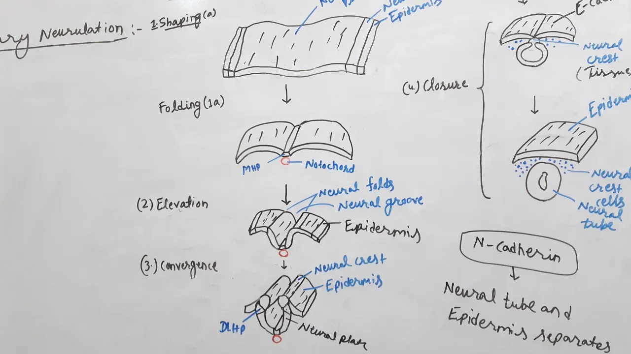





Neural tube formation (neurulation) is the process by which the neural plate, induced by the notochord to thicken at the midline, invaginates to form a neural groove and subsequently fuses along its edges to create a closed neural tube; this primary neurulation process begins at the cranial end and proceeds in a zipper-like fashion toward the caudal end, with the anterior neuropore closing around day 24-25 and the posterior neuropore closing around day 27-28, while secondary neurulation occurs in the tail bud region where mesenchymal cells form a medullary cord that hollows out and fuses with the primary neural tube.
Neurulation and Neural Tube Formation: Embryology Explained
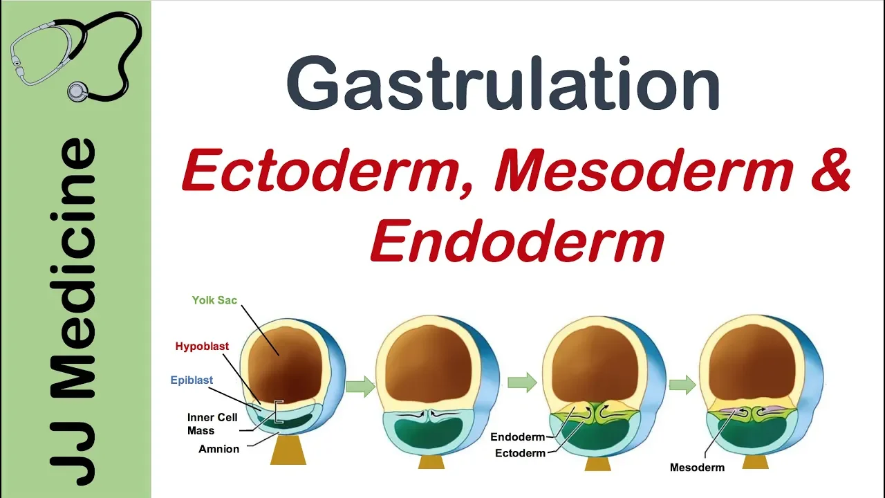
Added:The process of gastrulation and the establishment of the three primary germ layers: ectoderm, mesoderm, and endoderm.
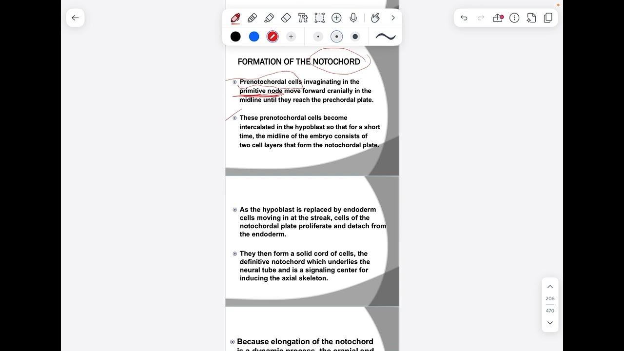
The structure and function of the embryonic notochord, specifically its role in secreting inductive molecular signals.
Fundamental cellular mechanisms of development, including apical constriction, cell adhesion changes, and cytoskeletal remodeling.
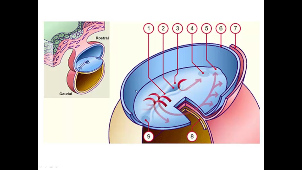
Basic anatomical terminology of embryonic body axes, such as anterior-posterior (rostral-caudal) and dorsal-ventral axes.
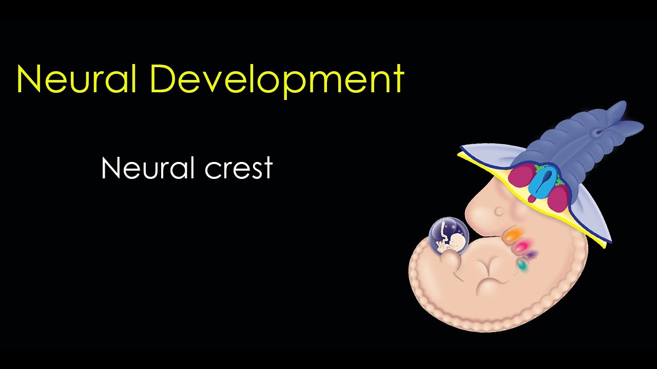
The formation, migration, and differentiation of neural crest cells (often referred to as the 'fourth germ layer').
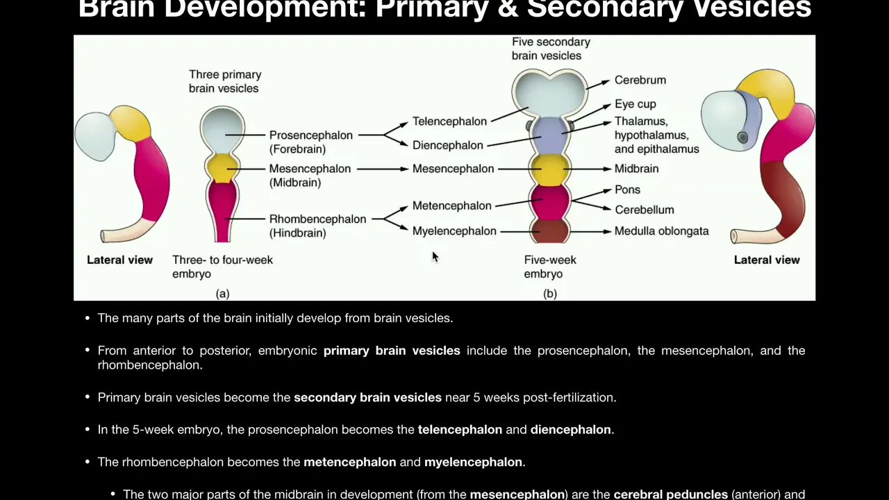
Primary and secondary brain vesicle development (prosencephalon, mesencephalon, rhombencephalon) and early brain regionalization.
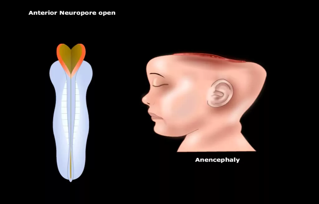
Clinical correlates of defective neurulation, including neural tube defects (NTDs) like spina bifida and anencephaly, and the preventative role of folic acid.
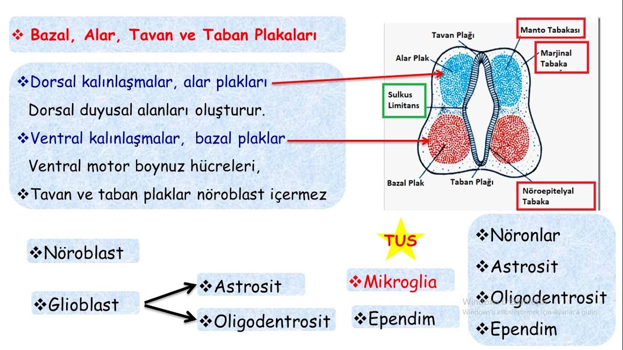
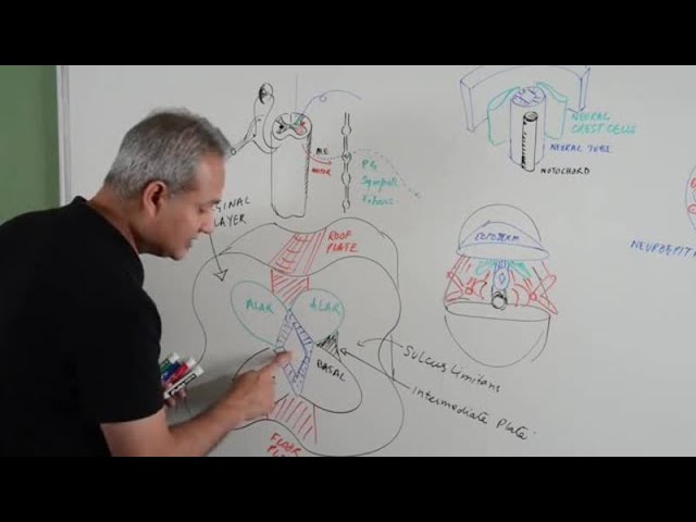
The cellular differentiation within the neural tube, specifically the dorsal-ventral patterning of the spinal cord into sensory (alar) and motor (basal) plates.
29.7K views805likes20:56Original Release: 2024-05-22
They will move like so. Here they are fusing together into the amniotic cavity. Looks like a dead body is lying here. Fold like this towards the periphery. Aggregate like this and they will fuse with the primary neural tube. Hello, I'm Dr. Aizaz from medicovisual.com and in today's visual lecture we will talk about development of neural tube. Basically, we will discuss the process of neurulation. So here we have the trilaminar germ disc surrounded by two cavities and within the trilaminar germ disc I will only show the ectoderm and endoderm.
I will not show the mesoderm because that is not relevant with our discussion today. I will show the notochord along with that I will show the ectoderm and endoderm and of course the ectoderm it is surrounded from all side by a cavity called amniotic cavity and here is the endoderm. Ectoderm was surrounded by amniotic cavity and here is the endoderm. It is surrounded by yet another cavity and this cavity is called yolk sac cavity. So this one is surrounded by amniotic cavity.
Amniotic cavity is surrounding the ectoderm and yolk sac cavity is surrounding the endoderm. Now what will happen here that let me show you. So let me first introduce you to these structures.
So here is the primitive node and this is the primitive streak. Actually, the cranial, cranial dilated end of primitive streak is the primitive node and here is the cloacal membrane and here is the oropharyngeal membrane. So at the sites of cloacal as well as oropharyngeal membrane there is ectoderm and endoderm only with little to no mesoderm in between these structures. Now let's discuss the process of neurulation. Now what will happen that if I go deep into the ectoderm we will see that here we have the notochord. So between the ecto and endoderm we have the notochord in the central midline. We have this notochord. This is not corn on the cob. This is actually notochord. Right! So here we have notochord. Now what will happen that this notochord, the central midline structure notochord it will release certain factors. It will release certain chemical factors and these chemical factors they will cause thickening of this midline ectoderm.
They will release certain chemicals which will cause thickening of midline ectoderm. So basically this midline ectoderm it will be converted into a platelike structure with the help of these inductive factors that will be released by notochord and along with that some textbooks they also claim that there is some role of paraxial mesoderm and there is some role of this primitive node and some other structures but mainly the main inducer of neurulation is the notochord.
Of course there are other structures as well that are involved but mainly it is the notochord which induces the thickening of midline ectoderm to form the neural plate. Let's see the animation. So here you can see that ectoderm is almost uniform at this time. Now at the midline it will thicken.
Here you see it will be thickened like this. Like this a thickened plate will be formed and this is basically a key like structure. You see this is shaped like a key and of course just like ectoderm it will also be surrounded by amniotic cavity. Please remember that. Now this structure that has just been formed it is called neural plate. Now at the margins of neural plate, at the lateral margins of neural plate here you can see I have painted this part with a different color. These are future neural crest cells. Neural crest cells, they will be forming the crest of this neural tube and then they will separate and they will form the neural crest cells. We will see this later. Now this is basically the neural plate. This thickened plate is called the neural plate. Now what will happen that of course the embryo will grow and as the embryo grows especially at the cranial end as the embryo grows this neural plate it will also grow and it will lengthen up especially at this uniform end. There is this dilated cranial end which will form the brain and here there is somewhat Uniformly-wide caudal end which will form the spinal cord. So this will form the brain this will form the spinal cord. Especially this caudal end it will lengthen up like this. As you can see the animation actually whole embryo is growing and now what will happen that this neural plate it will invaginate and its margin they will try to come close towards each other in the central midline and they will try to fuse with each other. Let's see the animation to understand it properly.
So here you can see let's stop it for a second that this neural neural plate is invaginating.
You can see it properly in this 3d model. Wow this is amazing. It's very easy to teach using these 3d models and 3d animations. So you see that they will move like this and they will come close together and they will try to meet in the midline. Now you can see that this neural plate is no more a plate it is now a curved structure and here a central groove central midline groove has been formed. Let me show you. So here this is a central midline groove which is naturally it is named as neural neural groove and here you can see I have painted this these margins with a different color and these are basically this color is Off, let me use the other color these are basically the future neural neural neural crest cells these are basically the future neural crest cells. Now what will happen neural groove has been formed neural crest cells are there and they are trying to meet with each other and of course same situation here as well and please remember again the whole this structure is surrounded from all side with this amniotic cavity. Now what will happen that they will begin to meet with each other and this process this fusion process it will start somewhat here in the future cervical region in the future cervical region this process will start that they will begin to fuse with each other like this. Let me show you the animation so here you can see let me stop it here that they are beginning to fuse like this in the in the cranial region basically cervical region and then from the cervical region this will move in a zip like fashion cranially as well as caudally. So this fusion process will move cranially that it will fuse cranially as well as it will move caudally that here in a zipper like fashion it will move caudally as well. So let's see the animation that what is going on here so here they are fusing together and before they are completely fused let me stop it here here you can see that we still have these openings this opening as well as this opening this is the cranial opening this is the cranial opening and this is the caudal opening the cranial opening is also called anterior opening and caudal opening is also called posterior opening or basically they are technically called as neuropores. So still when it is not completely fused we have these openings called anterior and posterior neuropores. So this is anterior or cranial neuropore why anterior why posterior we have discussed this in the lecture of gastrulation that it is a convention which is put by the mouse embryologists and please watch my lecture on the gastrulation to understand why it is still called anterior and posterior.
Technically it is not anterior it is cranial and it is not posterior it is caudal but still somehow we conventionally call it anterior as well as posterior. So anyhow the another point that I want to show here is that first of all let me tell you that these openings are into which cavity that's why I again and again focused on the on this cavity. So basically these are opening into the into the amniotic cavity. What is the significance? What is the importance of this? The reason is that they are opening into this cavity is that now basically at this stage the blood vessels of the neural tube the primitive neural tube its blood vessels are not properly formed so it gets its nutrition from the amnion from the amniotic fluid it gets some important factors for its development as well as its nutrition from the amniotic cavity. So that's why until the until here as well there is this neuropore so until the blood vessels are not completely formed the blood vessels of the neural tube until they are not completely formed these openings are required for nutrition and growth and certain other factors are required that are required for development of the neural tube they are provided by the amniotic cavity not amniotic cavity actually amniotic cavity is filled with a fluid that is the amniotic fluid and this fluid provides important nutritive and other factors that are required for the developing neural tube. So that is why there are these temporary openings but as this fusion process continues as this neurulation process continues these openings they close. Now here you can again see that the cranial neuropore it closes first and the caudal neuropore it closes later the reason is very logical you see it starts from here it does not start from the midline it does not start from somewhat here it start from the somewhat towards the cranial region so as it starts from the cranial region this fusion process as it starts somewhat cranially so it moves cranially as well as caudally so naturally it will reach this end this cranial end first as compared to the caudal end so first this fusion process will complete in the cranial end it will reach towards the cranial end first and later it will reach towards the caudal end. I hope you can understand this explanation that first this process cranial region is closed cranial neuropore is closed and then caudal neuropore is completely closed but ultimately both of them are closed. Now by the way let me tell you that the cranial neuropore it closes at about 24 to 25th day of gastrulation and the caudal neuropore it closes at about 27 to 28 day there is some discrepancy in the literature of course but usually it is 24 to 25th day and it is 27 to 28th day so you see it is somewhat earlier slightly earlier and it is somewhat later but anyhow both of them close and as the blood vessels develop so the these neuropores they are no more needed so they are closed it's very natural right so that's how this neural tube is formed if I go deeper don't worry I will show the show you the cross section as well so you understand you will understand it properly don't worry about that and here is this primitive node and primitive streak now let me show you the cross section so that you really understand that what is going on here I don't know why when I look at this structure it looks like a dead body is lying here you see dead body covered with the with a cloth but ironically the development of central nervous system is the sign of life, Vitality, and ultimately the consciousness, not the death.
Let's move to the cross section of the neural tube neurulation process and let's see how it happens so what I will do is that let me explain what I mean by the section basically you imagine that we cut from here we somewhat cut from here like this yeah like this so we cut from here like this and then so we've cut from here like this we remove this part right and then we are standing here we are standing here and we are looking towards this direction towards here so let's go to that section to understand it further so here is again the cut section and I hope you can understand all these structures so here is this notochord and this notochord it induces the formation of neural plate now what will happen that neural plate will fold like this and here you can see this is the groove called neural groove and here these are the future neural crest cells so it will fold like this and then it will fuse in the midline it will fuse like this and now what will happen that these future neural crest cells here they will separate and they will move towards the sides they will move towards the periphery and then they will go to different parts of the body and they will form some important structures basically these are like ambassadors of central nervous system to different parts of the body so they will form some of the very important structures for example they will form the peripheral nervous system ganglias and some components of skin and some component of different glands we will not discuss the details in this lecture but anyhow these are the neural crest cells and neural crest cells basically they separate from here from this developing neural tube and by the way as they as these folds as they fuse the surface ectoderm the surface ectoderm it will meet in the midline and it will become continuous again now here I want to tell you something very logical that as this neural plate is formed what happens that the ectoderm it is divided into two parts the ectoderm that is forming this neural plate it will be forming the nervous system so it is called neuroectoderm and rest of the ectoderm that remains it can be simply called ectoderm but it is specifically also called surface ectoderm so this is surface ectoderm and this is the neuroectoderm which will be forming the neural tube as well as the neural crest cells so this was about the process of neurulation but that's not all actually this is actually called primary neurulation there is something called secondary neurulation and which is not very important for humans but because it is mentioned in many important textbooks so that's why I am gonna tell you about the secondary neurulation as well in this visual lecture so here I have removed the amnion and here we have the section and this primary neural tube has been formed now here if I remove this part here there is a problem that the neural tube the notochord is up to this point up to the primitive node it starts from the oropharyngeal membrane which is not shown in this 3d model it starts from the oropharyngeal membrane and ends at here at the this primitive node now what happens that we need the neural tube beyond this point as well so how the neural tube beyond this point forms this part is called tail bud let me explain this thing to you so this part from the this is the primitive node from primitive node up to cloacal membrane this is the cloacal membrane cloacal membrane so from primitive node up to cloacal membrane this part is called tail bud or tail eminence tail bud now this tail bud it consists of some undifferentiated mesenchymal cells some undifferentiated irregularly shaped mesenchymal cells like this you see what is the origin of these mesenchymal cells just like all other cells they originate from the primitive streak they ingress through downwards during the process of gastrulation they ingress downward and they do not form a specific mesoderm like the other parts of the of the embryo that form the paraxial mesoderm lateral plate mesoderm intermediate mesoderm and so on it still remains undifferentiated like this and what will happen that these mesenchymal cells they will aggregate like this and they will form a solid cord they will form a solid cord of cell this is the solid cord of cell they will form the solid cord of cell in the tail bud region this is called medullary cord what it is called it is called medullary medullary cord medullary cord now what will happen to this medullary cord is that the cells in the central core or central midline of this medullary cord they will die they will undergo apoptosis process and a hollow cylinder will be formed and then this cylinder will meet with this primitive not primitive primary neural tube this secondary neural tube this is now called secondary neural tube and remember it does not form by the typical process of the formation of neural plate and neural fold and neural groove and so on it forms with the help of these mesenchymal cells and again it will fuse with the primary neural tube the secondary neural tube it will fuse with the primary neural tube and it will form this complete neural tube that starts from the oropharyngeal membrane and ends at the cloacal membrane now it is believed that this process this secondary neurulation it is not much relevant with the human embryos but some research indicates that the sacrococcygeal region of our spinal cord it forms as a result of secondary neurulation so that was about the process of neurulation i hope you learned something from this visual lecture thank you so much for watching this video
Up Next
5:02
Methylene Blue Health Benefits: Mitochondria, Dosage, and Safety
@Drberg
993.4K views•2023-04-04

48:47
Graphic Medicine: Comics for Collaborative Healthcare Communication
@nationalpatientadvocate
189 views•2023-12-04

10:28
Neuroanatomy: Central and Peripheral Nervous System Divisions Explained
@AKLECTURES
136.2K views•2014-09-20

3:05
Stages of Labor and Vaginal Birth | Childbirth Animation
@nucleusmedicalmedia
52.1M views•2017-08-18
Related Study Plans & Knowledge Roadmaps
Structured learning paths in Medicine
medicine
Abnormal LFTs: Diagnostic Workup & Pathology
01Liver Anatomy and Basic Physiology
02Introduction to Liver Function Tests (LFTs)
03Hepatocellular vs. Cholestatic Patterns
+ 3 more learning modules
12h6 modules
Explore Roadmap
medicine
Analyzing Blood Smears: RBC, WBC & Platelets
01Introduction to Blood Components & Smear Preparation
02Normal Morphology of Blood Cells
03Erythrocyte Pathologies & Morphological Variants
+ 2 more learning modules
12h5 modules
Explore Roadmap
medicine
Mechanical Ventilation: Waveforms & Alarms
01Anatomy and Physiology of Breathing
02Introduction to Mechanical Ventilation & Core Modes
03Ventilator Settings and Waveform Interpretation
+ 2 more learning modules
12h5 modules
Explore Roadmap
medicine
Neuro Exam: Localization & Stroke Syndromes
01Foundations of Neuroanatomy and Vascular Supply
02The Neurological Examination: Step-by-Step
03Principles of Neurological Localization
+ 3 more learning modules
18h6 modules
Explore Roadmap