Bacteriophages (phages) can be isolated from environmental samples such as bird feces by first homogenizing the sample, filtering it through a 0.22 micron filter to remove bacteria, then performing a double agar overlay assay where the filtrate is mixed with melted agar and bacterial culture, allowing phages to create visible plaques that can be individually isolated using a glass pipette for further purification and potential therapeutic use against antibiotic-resistant infections.
How to Isolate Bacteriophages from Environmental Samples
Added:Deep Dive
Basic biology of bacteriophages, including their structure and the distinction between lytic and lysogenic life cycles.

Lytic v. Lysogenic Cycles of Bacteriophages
799.8K
Bacteriophages (viruses that infect bacteria) reproduce through two distinct cycles. In the lytic cycle, the phage injects DNA into the bacterium, destroys host DNA, uses bacterial machinery to produce viral components, and causes the cell to burst, releasing new viruses. In the lysogenic cycle, phage DNA integrates into the bacterial chromosome as prophage DNA, replicating passively through cell divisions until environmental stress triggers induction, converting to the lytic cycle. The key difference is immediate versus delayed viral reproduction.
Click to watch →

Lytic cycle and lysogenic cycle of bacteriophage | virology lecture 5
103.6K
Bacteriophages replicate through two distinct cycles: the lytic cycle, where the virus hijacks the host bacterial cell machinery to produce new viral particles and then lyses (bursts open) the host cell to release progeny viruses; and the lysogenic cycle, where the viral DNA integrates into the bacterial chromosome as a prophage and remains dormant for multiple generations before potentially converting to the lytic cycle. Both cycles involve adsorption, penetration, biosynthesis, assembly, and maturation stages, but differ fundamentally in whether the virus immediately kills the host (lytic) or establishes a latent infection (lysogenic).
Click to watch →

Bacteriophage; Discovery, Structure, Life Cycle (Lytic Cycle Lysogenic) Economic importance
108.1K
Bacteriophages are viruses that exclusively infect bacteria, demonstrating extreme host specificity where each phage targets only particular bacterial species or strains. Structurally, they consist of nucleic acid (double-stranded circular DNA ~150nm) surrounded by protein capsids, featuring characteristic morphological components including heads, collars, base plates, and six tail fibers visible under electron microscopy. Classified into nine families and thirty-two subfamilies based on physiological properties, bacteriophages follow a complete lytic life cycle: attachment to bacterial receptors, penetration by DNA injection, biosynthesis of viral components using hijacked cellular machinery, maturation and assembly of new phage particles, and finally lysis of the bacterial cell to release progeny phages. This cycle ensures rapid propagation and represents the fundamental mechanism by which bacteriophages propagate within bacterial populations.
Click to watch →

Bacteriophage | Structure And Replication Of Bacteriophage In Lytic And Lysogenic Life Cycle
10.8K
Bacteriophages are viruses that infect bacteria, consisting of a protein head containing DNA and a tail with fibers for attachment. They have two life cycles: the lytic cycle, where the virus immediately takes over the bacterial cell to replicate and then destroys it, releasing new viruses; and the lysogenic cycle, where the viral DNA integrates into the bacterial chromosome as a prophage and remains dormant until triggered by adverse conditions (induction), at which point it switches to the lytic cycle.
Click to watch →

أحياء السادس العلمي | منهج 2026 | الفصل الثالث | 3| التكاثر في الفايروسات
74.5K
Bacteriophage is a specific type of virus that infects E. coli bacteria, with the term meaning 'bacteria eater' in Greek. Bacteriophage viruses have a distinctive structure consisting of three main parts: a head (containing genetic material), a neck (connecting the head to the tail), and a tail (with fibers for attachment). The lytic cycle of virus reproduction consists of five distinct stages: (1) Attachment - the virus attaches to the host cell surface using specialized tail fibers that bind to specific receptor sites on the bacterial cell wall, (2) Penetration - the virus injects its genetic material into the host cell by secreting enzymes that weaken the cell wall and create a hole, (3) Biosynthesis - the host cell's machinery is hijacked to produce viral components, (4) Maturation - new virus particles are assembled from the synthesized components, and (5) Release - new viruses are released from the host cell, often causing the cell to burst. A single bacteriophage virus can produce 100-200 new virus particles within approximately 25 minutes during the lytic cycle.
Click to watch →
Deep Dive
Fundamentals of aseptic technique to prevent contamination of environmental samples and bacterial cultures.

GCSE Biology Revision "Required Practical 2: Culturing Microorganisms" (Triple)
864.1K
Aseptic technique is essential when preparing bacterial cultures to prevent contamination by unwanted microorganisms naturally present in the environment. Key steps include: sterilizing petri dishes, bacterial nutrient broth, and inoculating loops by passing them through a Bunsen burner flame; using adhesive tape to secure agar plate lids; and placing plates upside down in an incubator at 25°C. This prevents moisture from dripping onto colonies and reduces the risk of harmful bacteria growing.
Click to watch →

Aseptic Techniques Required Practical 6 | A-Level Biology
1.6K
Aseptic techniques are essential methods for culturing bacteria that prevent contamination. Key techniques include: using sterile pipettes (pre-packaged, single-use, discarded in disinfectant); flame sterilizing inoculating loops until they glow red; flaming the neck of McCartney bottles before use; working near a lit Bunsen burner to create upward convection currents that carry airborne microorganisms away; boiling agar to kill contaminating microorganisms; keeping petri dish lids at an angle during transfer to minimize exposure time; taping lids securely but not sealing completely to allow oxygen for bacterial respiration. These techniques ensure only the intended bacterial strain grows without environmental contamination.
Click to watch →

The whole of CELL STRUCTURE. AQA Biology or combined science 9-1 revision for paper 1
197.5K
To maintain uncontaminated bacterial cultures, follow strict aseptic techniques: flame the loop to sterilize it before use; work near the flame to prevent airborne contamination; open containers minimally and flame the lid briefly; transfer samples quickly to minimize exposure; incubate at appropriate temperatures (37°C or 25°C). These procedures ensure pure cultures for experiments.
Click to watch →

Aseptic Technique
6.1K
Aseptic technique is essential for preventing contamination during microbiological procedures. Key practices include: (1) Always flame the inoculation loop until it glows red and let it cool before use, (2) Pass culture tube caps through the flame before opening, (3) Keep caps in hand throughout procedures to prevent contamination, (4) Flame the mouth of tubes after inoculation, (5) Never set sterilized loops on the bench. These practices ensure that only the intended bacteria are cultured and that the experimental results remain valid.
Click to watch →

Microbiology - GCSE Science Required Practical (Triple)
183.4K
Aseptic technique is essential when handling live bacterial cultures to prevent contamination. Key practices include: washing hands thoroughly before beginning, using a Bunsen burner to create a sterile area by directing blue flame at the workstation, minimizing exposure of open containers to the air, and working within the sterile zone created by the heat source. The instructor emphasizes that even though the E. coli strain used is non-pathogenic, all procedures must be conducted as if handling dangerous microbes.
Click to watch →
Deep Dive
Standard bacterial culturing methods, such as preparing agar plates, inoculating liquid media, and serial dilutions.

Culture method in hindi || Culture method in microbiology || Types of culture method microbiology
146.8K
Pour plate culture involves pouring molten agar (15 ml) into a Petri dish containing a diluted sample. The procedure requires melting agar in a tube, cooling in a water bath at 45-50°C, performing serial dilution of the specimen, adding 1 ml of diluted sample to the Petri dish, pouring molten agar over the sample, mixing well, and allowing solidification. The plate is incubated at 37°C overnight. This method estimates viable cell count in suspension and is used for urine culture quantification. Liquid culture is used for blood and body fluids, inoculated directly into liquid medium using a pipette or syringe. Bacterial growth is detected by observing turbidity or surface pellicle formation.
Click to watch →

Bacterial culture
50.1K
This comprehensive workflow covers bacterial culture fundamentals. Liquid media uses larger-diameter culture tubes, while solid media employs Petri plates containing agar as the solidifying agent. Inoculation transfers bacteria using an inoculating loop, with liquid media plugged using non-absorbent cotton. Three plating methods exist: streak plate (zigzag motions for isolated colonies), spread plate (glass spreader for homogeneous lawn), and pour plate (mixing culture with molten agar at ~45°C). Post-plating, incubate at appropriate temperatures—standard bacteria at 37°C for 24 hours, pathogens requiring enriched media and extended periods up to 5-7 days.
Click to watch →

2025 AQA GCSE Biology Paper 1: The final prep session
6.3K
Bacteria can be cultured on agar plates (solid) or in liquid broth. Agar allows distinct colonies to form for separation, while broth mixes everything together. Both require nutrients (glucose, amino acids) and permissive temperature (e.g., 37°C for E. coli). Sterilization methods include autoclaving (high pressure/heat) and passing inoculating loops through a Bunsen burner flame.
Click to watch →

MỘT SỐ PHƯƠNG PHÁP NGHIÊN CỨU VI SINH VẬT | SINH HỌC 10
4.5K
Bacterial cultivation employs three primary methods. For liquid media, sterilize the inoculation loop, transfer the sample by dipping the loop into the liquid without touching tube walls, and gently stir to transfer biomass. For slanted agar, streak in a zigzag pattern from bottom to top of the tube, rotating the plate during spreading. For spread plate method, dispense bacterial sample onto agar using a micro pipette, then spread evenly with a sterilized spreader while rotating the plate to ensure uniform distribution. After any method, invert the plate and incubate at appropriate temperature and time in an incubator. Each method serves different purposes in bacterial identification and study.
Click to watch →

Diluting Cultures
583
To prepare bacterial culture dilutions, transfer 1ml of culture into 99ml of sterile water to create a 10^-2 dilution, then transfer 1ml of this dilution into another 99ml of sterile water to create a 10^-4 dilution, and repeat to create a 10^-6 dilution; for plating, add 0.1ml of the 10^-4 dilution to the 10^-5 plate, 1ml of the 10^-6 dilution to the 10^-6 plate, and 0.1ml of the 10^-6 dilution to the 10^-7 plate, then pour molten agar from a 55°C water bath into each plate and gently swirl to distribute cells evenly before allowing the agar to solidify for approximately 15 minutes.
Click to watch →
Deep Dive
The concept of antibiotic resistance in bacteria and why alternative treatments like phage therapy are necessary.
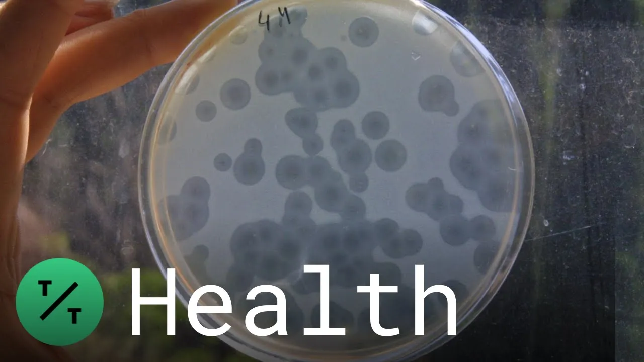
Is Phage Therapy the Answer to Antibiotic Resistance?
9.3K
Phage therapy uses bacteriophages—viruses that naturally evolved to attack bacteria—to combat antibiotic-resistant infections; these viruses are highly specific, targeting only harmful bacteria while preserving beneficial microbiota, making them a promising alternative or complement to traditional antibiotics which are becoming increasingly ineffective due to multi-drug resistance.
Click to watch →
Race Against Resistance: The Life And Death Struggle To Save Antibiotics | BBC StoryWorks
387.3K
Bacteriophages are naturally occurring viruses that specifically infect bacteria, serving as Earth's most prolific bacterial killers. Adaptive Phage Therapeutics develops personalized phage therapy by collecting environmental samples and screening for phages that match specific resistant bacteria. When conventional antibiotics fail, phage therapy offers a targeted alternative that can eliminate dominant resistant strains, allowing remaining bacteria to respond to standard treatments. This approach represents a potential solution for infections that have become untreatable with existing antibiotics.
Click to watch →
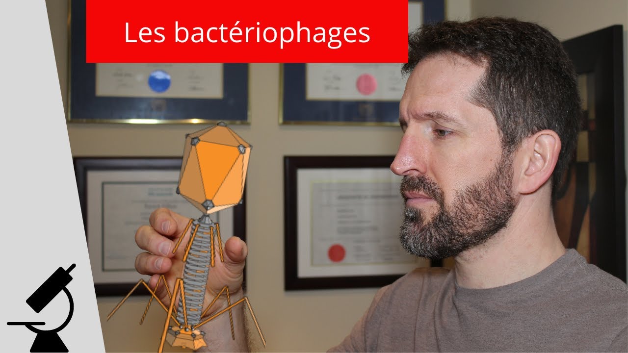
LES BACTÉRIOPHAGES : tueurs de bactéries
824
Bacteriophages have been used as antimicrobial agents since the 1920s, before antibiotics became widely available. However, antibiotics were preferred because they are easier to produce and have a broader spectrum of activity. With the rise of antibiotic-resistant bacteria, bacteriophage therapy has regained interest. Unlike antibiotics, bacteriophages are highly specific to particular bacterial strains. Antibiotic resistance develops through natural selection: when antibiotics are used, they kill susceptible bacteria but leave behind any bacteria with random mutations making them resistant. These resistant bacteria then survive and multiply, becoming the dominant population. This is why antibiotic overuse accelerates the development of resistant bacterial strains.
Click to watch →

New therapy could be solution to antibiotic resistance
1.5K
Phage therapy uses bacteriophages (viruses that infect and kill bacteria) as a natural solution to combat antibiotic-resistant superbugs; unlike antibiotics, phages are unaffected by bacterial resistance mechanisms and can target specific bacteria without harming beneficial microbes, offering a promising approach to address the growing global health threat of antimicrobial resistance.
Click to watch →

Using 1 germ to fight another when today's antibiotics fail
787
Phage therapy uses viruses called bacteriophages to fight bacterial infections by specifically targeting and killing particular bacterial strains through a mechanism different from traditional antibiotics—phages attach to bacteria, inject their DNA, replicate inside the bacterial cell, and then burst open the cell to destroy it, offering a potential solution for antibiotic-resistant infections while preserving beneficial gut bacteria.
Click to watch →
Prerequisite Knowledge
- Concept 01Basic biology of bacteriophages, including their structure and the distinction between lytic and lysogenic life cycles.
- Concept 02Fundamentals of aseptic technique to prevent contamination of environmental samples and bacterial cultures.
- Concept 03Standard bacterial culturing methods, such as preparing agar plates, inoculating liquid media, and serial dilutions.
- Concept 04The concept of antibiotic resistance in bacteria and why alternative treatments like phage therapy are necessary.
Subsequent Learning
- Step 01Phage purification and titration techniques, such as the double-layer agar plaque assay, to determine viral concentration.
- Step 02Host range determination and specificity testing to identify which bacterial strains the isolated phage can target.
- Step 03Phage genomics and bioinformatics, focusing on sequencing phage DNA to rule out lysogenic genes or toxin-encoding regions.
- Step 04Formulation, stability testing, and clinical applications of phage therapy for treating multidrug-resistant bacterial infections.
Phage Hunt
Assay Pick
Phage Hunt
0:05Playing Section
- 1
Describes finding phages in bird feces at a park targeting E. coli.
- 2
Details homogenizing sample in a 50 ml conical tube with buffer.
- 3
Centrifuging, filtering through 0.22 µm to remove bacteria.
Safety, Regulatory, and Resistance Limitations of Wild-Type Environmental Phages
While isolating bacteriophages from the environment is a foundational step in phage therapy research, relying on wild-type environmental isolates presents significant challenges compared to standardized treatments. Critiques of this approach highlight several key limitations: first, the risk of lysogenic (temperate) phages integrating into the host genome and transferring antibiotic resistance genes or toxins (transduction). Second, the highly narrow host range of wild phages makes empirical treatment difficult, often requiring custom isolation processes that are too slow for acute infections. Third, bacteria rapidly evolve resistance to specific phages. Consequently, many researchers and regulatory bodies advocate for genetically engineered phages, purified phage lytic enzymes (lysins), or highly characterized synthetic phage cocktails over raw environmental isolates to ensure safety, predictability, and broad-spectrum efficacy.
Deep Dive
Phage purification and titration techniques, such as the double-layer agar plaque assay, to determine viral concentration.
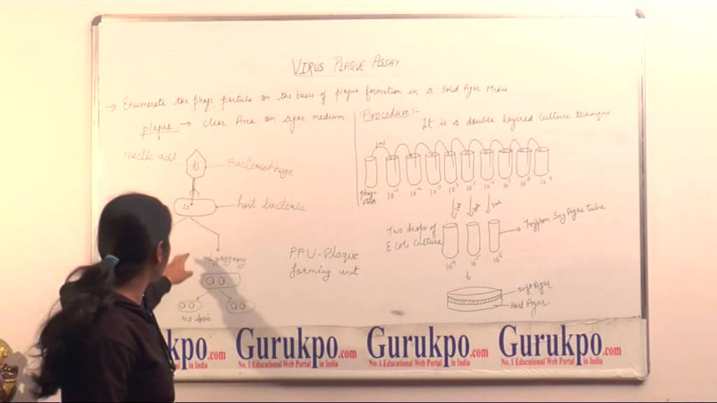
What is Virus Plaque Assay, BSc by Ms. Poonam Rajawat, Biyani Girls College, Jaipur
15.3K
Virus plaque assay is a double-layer technique used to enumerate bacteriophage particles by counting clear zones (plaques) formed on solid agar media when bacteriophages infect bacterial cells and cause lysis; each plaque represents one plaque-forming unit (PFU), and the original phage concentration is calculated by multiplying the plaque count by the dilution factor.
Click to watch →
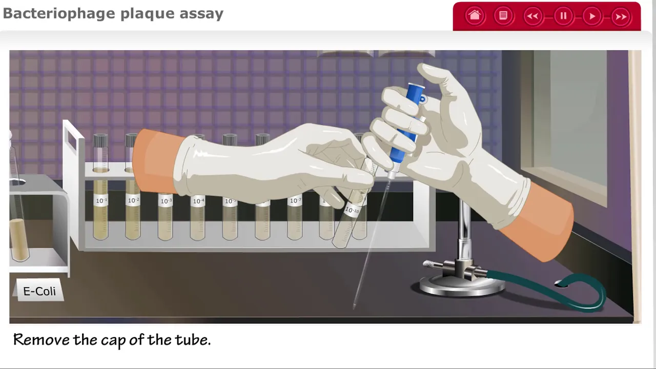
Bacteriophage Plaque Assay for Phage Titer UPDATED
49.7K
The bacteriophage plaque assay is a laboratory technique used to determine the concentration (titer) of bacteriophages in a sample by serially diluting the phage stock, mixing each dilution with a bacterial culture (E. coli) in soft agar, pouring onto petri plates, and incubating; the number of clear plaques formed indicates the original phage concentration, with more dilute samples yielding fewer plaques.
Click to watch →

Module 35 Determination of Phage Titre T4 by Plating Method Using E coli
145
The plaque assay is a fundamental microbiological technique used to determine the concentration of bacteriophages (such as T4) in a sample by counting clear zones (plaques) formed when phages infect and lyse bacterial cells (E. coli K12 or strain B) on agar plates; each plaque represents one or more phage particles that infected a single host cell, and the titre is calculated by dividing the number of plaques by the dilution factor and volume of sample used, expressed as plaque forming units per milliliter (PFU/mL).
Click to watch →
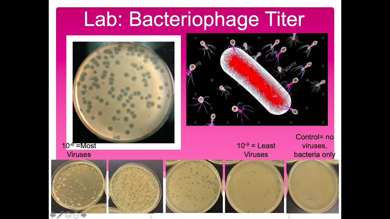
Bacteriophage Titer Lab.
20.2K
Bacteriophages are viruses that specifically infect bacteria, and their titer (concentration) can be determined using the double agar layer technique, which involves mixing bacteria with serial dilutions of phage, pouring the mixture onto agar plates, and counting the resulting clear spots called plaques that form when phages infect and lyse bacterial cells; the titer is calculated by dividing the number of plaques by the dilution factor to obtain plaque-forming units per milliliter.
Click to watch →

Lab 6-5: Plaque Assay
12.5K
A plaque assay determines viral titer by serially diluting a phage stock, mixing each dilution with E. coli host cells, and plating onto agar to form plaques (clear zones where viruses lyse bacterial cells); the titer is calculated as plaque-forming units (PFU) divided by the final dilution factor, with reliable counts obtained from plates containing 30-300 plaques.
Click to watch →
Deep Dive
Host range determination and specificity testing to identify which bacterial strains the isolated phage can target.
Webinar AMR 1: Mengatasi Resistensi Bakteri: Tantangan dan Solusi melalui Bakteriofaga di Indonesia
1K
Phage host range determines which bacterial strains a phage can infect. Studies show that most isolated phages can infect a broad range of host strains, with only about 13.5% of samples failing to infect any tested host. This specificity is determined by the compatibility between phage receptors and bacterial surface structures. Understanding host range is crucial for selecting appropriate phages for therapeutic applications.
Click to watch →

Jaden Bhogal Presents "Comparing Phage Host Range Determination Techniques" | The 2021 GHLC at JHU
298
Host range determination—the identification of which bacterial species a phage can infect—is critical but problematic due to multiple competing methods lacking standardization. This research compared four techniques (spot testing with/without dilutions, plaque testing, culture lysis) using quantitative meta-analysis of 22 studies. While overall material usage showed no significant differences, spot testing without dilutions used significantly less phage filtrate than plaque testing, making it more resource-efficient for high-throughput screening needed in therapeutic development.
Click to watch →
![[Frédérique Le Roux] Des virus contre les bactéries : une alternative aux antibiotiques](https://i.ytimg.com/vi/RgcqA9gET7s/maxresdefault.jpg)
[Frédérique Le Roux] Des virus contre les bactéries : une alternative aux antibiotiques
399
Phages are isolated by filtering environmental samples and growing bacteria on selective media. Phages are identified by creating plaques (clear zones where bacteria have been lysed). Phage specificity is determined by receptor binding - each phage can only attach to bacteria with specific surface proteins. This mechanism is analogous to how viruses bind to host cell receptors.
Click to watch →

Андрей Летаров: "Я назову тебя лямбда – самое изученное существо на планете"
10.7K
Different lambda phage strains have different tail fiber variants that determine their host range. The gpJ protein contains a receptor-binding domain that recognizes specific bacterial receptors. Some lambda phage strains have evolved modified tail fibers that recognize different receptors, allowing them to infect different bacterial strains. This evolution of host range is an important aspect of phage ecology and evolution.
Click to watch →

VIP Webinar Series (13th)- Phage Isolation, Culture, and Characterization
1.2K
Comprehensive phage characterization includes: Efficiency of Plating (EOP)—serial dilution to find highest dilution producing confluent lysis; Routine Test Dilution—the highest dilution failing to produce confluent lysis (standard for future testing); Single Step Growth Curve—analyzing sigmoid curves to determine latent period (time to maximum growth rate) and burst size (phages released per cell); Host Range Determination—testing coverage across bacterial species within genus or family; PFGE for genome sizing; Electron microscopy for morphological identification. Different phages show varying lytic activities: clear confluent lysis, opalescent lysis, no reaction, individual plaques, or resistant mutants. For difficult-to-culture bacteria like Bacteroides, metagenomic approaches are used: extract all genetic material from the sample, sequence it, and use bioinformatics to distinguish phage sequences from host and human DNA. The CRASS phage was discovered this way in human fecal metagenomes—it is 97 kb and six times more abundant than all other known phages combined. Temperate phages can integrate their DNA into bacterial genomes (lysogeny), avoiding lysis. To detect lysogenic phages: Mitomycin C induction—add 4-8 μg/ml to bacterial cultures, incubate 2 hours, then incubate overnight with shaking; UV irradiation—expose diluted bacterial cultures (OD 0.2-0.3) to 6-30 mJ/cm² UV-C, then incubate overnight. Both methods induce the lytic cycle, allowing detection of previously latent phages through plaque formation on host lawns.
Click to watch →
Deep Dive
Phage genomics and bioinformatics, focusing on sequencing phage DNA to rule out lysogenic genes or toxin-encoding regions.
Phage Genome Annotation with Deborah Jacobs-Sera | IBRC & Phage Directory
2.5K
Phages are viruses that infect bacteria, with genomes adapted to specific hosts and lifestyles characterized by short genes and limited redundant DNA. The International Bacteriophage Research Consortium and Phage Directory co-host this webinar series to address critical needs in standardized phage bioinformatics training. Phage research serves both basic science (understanding viral biology) and applied science (therapeutic applications like phage therapy). The field attracts researchers because phages are incredibly abundant—estimated to exceed stars in the sky—with numbers so vast that analogies compare them to beetles covering Earth's surface or requiring the entire human population to balance them on a scale. Phage genome sequencing begins with DNA extraction from environmental samples, followed by Illumina sequencing for adequate coverage. Phage genomes exist as linear DNA molecules inside viral heads, even though they become circular during host infection. Proper orientation requires identifying genome termini, which can be direct terminal repeats, circular permutation, or 5' or 3' overlaps. The convention is to orient genomes so structural genes read left-to-right, following historical precedent from early phage research. Gene order typically follows a pattern: terminase/portal genes at ends, scaffolding/protease for capsid assembly, capsid genes, tail genes, lysis cassette, immunity cassette, and host DNA interaction genes.
Click to watch →

DMC Natural Sciences: Bacteriophage Applications in Agriculture
391
Advanced bioinformatics tools enable comprehensive phage genome analysis: DNA Master predicts gene locations using probability algorithms; Famirator compares genomes to identify conserved gene clusters and evolutionary relationships; HHpred assigns protein functions and predicts 3D structures. Analysis of Goose phage revealed 87 genes, including a stolen bacterial endonuclease gene repurposed for host attack and an integrase gene lost due to dispensability. The tape measure protein determines tail length through precise molecular counting during assembly.
Click to watch →

Friday Lecture 1 Phage Genome Organization
149
Phage genome organization is conserved within families (synteny). Cytovirid genomes have left arm encoding structural genes (terminase, portal, lysis cassette) and right arm encoding early lysogenic genes. Lysogenic cassette (attP, integrase, immunity repressor) is central. Protoid genomes have different organization with terminal protein at 5' end. Terminal proteins serve as primers for DNA replication, with serine hydroxyl group attaching first nucleotide. This conservation allows gene function prediction based on position—for example, genes following tape measure protein are likely minor tail proteins. Phage structural proteins include major capsid protein, portal protein, terminase, capsid maturation protease, scaffolding protein, and minor tail proteins. Understanding genome organization and packaging mechanisms is essential for genome annotation and comprehending phage biology.
Click to watch →

Foro de Investigación Subgraduado | Departamento de Ciencias y Tecnología - Inter Investiga
111
Phage genomes contain genes organized in functional regions: structural genes (capsid, tail) at the beginning, and genes for host interaction (injection, lysis, lysogeny) at the end. The region containing immunity repressors and integrases determines whether the phage causes bacterial death (lytic cycle) or integrates into the host genome (lysogenic cycle). Isolated bacteriophages are deposited in public databases (such as the Actinobacteriophage Database) for global scientific access. Phages are also preserved in ultra-freezers at -80°C for long-term storage and potential clinical applications.
Click to watch →
Lytic cycle and lysogenic cycle of bacteriophage | virology lecture 5
103.6K
The lysogenic cycle is a temperate state where bacteriophage DNA integrates into the bacterial chromosome as a prophage, remaining dormant without producing new particles. This allows passive propagation through bacterial binary fission, exponentially increasing phage DNA copies. The prophage can remain integrated for many generations until environmental triggers induce phage conversion to the lytic cycle. This dual-cycle strategy provides evolutionary advantages: lysogeny ensures survival during unfavorable conditions while maintaining latent infectivity, while lytic phase enables explosive reproduction when conditions permit.
Click to watch →
Deep Dive
Formulation, stability testing, and clinical applications of phage therapy for treating multidrug-resistant bacterial infections.
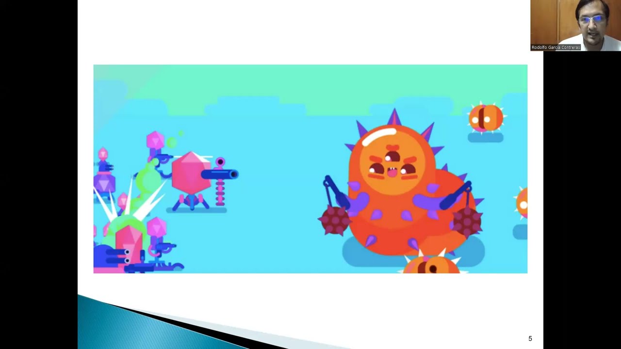
USO DE BACTERIÓFAGOS PARA COMBATIR INFECCIONES POR BACTERIAS MULTIDROGO RESISTENTES
681
Bacteriophage therapy offers several advantages over antibiotics: they are easy to isolate from various sources including water, soil, and clinical samples; they are highly specific to target bacterial strains; they self-replicate in the presence of target bacteria, increasing concentration proportionally with bacterial load; and they can be used against multidrug-resistant infections when antibiotics fail. The FDA has approved phage therapy for compassionate use in patients with multidrug-resistant infections when no other treatment options exist. This has successfully treated septicemia, infected valve infections, and other life-threatening bacterial infections. The case of Stephanie Strachan, who survived a multidrug-resistant Citrobacter baumannii infection through phage therapy after conventional treatments failed, contributed to the formation of an institution dedicated to studying bacteriophages.
Click to watch →
EP-051 - Industry Insight Bacteriophage Therapies
260
Phage therapy offers multiple advantages over antibiotics. Phages are self-amplifying—they multiply at infection sites, manufacturing themselves where needed and disappearing when the job is done. They can penetrate biofilms that antibiotics cannot, dissolving protective bacterial shields or replicating inside structures. Phages preserve the microbiome by targeting only specific bacteria, avoiding the indiscriminate killing that causes C. diff infections and yeast overgrowths. Phages and antibiotics work synergistically, with studies showing over 90% bacterial eradication when combined versus 77% improvement alone. CRISPR-enhanced phages can reprogram bacteria to become antibiotic-sensitive or self-destruct. However, treating multidrug-resistant infections requires personalized phage cocktails: isolate and identify exact bacterial strains, screen hundreds of phages from libraries, combine three to eight different phages, amplify each separately, purify, test for contaminants, and combine in specific ratios. Multiply this by hundreds of patients—impossible without digital systems. Cloud-based manufacturing execution systems enable real-time tracking of phage libraries, production history, stability data, and efficacy profiles. Temperature control is critical—some phages need refrigeration while others are stable at room temperature. IoT sensors monitor conditions and trigger alerts for failures. Electronic batch records replace paper-based GMP documentation. Approximately 90 clinical trials are ongoing worldwide, with the Belgian consortium showing 77.2% clinical improvement and 61.3% bacterial eradication. The NIH is running trials for cystic fibrosis patients.
Click to watch →

Traitement des infections à bacilles Gram négatifs multirésistants: quelles perspectives?
582
Bacteriophages are viruses that specifically infect and kill bacteria, representing the most abundant living organisms on Earth. They have been used in Eastern European countries for decades but fell into disuse due to lack of rigorous clinical studies. Phage therapy offers targeted treatment with minimal disruption to the microbiome, as phages only affect their specific bacterial hosts. Applications include intravenous administration, aerosol delivery, local application to wounds, and intravesical instillation. Phage therapy shows synergistic effects with antibiotics, restoring susceptibility to some antibiotics. Susceptibility testing (phagotyping) is performed by sending bacterial isolates to specialized laboratories. Clinical experience at CHIREC hospital with 21 patients treated for MDR Gram-negative infections shows predominantly OXA-48-producing Enterobacteriaceae, Pseudomonas with porin mutations and efflux pumps, and VIM-producing organisms.
Click to watch →

La phagothérapie en Belgique
315
Phage resistance develops primarily through bacterial receptor mutations, with larger phage genomes having greater capacity to overcome resistance. The coevolutionary 'arms race' between phages and bacteria involves trade-offs between resistance costs and infectivity. Phage-antibiotic synergy occurs through multiple mechanisms: antibiotics cause bacterial filamentation increasing phage sensitivity, phages accelerate lysis and virion production, and phages degrade bacterial capsules facilitating antibiotic penetration into biofilms. This synergy can restore antibiotic sensitivity in resistant bacteria. Phage susceptibility testing uses the double agar overlay method, calculating efficiency of plating (EOP) as the ratio of plaque-forming units on test versus reference bacteria. An EOP of at least 0.1 is considered therapeutic. However, some antibiotics (like rifampicin affecting RNA polymerase) can antagonize phage replication, requiring careful synergy testing.
Click to watch →

An Introduction to Bacteriophages Basic Science and Clinical Applications June 30, 2025
271
Successful phage therapy implementation requires coordinated efforts across clinical, laboratory, and regulatory domains. Physicians identify difficult-to-treat infections, microbiology labs characterize resistant organisms, and specialized phage centers conduct susceptibility screening. FDA oversight through IND applications enables compassionate use while ensuring safety. Two major bottlenecks exist: finding appropriate phages (some organisms require extensive environmental sampling) and producing clinical-grade phages meeting pharmaceutical standards. Recent clinical trials provide compelling evidence: aerosolized phage cocktails reduced Pseudomonas counts by 2 logs in cystic fibrosis patients; phage plus antibiotic achieved >80% microbiological success in resistant UTIs; combined intravenous/topical phage accelerated healing in diabetic foot osteomyelitis; and phage therapy outperformed antibiotics alone in complicated Staphylococcus sepsis. These studies establish proof-of-concept and frameworks for larger trials, positioning phage therapy at an inflection point for mainstream integration.
Click to watch →
Phage Hunt
Assay Pick
Phage Hunt
0:05- 1
Describes finding phages in bird feces at a park targeting E. coli.
- 2
Details homogenizing sample in a 50 ml conical tube with buffer.
- 3
Centrifuging, filtering through 0.22 µm to remove bacteria.
Safety, Regulatory, and Resistance Limitations of Wild-Type Environmental Phages
While isolating bacteriophages from the environment is a foundational step in phage therapy research, relying on wild-type environmental isolates presents significant challenges compared to standardized treatments. Critiques of this approach highlight several key limitations: first, the risk of lysogenic (temperate) phages integrating into the host genome and transferring antibiotic resistance genes or toxins (transduction). Second, the highly narrow host range of wild phages makes empirical treatment difficult, often requiring custom isolation processes that are too slow for acute infections. Third, bacteria rapidly evolve resistance to specific phages. Consequently, many researchers and regulatory bodies advocate for genetically engineered phages, purified phage lytic enzymes (lysins), or highly characterized synthetic phage cocktails over raw environmental isolates to ensure safety, predictability, and broad-spectrum efficacy.
Basic biology of bacteriophages, including their structure and the distinction between lytic and lysogenic life cycles.
Fundamentals of aseptic technique to prevent contamination of environmental samples and bacterial cultures.
Standard bacterial culturing methods, such as preparing agar plates, inoculating liquid media, and serial dilutions.
The concept of antibiotic resistance in bacteria and why alternative treatments like phage therapy are necessary.
Phage purification and titration techniques, such as the double-layer agar plaque assay, to determine viral concentration.
Host range determination and specificity testing to identify which bacterial strains the isolated phage can target.
Phage genomics and bioinformatics, focusing on sequencing phage DNA to rule out lysogenic genes or toxin-encoding regions.
Formulation, stability testing, and clinical applications of phage therapy for treating multidrug-resistant bacterial infections.
40.7K views653likes3:47@BCMTAILORLabsOriginal Release: 2021-01-06
hi everyone today i'm going to show you how to isolate a phage so phage hunting can be really fun you can go anywhere to find phages you can go to the ocean you can go to a nice lake you could go to the park one of my first experiences phage hunting was actually at a park so what we did is we turned to geese and duck poop in order to find phages i was studying e coli at the time that can infect and kill people but it's also found in birds and so what we thought is that if we could find these bird samples where these e coli were the phages would probably be there too and we could isolate those phages and then purify them and use them as a treatment for this e coli infection and so what we did is we went to the park we had 50 mil conicals and spatulas and that's really all you need in order to find a phage so first you homogenize your sample this is about half fecal half buffer then you centrifuge you take the supernatant and you filter this is a 0.22 micron filter and this is to remove any bacteria i use a glass vial sometimes to keep my lysates and my phage sometimes you may need to centrifuge a second time to get it through that filter but don't worry eventually you will get it next you take your plate and your melted auger i use about three ml of melted auger and to that i add about a hundred microliters of an overnight culture of bacteria then i add my fecal filtrate you can add anywhere from 100 to 500 microliters to that same vial and i mix and i plate this is a double auger assay after an overnight incubation at 37 you look for plaques sometimes you can look at your plate horizontally to see if there are indentions that's usually indicative of a bubble and not a plaque once you find your plaque you pick it i use a glass pipet and some buffer about 500 microliters per plaque do you see the plaque i pierce right through that plaque and i pipet as much as i can and then i pipet up and down into the buffer and that's plaque isolation you can use that to replate and make a light
Up Next

6:56
Nernst and Goldman Equations: Calculating Equilibrium Potential
@NonstopNeuron
41.2K views•2023-08-07

50:12
Bacterial Communication: Quorum Sensing and Biofilm Formation Explained
@JHUAAP
12.6K views•2011-06-28

5:35
Enteric Nervous System Explained: The Gut's Brain | Neurobiology Lecture
@alumniu6029
438 views•2018-09-12

7:08
Bacteriophages: Earth's Deadliest Killers and Future Antibiotics
@kurzgesagt
34.6M views•2018-05-13
Related Study Plans & Knowledge Roadmaps
Structured learning paths in Biology
biology
RNAi: Biogenesis, RISC & Gene Silencing
01Molecular Biology Foundations: DNA to Protein
02Introduction to RNA Interference (RNAi)
03Molecular Biogenesis of miRNA and siRNA
+ 2 more learning modules
10h5 modules
Explore Roadmap
biology
Mycorrhizae: Signaling, Biophysics & Ecology
01Foundations of Mycorrhizal Symbiosis
02Molecular Signaling and Root Colonization
03Biophysics and Bioenergetics of Nutrient Exchange
+ 1 more learning modules
11h4 modules
Explore Roadmap
biology
Circadian Clocks: TTFL, SCN & Periphery
01Foundations of Molecular Biology & Gene Expression
02Introduction to Chronobiology and Actograms
03The Core TTFL: Molecular Architecture of the Clock
+ 3 more learning modules
14h6 modules
Explore Roadmap
biology
Population Genetics: Math, Drift & Selection
01Genetic & Probability Foundations
02The Hardy-Weinberg Principle and Baseline Models
03Mathematical Modeling of Natural Selection
+ 3 more learning modules
18h6 modules
Explore Roadmap