











![Tomographic Image Reconstruction: Introduction (Part 1) [L28]](https://i.ytimg.com/vi/KjYnjlFLzvA/maxresdefault.jpg)





![PET Imaging: Introduction (Part 1) [L33]](https://i.ytimg.com/vi_webp/tkTxNEFyO7Y/maxresdefault.webp)



![What is the Standard Uptake Value (SUV) in PET and SPECT Imaging? [L38]](https://i.ytimg.com/vi_webp/nmT9RrNxzK0/maxresdefault.webp)
![[18F]FDG-PET Workshop - Computation of Standardized Uptake Value Images](https://i.ytimg.com/vi/9_GKP0-pQRk/maxresdefault.jpg)



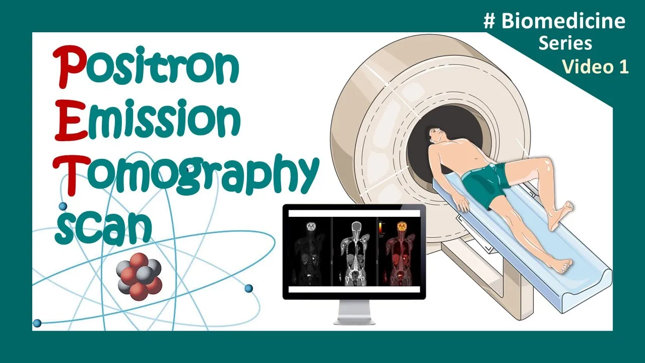
SPECT (Single Photon Emission Computed Tomography) and PET (Positron Emission Tomography) are the two main modalities for 3D tomographic imaging in nuclear medicine. SPECT uses single gamma-ray emitting radioisotopes with mechanical collimation to acquire projections, while PET uses positron-emitting radioisotopes with electronic collimation through coincidence detection of back-to-back gamma photons. PET offers higher sensitivity (up to 100 vs. 1 in 10,000 for SPECT) and better resolution (~5mm vs. ~10mm for SPECT), but SPECT is more cost-effective using isotopes like Technetium-99m, making it still widely used for applications like bone metabolism imaging.
SPECT vs PET Imaging: Basic Concepts in Nuclear Medicine
Added:Basic principles of nuclear physics, specifically radioactive decay, radionuclides, and the difference between gamma emission and positron-electron annihilation.
The fundamental distinction between structural imaging (like CT or MRI) and functional/molecular imaging.
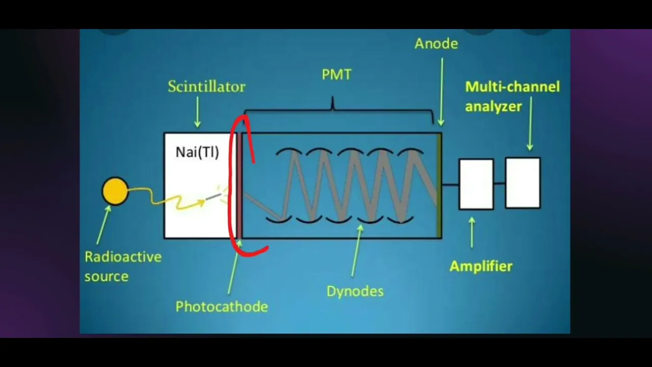
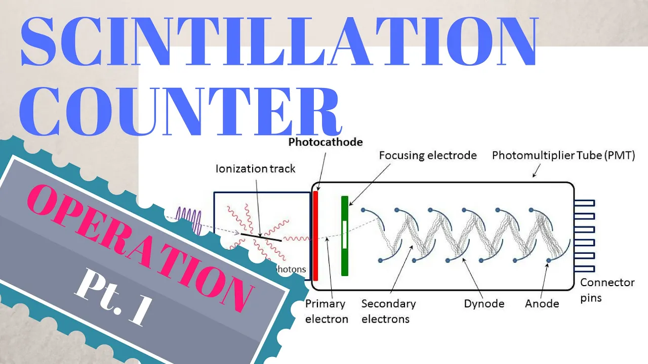
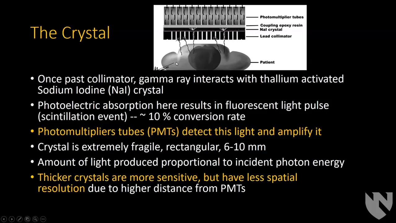
Standard radiation detection concepts, including how scintillation crystals and photodetectors convert gamma-ray photons into electrical signals.
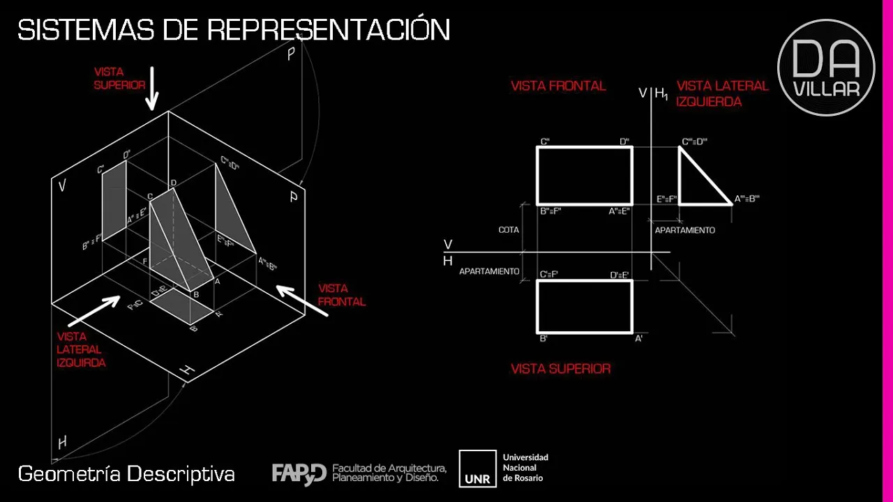
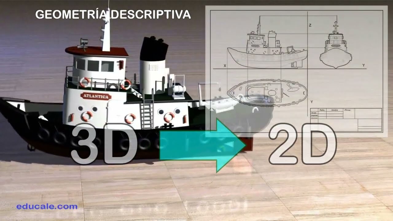
Elementary geometry and physics of projections, representing how a 3D object's properties are captured on a 2D detector plane.
Advanced mathematical image reconstruction algorithms, comparing Filtered Backprojection (FBP) with Iterative Reconstruction methods (e.g., OSEM).
The design and clinical benefits of hybrid imaging modalities, such as PET/CT, SPECT/CT, and PET/MRI, which combine anatomical and functional data.
The biochemistry of key radiopharmaceuticals, such as Fluorodeoxyglucose (18F-FDG) and Technetium-99m, and how they target specific metabolic pathways.
Quantitative analysis techniques in molecular imaging, including the calculation of Standardized Uptake Values (SUV) in oncology.
Radiation safety, dosimetry calculations, and ALARA (As Low As Reasonably Achievable) protocols for clinical nuclear medicine procedures.
653 views9likes12:36Original Release: 2021-04-03
welcome to another short video on imaging in nuclear medicine in this video we want to have a brief look at the two main modalities that we use for 3d tomographic imaging in nuclear medicine so before diving into those two main modalities let's quickly refresh our memory what we need to do to do tomography and um a crucial term in tomography is the so-called projection or the parallel projection so what is the projection so let's say we want to image the 3d distribution of a quantity of interest so nuclear medicine that would be the activity or the tracer concentration of our radiotracer let's say we have a brain here so we have 3d distribution of the tracer in the brain so a projection would be if you would take this 3d distribution and we would project it onto one plane we can do that by calculating the line integrals along a certain direction so we have many line integrals all for example here in this example going in the vertical direction so from top to bottom so if we do that we project the 3d distribution into a 2d plane and of course i can do that we can do that in different directions so here for example the 45 degree projection then a 90 degree projection so this would be basically looking through the brain from left to right so you would get that projection you can do it for 135 degrees and of course also 180 degrees so in the end we obtain a series so from a 3d distribution we can obtain a series of 2d projection images and then actually it was shown already in the beginning of the 20th century so around 1917 by the austrian mathematician that if you collect many of those projections of those line integrals and they are well sampled you can actually reconstruct back the 3d distribution of your quantity of interest so in our case it would be the activity distribution or the tracer distribution so the key to do tomographic image reconstruction is to somehow acquire those projections so how do we acquire those projections in nuclear medicine as i told you already there are two main ways of doing that the first one is called single photon emission computer tomography or also spect inspect use this radioisotopes that emit single gamma rays so single photons so let's think of a toy example so we have a patient indicated by the grey ellipse here and let's assume in that patient we have four regions that accumulated the radio tracer so we get actually single gamma emissions from four different spots so from the four blue spots and of course a single gamma emitter emits gamma rays not only in one direction but in all directions soon and all four pi right um so and i've colored those emissions in a different direction with different colors so red is the vertical direction green is horizontal and black is the 45 degree direction so you could naively think that okay what would happen if you just take a single detector that can detect single gamma rays if i place it at a certain position what is the detector actually seeing and if you look at that arrangement you can actually see unfortunately of course there is no end so there is no angular information in the detected events so of course this detector will see photons coming from all directions right so it will see the red photons coming from that emission point but of course we'll also see the black photons coming from that emission point so if we simply do that we don't acquire a projection but there's an easy way to fix that so we can just use what's called mechanical collimation so if you put a bit of metal here at the sides of the detector so to create basically a narrow hole or a narrow tunnel we can black and we can sorry we can block the photons that come here from the oblique direction so in that way now the detector is only sensitive to photons coming from basically this line or this volume here which is actually proportional to the line integral of all the activity along that line so this is exactly what we need then of course instead of using one collimated detector you can use many collimated detectors and if you do that of course then you acquire many of those projection lines and you can see an example of such a system actually here so this is a clinical uh spec system as you can find it in many in many hospitals and so you can see here this is a detector head containing many detectors and a collimator so the collimate actually is shown here so keep in mind that the way i've shown it here is not really how a detector had and a speck system works but for just understanding the basic principle that's that's enough and actually if you look at the camera you can see there's not only one detector head but two detector heads so there's another one here so there would be another detector right here and those detector heads they can first of all move in the vertical direction so you can move them you can bring them very close to the patient we will see later why this is important and moreover you can also rotate the detector heads around the patient so we can acquire projections from all different angles what we need for tomographic image reconstruction so in that way we can acquire all our projection images and we can do image reconstruction the second modality is called pet or positron emission tomography and pet life is a bit simpler so if you remember in pets we use radioisotopes that emit positrons and then the positrons after decay or the emitted positrons they annihilate with an electron into two five 11 kv gamma photons unfortunately they are emitted back to back so the angle between them is to very good approximation it's 180 degrees it's not exactly 180 degrees but it's very close to 180 degrees so they're emitted back to back so if we now actually put a complete ring of small detectors around our patient if you look at the individual detectors of course an individual detector still sees photons from all the directions right so if you would focus on that detector that will see photons from that emission point but also photons from that emission point and of course also photo from photons from that emission point but if you use a trick so if you look at what we call coincidence detection so basically we look which or we look at pairs of detectors that saw or the detected photons within a very short amount of time which is usually called the coincidence window so that window is a few nanoseconds so let's say for example we saw that this detector saw a photon and then within a short amount of time this detector here saw a photon as well then we can assume that those two photons came from the same positron electron annihilation and that means of course since the photons are emitted back to back that we know that the positron electron annihilation must have happened somewhere on the line connecting those detectors and the same is of course true for any pair of detectors so you can connect those two detectors so any coincidence detections that we see between those detectors must have happened somewhere on the line between the detectors and of course i can do that for all the vertical lines and all for the oblique lines so by basically recording all those coincidence detections we measure all the projections that we need and the important difference compared to spec is that here in the pet scanner we don't need to collimate our detectors and this is nice because of course a collimator blocks a huge part of the incumbent photons so that actually leads to a loss of of sensitivity as an example you can see here a clinical pet system actually you can't see too much this is actually a pet ct combination so there's a ct scanner here and then somewhere behind there's a ring of pet detectors not too interesting to see but if you're ever in a hospital you will see those devices let's have a quick look at the comparison between the two modalities so as i told you inspect the principle is that we use single gamma emitters we have collimated detectors in a detector head and we rotate that detector head or multiple of those heads around the patient to acquire all the projections in contrast and pads we use what is sometimes called electronic collimation so we look at coincidence detections between two detectors to get the all the projections that we need um if you compare the sensitivity so that's basically how many of the emitted photons do we really detect that is quite low in spect um depends a bit on the collimator that you use but basically the collimator blocks most of your incoming photons so the sensitivity can be around one in in ten thousand in pets because we don't need a collimator the sensitivity is actually way higher so that can be to 100 or even higher now depends a bit on how long your your pet system is so sensitivity impact is way higher than in inspect in terms of resolution so if we look at standard whole body scanners as used in every hospital we also see that the resolution of the reconstructed images is way higher so in modern pet scanners that's around five millimeters now in modern spec scanners it depends a bit on the the spec system and especially on the column meter that you use but typically a typical benchmark right now is around 10 millimeters so also the resolution and pad of the pad images is usually way higher than the resolution of spec images in whole body human imaging um the most common isotopes that are used inspect is by far 99 meter stable tempnessium and in pet it's while the classical famous positron images are f-18 carbon 11 and also gallium 68 lately an important difference is also if you calculate the costs that are needed to basically do a spect or pet examination so the costs of a spec scan are much lower than the costs of a pet scan so that's why if you go to any nuclear medicine department you will see still see many spec scanners that are used because yeah they're just more cost effective so for example we know that we can image a bone metabolism with spectrosis so for example phosphonates labeled by technetium but we can also do we can also image the bone metabolism by sodium fluoride so that's a pet tracer and from the image quality point of view of course it's way better to do expect sorry to do a pet scan but from the cost point of view of course spec is way more cost efficient and that's why nowadays aspect is still used a lot to do actually bond with so to image the bone metabolism
Up Next

43:17
Radiolabelling Basics for Nuclear Medicine Physicians
@nuclearmedicinesnacks4682
1.6K views•2023-08-09

48:47
Graphic Medicine: Comics for Collaborative Healthcare Communication
@nationalpatientadvocate
189 views•2023-12-04

10:28
Neuroanatomy: Central and Peripheral Nervous System Divisions Explained
@AKLECTURES
136.2K views•2014-09-20

3:05
Stages of Labor and Vaginal Birth | Childbirth Animation
@nucleusmedicalmedia
52.1M views•2017-08-18
Related Study Plans & Knowledge Roadmaps
Structured learning paths in Medicine
medicine
Abnormal LFTs: Diagnostic Workup & Pathology
01Liver Anatomy and Basic Physiology
02Introduction to Liver Function Tests (LFTs)
03Hepatocellular vs. Cholestatic Patterns
+ 3 more learning modules
12h6 modules
Explore Roadmap
medicine
Analyzing Blood Smears: RBC, WBC & Platelets
01Introduction to Blood Components & Smear Preparation
02Normal Morphology of Blood Cells
03Erythrocyte Pathologies & Morphological Variants
+ 2 more learning modules
12h5 modules
Explore Roadmap
medicine
Mechanical Ventilation: Waveforms & Alarms
01Anatomy and Physiology of Breathing
02Introduction to Mechanical Ventilation & Core Modes
03Ventilator Settings and Waveform Interpretation
+ 2 more learning modules
12h5 modules
Explore Roadmap
medicine
Neuro Exam: Localization & Stroke Syndromes
01Foundations of Neuroanatomy and Vascular Supply
02The Neurological Examination: Step-by-Step
03Principles of Neurological Localization
+ 3 more learning modules
18h6 modules
Explore Roadmap