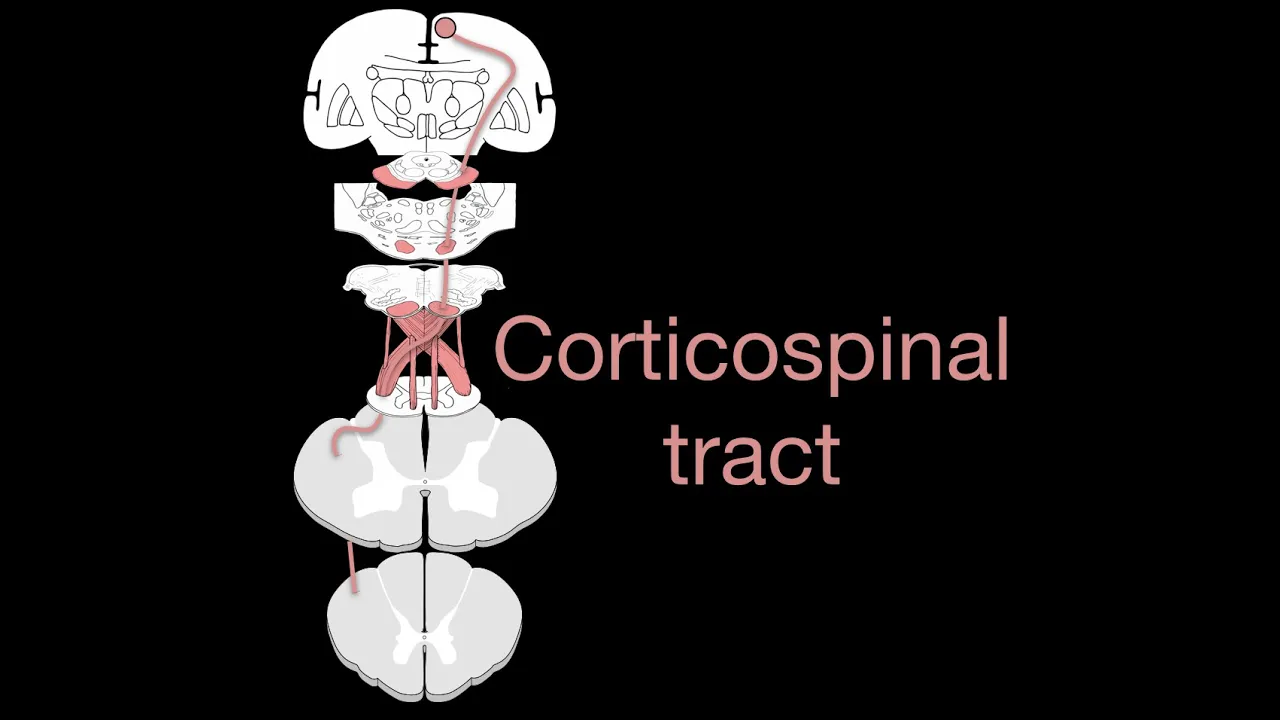
The corticospinal tract is a major neural pathway carrying movement-related information from the motor cortex to the spinal cord, consisting of upper motor neurons that connect to lower motor neurons controlling muscle contraction; approximately 90% of fibers decussate at the pyramidal decussation to form the lateral corticospinal tract (controlling distal muscle movements like hand movements), while the remaining 10% form the anterior corticospinal tract (controlling proximal trunk muscles); damage to this tract causes upper motor neuron syndrome with symptoms including weakness, hyperactive reflexes, and impaired fine motor control, particularly affecting writing and typing abilities.
Corticospinal Tract | Upper Motor Neuron Pathway Explained
Added:Deep Dive
Basic neuroanatomy, including the division between the central nervous system (CNS) and peripheral nervous system (PNS).
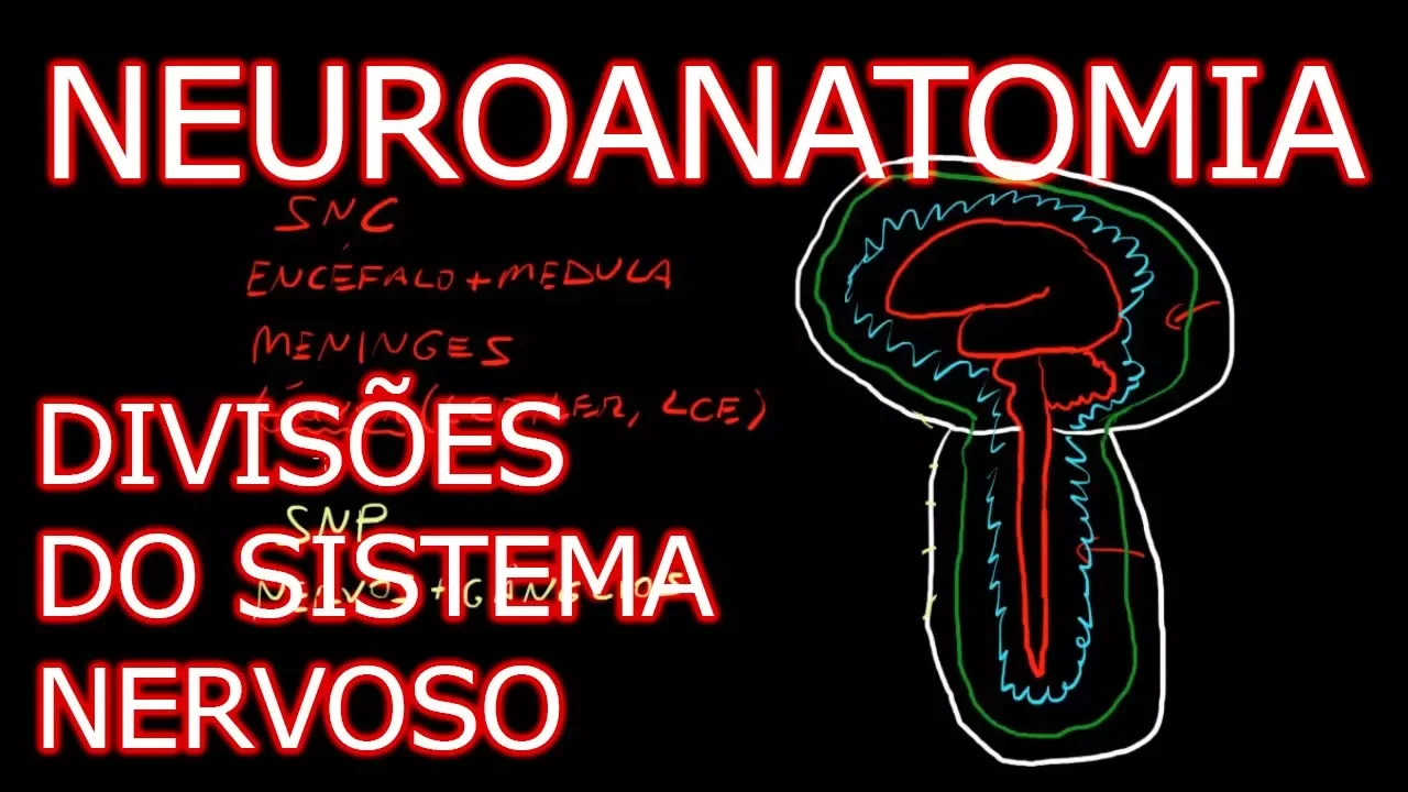
Aula: Introdução à Neuroanatomia - Divisões do Sistema Nervoso | Neuroanatomia Humana #1
222K
The nervous system is divided anatomically into the Central Nervous System (CNS) and Peripheral Nervous System (PNS). The CNS includes the brain (telencephalon, diencephalon, brainstem, cerebellum) and spinal cord, protected by bone. The PNS consists of nerves connecting CNS to effector organs (muscles, receptors), including spinal and cranial nerves. Ganglia are PNS structures, while nuclei are CNS structures. Both systems contain myelin, but produced by different cells (oligodendrocytes in CNS, Schwann cells in PNS).
Click to watch →
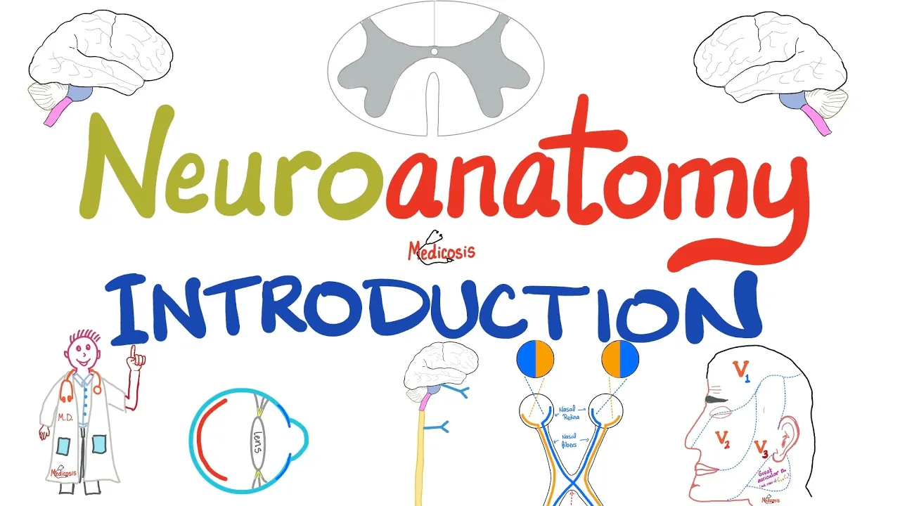
Introduction to Neuroanatomy - Learn the Basics - Neuroanatomy Playlist
377.9K
The nervous system is organized into the Central Nervous System (brain and spinal cord) and Peripheral Nervous System (all other nerves including cranial and spinal nerves). Motor nerves are efferent (carrying signals away from the brain) while sensory nerves are afferent (carrying signals toward the brain). The brainstem consists of midbrain, pons, and medulla, with the spinal cord having 31 segments. Myelinated fibers appear white (white matter) while unmyelinated appear gray (gray matter), with the spinal cord having gray matter internally and white matter externally, while the brain has gray matter externally and white matter internally. The autonomic nervous system includes sympathetic, parasympathetic, and enteric divisions, with preganglionic fibers being myelinated (appearing white) and postganglionic fibers being unmyelinated (appearing gray).
Click to watch →
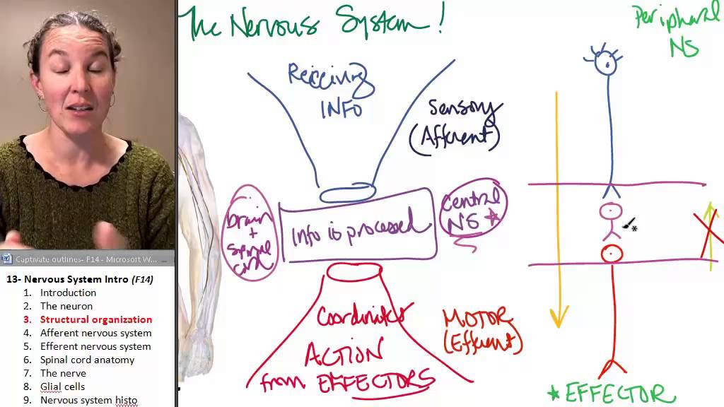
Nervous system 3- Structure
33.4K
The nervous system is divided into the central nervous system (CNS) and peripheral nervous system (PNS). The CNS includes all neurons entirely within the brain and spinal cord. The PNS includes all neurons not in the brain or spinal cord, even if they have parts that touch or enter the CNS. A bundle of axons in the PNS is called a nerve, while the same structure in the CNS is called a tract. A bundle of cell bodies in the PNS is called a ganglion, while the same structure in the CNS is called a nucleus.
Click to watch →

Neural Control and Coordination in One Shot | Antim yudh 3.O | NEET 2023 Crash Course
48.1K
The nervous system is divided into central (CNS) and peripheral (PNS) systems. The CNS includes the brain and spinal cord, serving as the main processing center. The brain is divided into forebrain (cerebrum, thalamus, hypothalamus), midbrain, and hindbrain (cerebellum, pons, medulla). The PNS includes somatic nervous system (voluntary skeletal muscle control) and autonomic nervous system (involuntary organ control). This organization allows efficient processing and coordination of body functions.
Click to watch →

SISTEMA NERVIOSO || CURSO PRE-MEDICINA
68.6K
The nervous system is divided anatomically into the Central Nervous System (CNS) and Peripheral Nervous System (PNS). The CNS consists of the brain and spinal cord, located centrally within the body. The PNS includes all nerves and ganglia extending to the periphery. The brain is composed of the cerebrum, cerebellum, and brainstem. The brainstem consists of the midbrain, pons, and medulla oblongata.
Click to watch →
Deep Dive
The anatomical structure of the cerebrum, specifically the location and function of the primary motor cortex (precentral gyrus) and the motor homunculus.
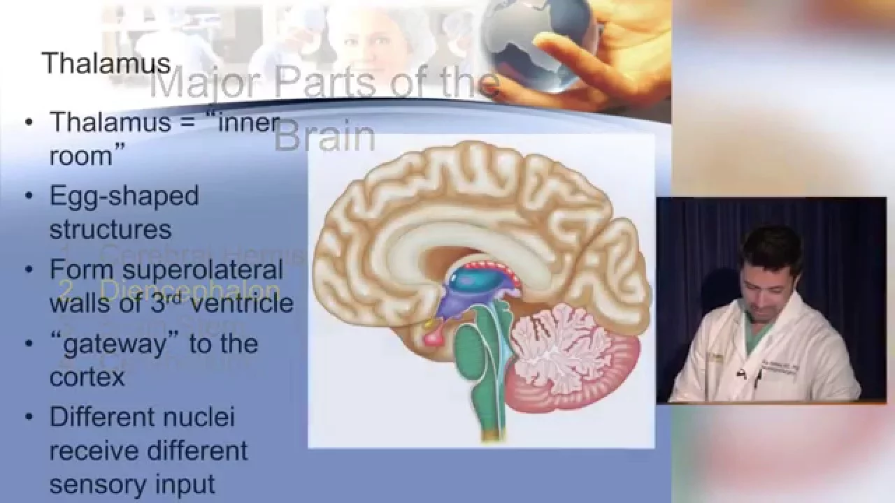
Neuroanatomy made ridiculously simple
1.2M
The primary motor cortex (Brodmann area 4) is located in the precentral gyrus, right behind the central sulcus at the back of the frontal lobe. It contains large pyramidal cells and has a somatotopic organization known as the motor homunculus. In this map, more lateral areas control hand and face movements, while more medial and superior areas control leg movements. This organization explains why strokes affecting different locations produce different deficits—for example, ACA strokes may cause leg weakness while sparing arm and hand function.
Click to watch →
Corticospinal tract
184.9K
The primary motor cortex is located in the precentral gyrus, which lies immediately anterior to the central sulcus on the lateral surface of the brain. The motor homunculus is a map within the precentral gyrus that represents motor control areas, with disproportionate representation where more complex body regions like the hands occupy larger cortical areas compared to simpler regions like the trunk or lower limbs.
Click to watch →
Nervous System || Important Questions || NORCET || AIIMS || By Raju Sir
53.6K
The primary motor cortex is located in the frontal lobe, specifically in the precentral gyrus. It controls voluntary movements of skeletal muscles. The motor homunculus maps different body parts to specific areas of the motor cortex, with body parts requiring fine motor control having larger representations.
Click to watch →

Vía Piramidal en vivo
18.8K
Brodmann area 4 (precentral gyrus) is the primary motor cortex controlling voluntary body movements. The lower part controls head, tongue, lips, and masticatory muscles, while the upper part controls upper limb, trunk, and lower limb. The motor homunculus represents body parts with disproportionately large cortical areas for those requiring fine motor control (face, tongue, lips, hands, feet). This contralateral representation means lesions cause paralysis on the opposite side of the body.
Click to watch →
![Lobus frontalis / frontal lob [DETAYLI KONU ANLATIMI]](https://i.ytimg.com/vi/zRfaITmTZC4/hqdefault.jpg)
Lobus frontalis / frontal lob [DETAYLI KONU ANLATIMI]
769
The primary motor cortex (Brodmann area 4) is located in the precentral gyrus between the central and precentral sulci. It controls voluntary movements and contains the motor homunculus, a body map where different body parts occupy specific areas. The face and tongue occupy a large area, followed by the hand and thumb, then the trunk, and finally the lower extremity. The medial surface controls the lower extremity, while the lateral surface controls the face, hand, and trunk. Motor commands travel through the corticospinal tract to control body movements. Lesions in different areas cause specific motor deficits: face and tongue weakness, hand paralysis, or leg paralysis depending on the location.
Click to watch →
Deep Dive
Cross-sectional anatomy of the spinal cord, distinguishing between grey matter (anterior/posterior horns) and white matter tracts.

Cross Section Of Spinal Cord 1 || anatomy
47K
The spinal cord cross-section reveals a butterfly-shaped gray matter surrounded by white matter, with gray matter containing neuronal cell bodies organized into ventral (lateral, central, medial groups), intermediate, and dorsal columns, while white matter contains ascending tracts (fasciculus gracilis, fasciculus cuneatus, spinothalamic tracts, spino-olivary tract, spino-tectal tract) and descending tracts (corticospinal tracts, rubrospinal tract, olivospinal tract, vestibulospinal tract, tectospinal tract).
Click to watch →

Anatomy & Physiology Of Nervous System | Nervous System | BSc Nursing 1st Year | Lifeline Batch #7
38.3K
The spinal cord cross-section reveals gray matter in the center (butterfly/H-shape) and white matter peripherally. The gray matter has anterior (ventral) horns containing motor neurons and posterior (dorsal) horns containing sensory neurons. The white matter is divided into anterior, posterior, and lateral funiculi. Ascending tracts carry sensory information to the brain, while descending tracts carry motor commands. The lateral spinothalamic tract carries pain and temperature sensations from the opposite side of the body.
Click to watch →
Spinal Cord Anatomy (1)
10.1K
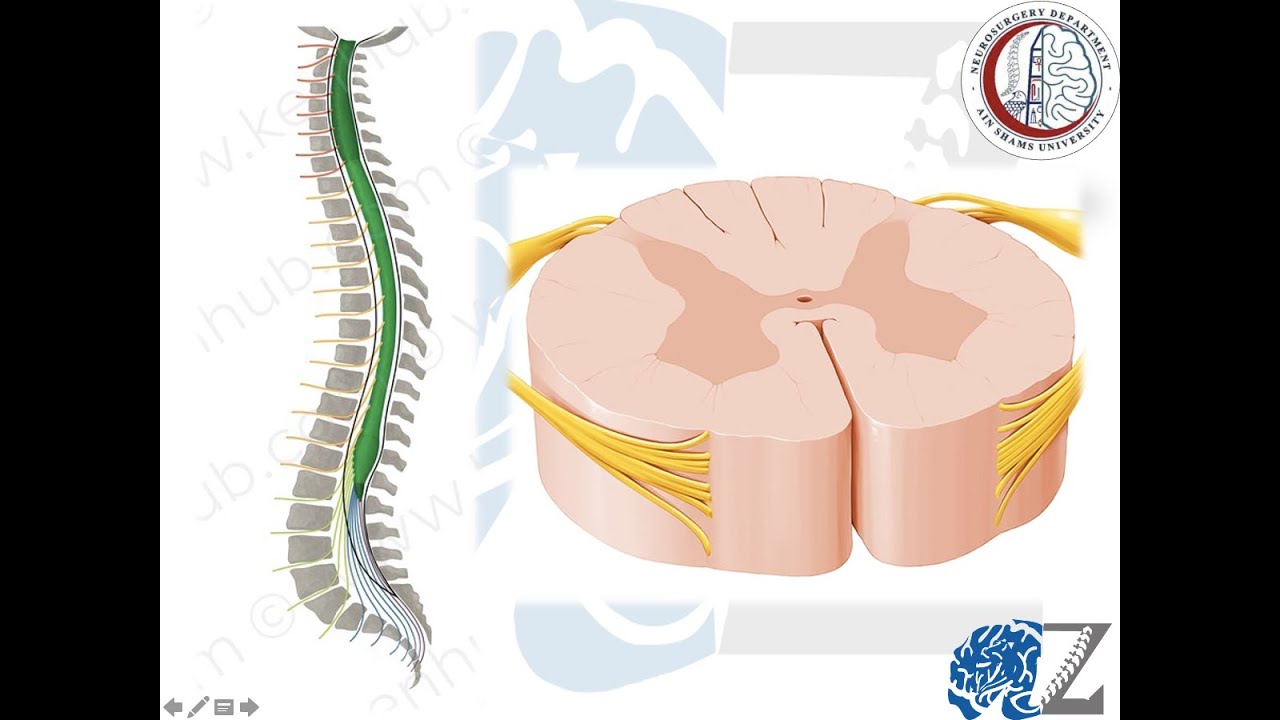
The spinal cord appears as a circle in cross-section with symmetrical right and left sides. The central part contains gray matter (H-shaped organ), while the outer part contains white matter. The gray matter is divided into anterior and posterior horns: the posterior horn contains sensory cells, and the anterior horn contains motor cells. The central canal is a hole in the middle connected to the brain's ventricular system. The spinal cord has an anterior medial fissure (where ventral roots exit) and a posterior medial fissure (where dorsal roots exit). The white matter surrounds the gray matter and contains axons extending outward on both sides.
Click to watch →
Internal Spinal Cord (Gray Matter, White Matter, Funiculus) - Anatomy
230.6K
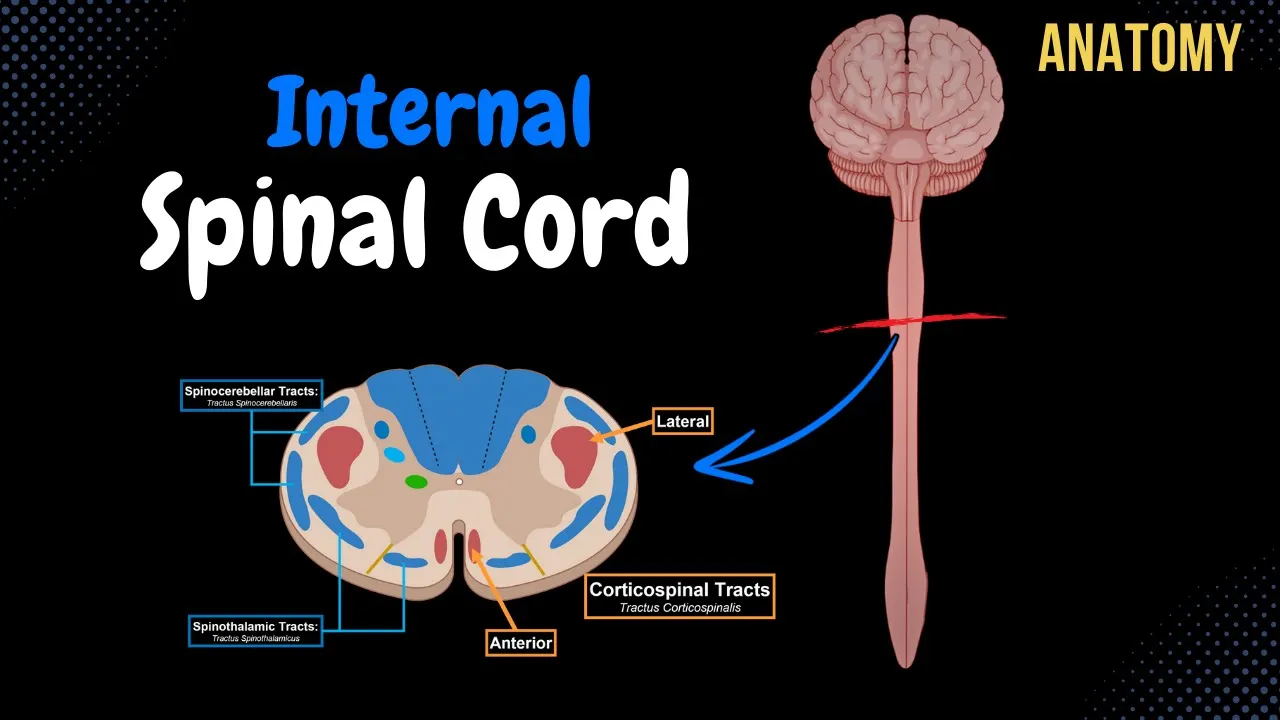
The spinal cord's internal surface is composed of grey matter (containing nerve cell bodies and dendrites) and white matter (containing myelinated axons). The grey matter consists of three horns: the anterior horn contains motor nuclei, the posterior horn contains sensory nuclei including the marginal nucleus, gelatinous substance, nucleus proprius, and posterior thoracic nucleus, and the lateral horn contains sympathetic and parasympathetic nuclei between C8-L2 and S2-S4 respectively. The white matter is divided into three funiculi: posterior, lateral, and anterior, which contain ascending and descending tracts. Ascending tracts are classified as unconscious (going to cerebellum for balance and posture) or conscious (going to cerebral cortex for pain, temperature, touch, and proprioception), while descending tracts are classified as voluntary (pyramidal tracts from primary motor cortex) or involuntary/extrapyramidal (from other brain regions).
Click to watch →

Neurology | Gross Anatomy of the Spinal Cord and Spinal Nerves
1.5M
In cross-section, the spinal cord shows distinct anatomical features: the anterior median fissure divides the anterior portion containing the ventral gray horn, while the posterior median sulcus divides the posterior portion containing the posterior gray horn. Between these horns are the lateral gray horns, found only between T1-L2, which contain preganglionic sympathetic motor neurons. The white matter contains three columns: dorsal funiculus (posterior), lateral funiculus, and ventral funiculus. As you move from the cervical to the coccygeal segments, white matter decreases while gray matter increases. White matter carries ascending sensory information and descending motor information; since sensory and motor tracts must travel longer distances from lower body regions to reach the brain, more white matter is needed in the cervical region. Conversely, larger muscle masses in the lower extremities require more motor neurons (gray matter) in the lumbar and sacral regions. The posterior gray horn contains sensory neuron cell bodies, while the ventral gray horn contains somatic motor neuron cell bodies. In the CNS, a bundle of neuron cell bodies is called a nucleus, while a bundle of axons is called a tract.
Click to watch →
Deep Dive
Fundamental concepts of neural signaling, including action potentials, synaptic transmission, and neurotransmitters.

أقوى مراجعة للبكالوريا... ليلة الرعد ( الجزء 2 )⛈️ 🔥
75.6K
Neural signaling involves resting membrane potential (-70 mV) from unequal ion distribution. Action potentials result from sodium influx followed by potassium efflux. Synaptic transmission uses excitatory (acetylcholine, EPSP) and inhibitory (GABA, IPSP) neurotransmitters. Synaptic potentials summate spatially and temporally to determine action potential generation. Synapses are classified as excitatory or inhibitory based on their effect on postsynaptic membrane potential.
Click to watch →

Nervous System Part 2
259
This comprehensive section covers the fundamental mechanisms of neural communication. Action potentials are electrical signals enabling long-distance communication in neurons and muscle cells, occurring only in axons and muscle cells (skeletal, smooth, cardiac). Key players are voltage-gated sodium and potassium channels. Depolarization begins when sodium channels open, creating positive feedback until threshold (-50 millivolts) is reached. Repolarization and hyperpolarization follow, with refractory periods ensuring one-way transmission. The all-or-none phenomenon ensures reliable signal transmission. Propagation occurs through local currents, with conduction velocity depending on axon diameter and myelination. Saltatory conduction in myelinated axons allows action potentials to 'jump' between nodes of Ranvier, making conduction 30 times faster. Synapses mediate information transfer between neurons or neurons and effector cells. Chemical synapses involve neurotransmitter release into the synaptic cleft, providing control and unidirectional communication. Neurotransmitters are classified by chemical structure: acetylcholine (neuromuscular junctions, autonomic), biogenic amines (catecholamines from tyrosine, indoleamines from tryptophan/histidine), amino acids (glutamate excitatory, GABA/glycine inhibitory), neuropeptides (substance P, endorphins), purines (ATP, adenosine), and gases/lipids (nitric oxide, endocannabinoids).
Click to watch →
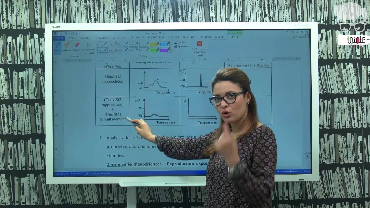
Bac SVT Communication Nerveuse Revision
3.1K
This comprehensive section covers the core principles of neurophysiology: (1) Experimental methods including microelectrodes, oscilloscopes, and stimulation techniques for recording neural activity; (2) Resting membrane potential at approximately -70 mV maintained by ion distribution; (3) Excitatory postsynaptic potentials (EPSPs) that depolarize the membrane toward threshold; (4) Temporal summation where multiple EPSPs add up algebraically to trigger action potentials; (5) Action potentials as all-or-nothing signals that propagate along axons; (6) Excitatory and inhibitory synaptic transmission using neurotransmitters like acetylcholine and GABA; (7) Inhibitory postsynaptic potentials (IPSPs) that hyperpolarize the membrane; (8) Local potentials that decay with distance versus action potentials that propagate without decay.
Click to watch →

AP Psychology Unit 2 Summary
284.1K
Neurons communicate using electrical and chemical signals. Resting potential means neurons have more negative ions inside. Depolarization occurs when sufficient stimulus meets threshold, triggering an action potential (all-or-nothing firing). Repolarization returns neurons to resting state. The refractory period prevents immediate re-firing. At synapses, neurotransmitters cross the synaptic gap: presynaptic terminals release them, postsynaptic terminals receive them. Excitatory neurotransmitters increase firing likelihood through depolarization; inhibitory neurotransmitters decrease likelihood through hyperpolarization. Major neurotransmitters include acetylcholine (muscle/action/memory), dopamine (movement/emotion), serotonin (mood/sleep), endorphins (pain), epinephrine/norepinephrine (fight-or-flight), glutamate (learning), and GABA (calming).
Click to watch →

Physiology L1
219
The nervous system serves as the body's communication network, receiving and processing information to coordinate responses. Neurons are the fundamental functional units, specialized cells with dendrites (signal reception), cell body (processing), and axons (signal transmission). Synaptic transmission is the process by which neurons communicate across synapses through neurotransmitters. Major neurotransmitters include acetylcholine (muscle contraction and memory), norepinephrine and epinephrine (stress response), histamine (immune responses), GABA (inhibitory signaling), glutamate (excitatory signaling), serotonin (mood regulation), and dopamine (reward and movement). Synaptic receptors bind neurotransmitters and can open ion channels, allowing sodium or potassium ions to flow across the membrane, changing electrical potential. This creates membrane potential changes that determine whether neurons will generate action potentials.
Click to watch →
Prerequisite Knowledge
- Concept 01Basic neuroanatomy, including the division between the central nervous system (CNS) and peripheral nervous system (PNS).
- Concept 02The anatomical structure of the cerebrum, specifically the location and function of the primary motor cortex (precentral gyrus) and the motor homunculus.
- Concept 03Cross-sectional anatomy of the spinal cord, distinguishing between grey matter (anterior/posterior horns) and white matter tracts.
- Concept 04Fundamental concepts of neural signaling, including action potentials, synaptic transmission, and neurotransmitters.
Subsequent Learning
- Step 01Clinical differentiation of Upper Motor Neuron (UMN) versus Lower Motor Neuron (LMN) lesions, including assessing reflexes, muscle tone, and the Babinski sign.
- Step 02The role of the basal ganglia and cerebellum in modulating, refining, and coordinating voluntary motor signals initiated by the corticospinal tract.
- Step 03Anatomy and function of the extrapyramidal descending motor pathways, such as the rubrospinal, reticulospinal, and vestibulospinal tracts.
- Step 04Pathophysiology of clinical conditions affecting these pathways, such as Amyotrophic Lateral Sclerosis (ALS), stroke syndromes, and spinal cord hemisection (Brown-Séquard syndrome).
Tract Overview
Pathway Descent
Fiber Decussation
Uncrossed Fibers
Functional Split
Damage Effects
Recovery Limits
Tract Overview
0:05Playing Section
- 1
Corticospinal tract carries movement info.
- 2
Links motor cortex to spinal cord.
- 3
Upper motor neurons contact lower ones.
The Distributed Network Model and Extrapyramidal Contribution
The traditional hierarchical model of motor control posits the corticospinal tract (CST) as the primary pathway for voluntary movement, teaching that its disruption causes classic 'upper motor neuron (UMN) syndrome' (including spasticity). However, modern neuroscience challenges this CST-centric view. Studies reveal that isolated lesions of the CST in primates lead to a loss of fine, fractionated finger movements, but not the severe spasticity or total paralysis traditionally attributed to UMN damage. Instead, clinical UMN syndrome is now understood to result largely from damage to co-traveling extrapyramidal pathways, particularly the reticulospinal tract. Furthermore, alternative frameworks like the 'Distributed Network Model' argue that motor control is not a simple top-down command chain. Instead, movement emerges from parallel, self-organizing loops involving the brainstem, basal ganglia, cerebellum, and spinal interneuronal networks. This alternative perspective shifts the focus from a single dominant tract to a highly plastic, redundant system where non-corticospinal pathways play a major role in both everyday motor execution and post-stroke recovery.
Deep Dive
Clinical differentiation of Upper Motor Neuron (UMN) versus Lower Motor Neuron (LMN) lesions, including assessing reflexes, muscle tone, and the Babinski sign.

Neurology | High Yield Medicine for FMGE | Medicine | Dr. Santhosh
9.2K
Upper motor neuron damage causes hypertonia, hyperreflexia, and extensor plantar response (Babinski sign). Lower motor neuron damage causes hypotonia/flaccidity, hyporeflexia/areflexia, and flexor plantar response. Muscle atrophy and fasciculations indicate lower motor neuron damage. These three features—tone, reflexes, and plantar response—provide a reliable framework for localizing neurological lesions in clinical examination.
Click to watch →

Upper and lower motor neuron (UMN, LMN) lesion | Central nervous system (CNS) physiology | Neurology
4.5K
Both UMN and LMN lesions cause weakness, but other features distinguish them. UMN lesions show normal muscle bulk initially (disuse atrophy later), hypertonia, exaggerated deep tendon reflexes, and positive Babinski sign. LMN lesions show muscle atrophy, hypotonia, lost reflexes, and fasciculations. Power is assessed by voluntary contraction. Muscle bulk by girth measurement. Tone by passive movement. Deep tendon reflexes are monosynaptic; superficial reflexes are polysynaptic and lost in both lesion types.
Click to watch →

Upper Motor Neuron Vs Lower Motor Neuron Lesion
6.6K
This comprehensive lecture covers the complete differentiation between upper motor neuron (UMN) and lower motor neuron (LMN) lesions. The instructor defines UMN as neurons from the CNS synapsing with anterior horn cells, and LMN as neurons from anterior horn cells directly innervating skeletal muscles. Using a teacher-student analogy, UMN is explained as the controller of LMN. The lecture systematically compares clinical features: muscle group affected (multiple in UMN vs single in LMN), muscle tone (hypertonia in UMN vs hypotonia in LMN), paralysis type (spastic in UMN vs flaccid in LMN), atrophy (absent in UMN vs present in LMN), fibrillation/fasciculation (absent in UMN vs present in LMN), reflexes (hyperreflexia in UMN vs areflexia in LMN), and Babinski sign (positive in UMN vs normal in LMN). These features form the clinical basis for localizing motor neuron lesions.
Click to watch →

Characteristic findings between UMN AND LMN LESIONS
104
Upper Motor Neuron (UMN) lesions are characterized by muscle mass decrease of 15-20%, spastic paralysis, hypertonia (increased muscle tone), hyperreflexia (exaggerated deep tendon reflexes), and presence of pathological reflexes (Babinski sign, pronator drift, Hoffman's sign), while Lower Motor Neuron (LMN) lesions show muscle mass decrease up to 80%, flaccid paralysis, hypotonia (decreased muscle tone), hyporeflexia (decreased reflexes), and presence of fasciculations and fibrillations.
Click to watch →

Neurology localization part 1
141.3K
Neurological localization relies on distinguishing upper motor neuron (UMN) from lower motor neuron (LMN) lesions through characteristic clinical findings: UMN lesions cause spastic weakness with brisk reflexes, velocity-dependent resistance, and Babinski sign, while LMN lesions produce flaccid weakness with absent reflexes, profound atrophy, fasciculations, and fibrillation potentials; UMN pathways include the corticospinal tract (cortex to spinal cord) and corticobulbar tract (cortex to brainstem motor nuclei), whereas LMN pathways involve anterior horn cells, nerve roots, plexuses, and peripheral nerves.
Click to watch →
Deep Dive
The role of the basal ganglia and cerebellum in modulating, refining, and coordinating voluntary motor signals initiated by the corticospinal tract.
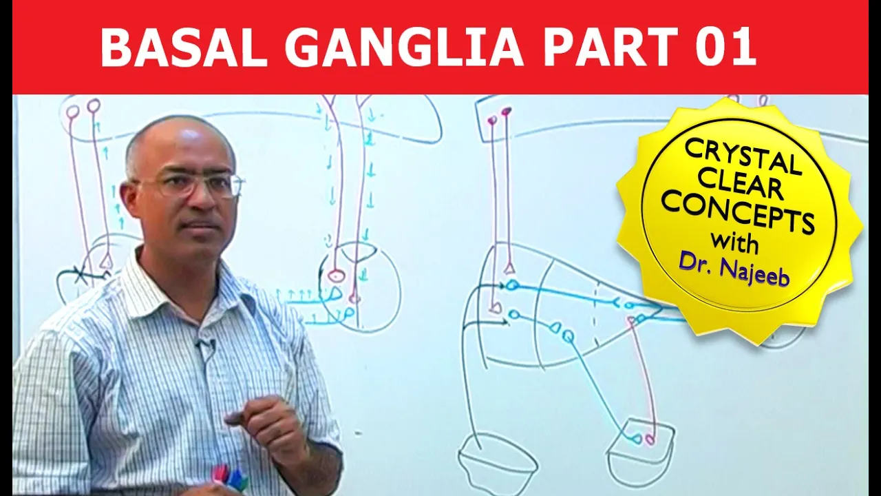
Basal Ganglia | Neuroanatomy | Part 1/3
679.5K
This extensive section explains how basal ganglia control movement through neural circuits. The motor system involves upper motor neurons (corticospinal and cortical nuclear fibers) descending from cerebral cortex to control lower motor neurons in spinal cord and brainstem. Motor plans are stored in basal ganglia and cerebellum. When initiating movement, the idea must first consult basal ganglia for planning and refinement before descending to lower motor neurons. Basal ganglia electrical activity begins even before movement execution. A cortical-basal ganglia-thalamic loop allows consultation, refinement, and execution coordination. Movement initiation begins in prefrontal cortex, proceeds to supplementary motor area and premotor area, consults basal ganglia, receives thalamic feedback, and activates primary motor and somatosensory areas before descending to lower motor neurons.
Click to watch →
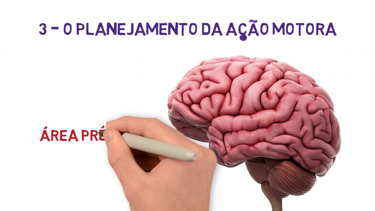
4 - Os Movimentos Voluntários - Córtex, Cerebelo e Núcleos da Base
58.3K
Two critical brain structures collaborate with the motor cortex for movement planning. The cerebellum provides information from previous movement attempts, helping the brain identify useful versus incorrect strategies. It modulates planned movement information by filtering out errors and reinforcing successful approaches. The basal ganglia, located deep in the brain, regulate information flow and energy distribution. They synthesize multiple fragmented signals into fewer, more powerful commands, consolidating information to improve energy efficiency. They also route only essential information to planning areas while filtering out irrelevant signals.
Click to watch →

Motor Systems: Neuroanatomy Video Lab - Brain Dissections
33K
This lecture introduces three major motor systems: the corticospinal tract (pyramidal system), cerebellar connections, and basal ganglia circuitry. The corticospinal tract serves as the final common pathway for voluntary movements, originating from the precentral gyrus, somatosensory cortex, and premotor areas. It travels through the internal capsule and cerebral peduncle, decussating at the spinal cord level to control the opposite body side. The cerebellum connects via three peduncles and has hemispheres and vermis regions. The basal ganglia include the caudate, putamen (striatum), globus pallidus, subthalamic nucleus, and substantia nigra, working through the thalamus to influence the corticospinal tract. These three circuits work together to produce coordinated voluntary movement.
Click to watch →

Neurology | Midbrain Anatomy & Function
589.9K
Upper motor neurons in the cortex send three types of descending fibers: corticospinal fibers to the spinal cord, corticonuclear fibers to cranial nerve nuclei, and corticopontine fibers to the cerebellum via the middle cerebellar peduncle. The substantia nigra pars compacta sends the nigrostriatal pathway to the striatum, releasing dopamine onto neurons in the direct and indirect pathways of the basal ganglia. Dopamine enhances motor activity in the direct pathway and reduces inhibition in the indirect pathway; damage leads to Parkinson's disease. The deep cerebellar nuclei send output through cerebello-rubral fibers (crossing at the superior cerebellar peduncle decussation to the red nucleus) and cerebello-thalamic fibers (crossing to the thalamus), transmitting motor plans from the cerebellum to the cortex for coordinated movement.
Click to watch →

14. Лімбічна система - Роберт Сапольскі
50.3K
The basal ganglia consist of interconnected nuclei including the caudate nucleus, putamen, globus pallidus, and subthalamic nucleus. They regulate voluntary movement through direct and indirect pathways: the direct pathway facilitates movement while the indirect pathway inhibits unwanted movements. The pyramidal tract carries motor commands from cortical neurons to spinal motor neurons. Dysfunction in these circuits causes movement disorders like Parkinson's disease. The cerebellum coordinates motor timing and precision.
Click to watch →
Deep Dive
Anatomy and function of the extrapyramidal descending motor pathways, such as the rubrospinal, reticulospinal, and vestibulospinal tracts.
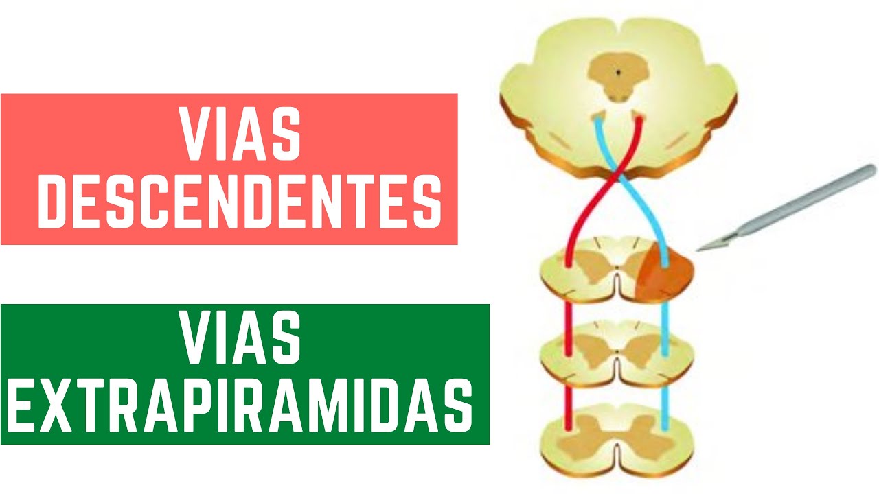
NEUROANATOMIA VIAS DESCENDENTES - VIAS EXTRAPIRAMIDAS
4.1K
The extrapyramidal motor pathways are involuntary descending tracts that do not pass through the pyramids of the medulla and include five main tracts: the rubrospinal tract (originating from the red nucleus, controlling upper limb flexion and inhibiting extension), the reticulospinal tract (from the reticular formation, controlling posture, balance, and autonomic regulation), the tectospinal tract (from the superior colliculus, coordinating involuntary head and eye movements in response to visual stimuli), the vestibulospinal tract (from vestibular nuclei, maintaining posture and equilibrium), and the olivospinal tract (from the inferior olivary nucleus, involved in motor coordination). These pathways are distributed in the lateral and anterior funiculi of the spinal cord, with the tectospinal tract only extending to the cervical region.
Click to watch →

Vías descendentes o motoras
7.8K
The descending motor pathways control voluntary and involuntary movements through two main systems: the pyramidal system (corticospinal and corticobulbar tracts) which controls voluntary movements of extremities, trunk, and head/neck, and the extrapyramidal system (rubrospinal, tectospinal, vestibulospinal, and reticulospinal tracts) which regulates involuntary movements, posture, balance, and muscle tone. The pyramidal system originates from the cerebral cortex (areas 4, 6, and sensory areas 1-3) and descends through the brainstem to synapse with lower motor neurons in the spinal cord anterior horn. The extrapyramidal system originates from the premotor cortex and brainstem nuclei, with tracts like the rubrospinal tract activating flexor muscles and inhibiting extensors, while the vestibulospinal and reticulospinal tracts facilitate extensor muscles. Lesions in upper motor neurons cause spastic paralysis with hyperreflexia and Babinski sign, while lower motor neuron lesions cause flaccid paralysis with hyporeflexia.
Click to watch →
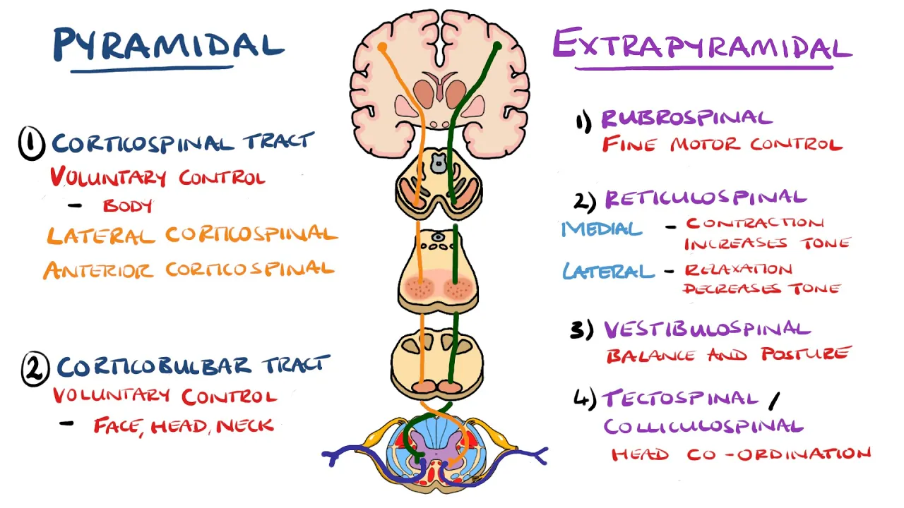
Extrapyramidal and Pyramidal Tracts - Descending Tracts of the Spinal Cord | (Includes Lesions)
798.9K
The descending tracts of the spinal cord are divided into pyramidal tracts (voluntary motor control) and extrapyramidal tracts (involuntary motor control). The pyramidal tracts include the corticospinal tract (controlling body musculature with 75% crossing at the medulla to form the lateral tract and 25% remaining as the anterior tract) and the corticobulbar tract (controlling facial and head muscles with bilateral innervation except for cranial nerves VII and XII). The extrapyramidal tracts include the rubrospinal tract (fine motor control, contralateral), reticulospinal tracts (medial excites extensors/increases tone, lateral inhibits voluntary movement/decreases tone), vestibulospinal tract (balance and posture, ipsilateral), and tectospinal tract (head-eye coordination, contralateral). Lesions in pyramidal tracts cause upper motor neuron signs including hypertonicity, hyperreflexia, weakness, and Babinski's sign, while extrapyramidal lesions result in parkinsonism, chorea, athetosis, and dystonia.
Click to watch →
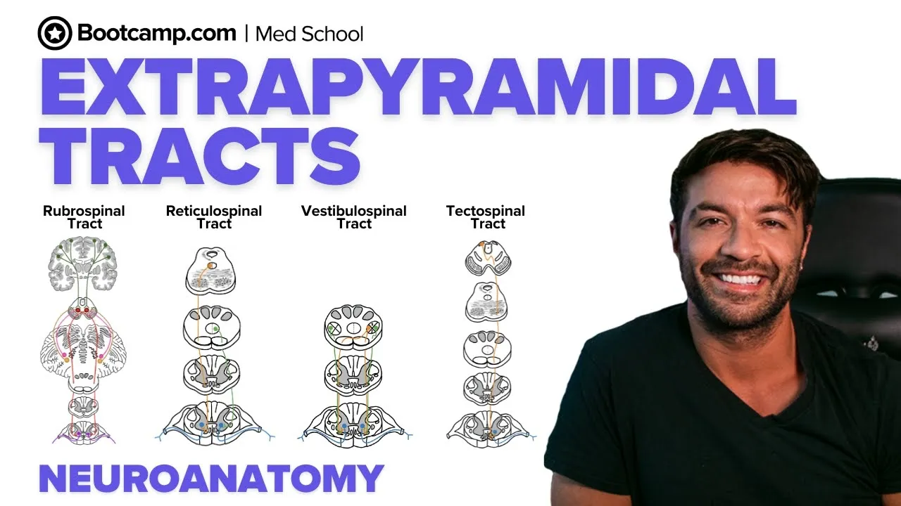
Extrapyramidal Tracts & Clinical Signs | Rubrospinal, Reticulospinal, Vestibulospinal & Tectospinal
1.2K
The extrapyramidal tracts are descending motor pathways that regulate posture, balance, and reflexive movements without passing through the medullary pyramids; they include the rubrospinal tract (originating from the red nucleus, maintaining flexor tone and fine motor control of upper limbs), reticulospinal tracts (medial from pons facilitating extensor tone and lateral from medulla inhibiting voluntary movement), vestibulospinal tracts (medial with bilateral projections and lateral with ipsilateral projections, controlling anti-gravity muscle tone based on vestibular input), and tectospinal tract (from superior colliculus enabling reflexive head, eye, and trunk movements to stimuli); damage to these pathways can result in clinical conditions such as decerebrate rigidity, characterized by continuous extensor muscle contraction.
Click to watch →
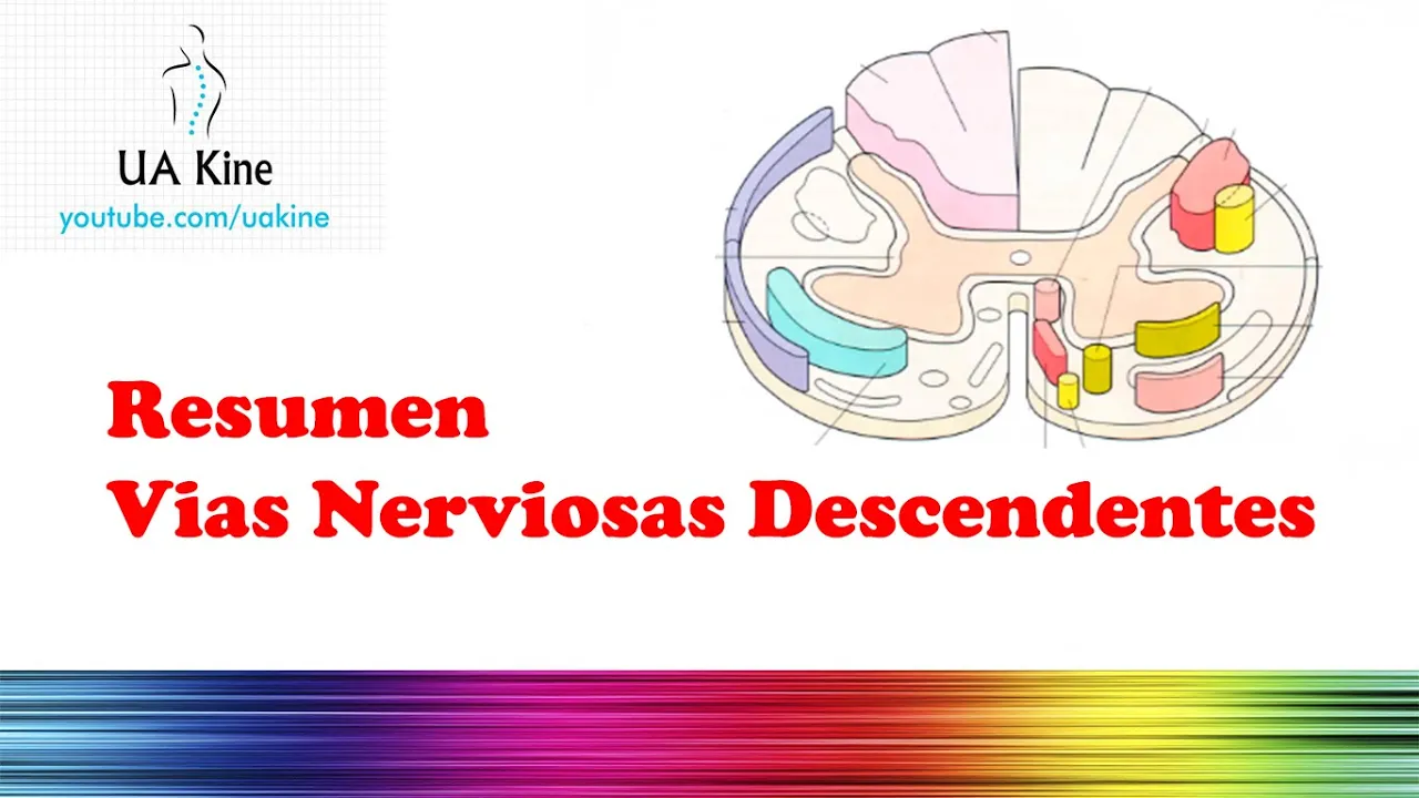
Resumen Vías Nerviosas Descendentes (Tractos)
13.3K
The descending motor pathways of the central nervous system are divided into pyramidal (corticospinal and corticonuclear tracts) and extrapyramidal systems (tectospinal, reticulospinal, vestibulospinal, and rubrospinal tracts). The pyramidal system, originating from the cerebral cortex, primarily stimulates flexor muscles and inhibits extensor muscles in the extremities, neck, and trunk. The extrapyramidal system, arising from structures like the midbrain, pons, and medulla, maintains muscle tone, automatic movements, and associated movements. The extrapyramidal tracts include: tectospinal (midbrain, facilitates contralateral muscles), reticulospinal medial (pons, facilitates axial and extensor muscles), reticulospinal lateral (medulla, facilitates flexor muscles), vestibulospinal lateral (facilitates extensor muscles), vestibulospinal medial (controls head position and facilitates flexors), and rubrospinal (red nucleus, activates flexor muscles). These pathways are classified into a medial system (vestibulospinal, tectospinal, reticulospinal) that facilitates extension, and a lateral system (corticospinal, rubrospinal) that facilitates flexion.
Click to watch →
Deep Dive
Pathophysiology of clinical conditions affecting these pathways, such as Amyotrophic Lateral Sclerosis (ALS), stroke syndromes, and spinal cord hemisection (Brown-Séquard syndrome).
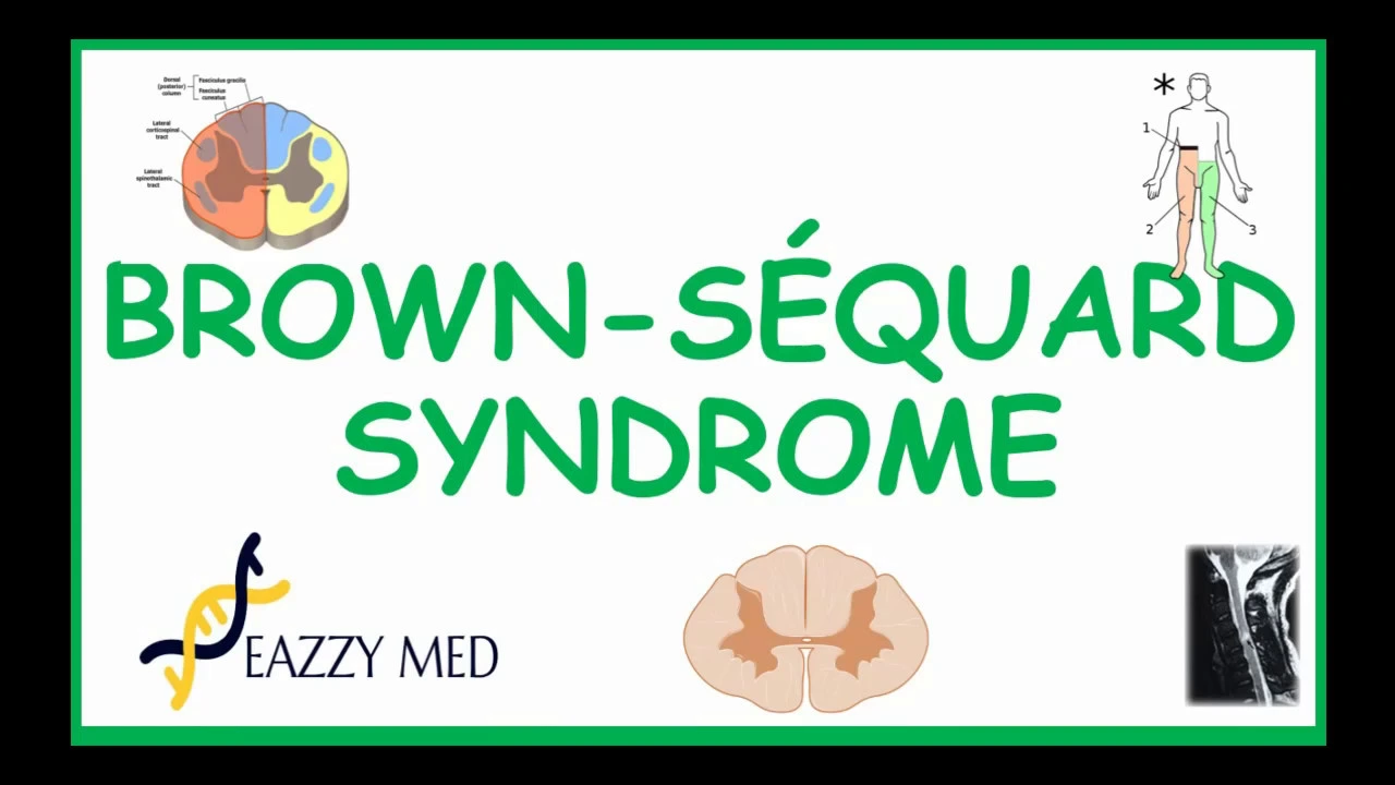
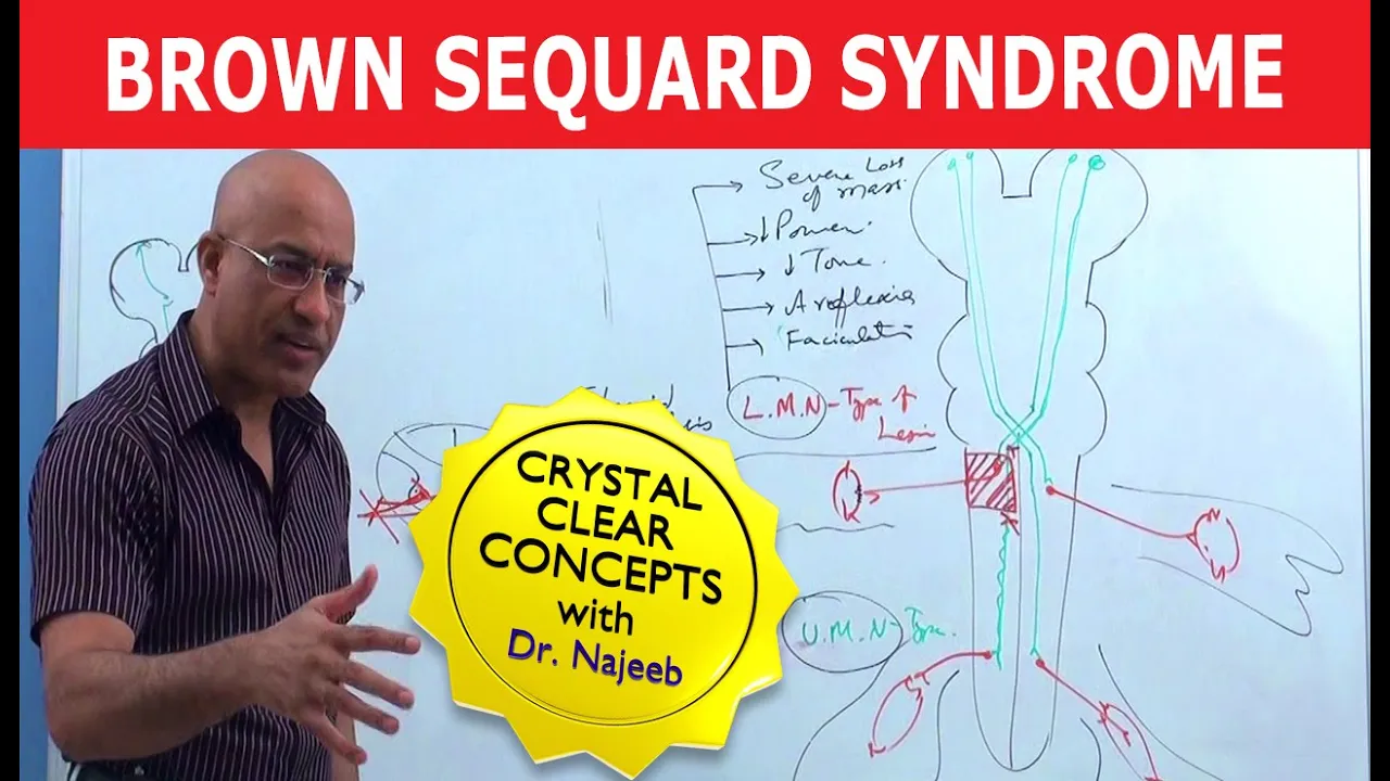
Brown-Sequard Syndrome causes, pathophysiology, symptoms diagnosis and treatment
74.1K
Brown-Séquard syndrome results from unilateral spinal cord hemisection, causing ipsilateral spastic paralysis and loss of proprioception due to corticospinal tract and dorsal column damage, while contralateral loss of pain and temperature sensation occurs due to spinothalamic tract disruption; diagnosis is confirmed via MRI, and causes include trauma, tumors, ischemia, or inflammatory diseases.
Click to watch →
Brown Sequard Syndrome | Internal Medicine 💊
261K
Brown Sequard syndrome results from hemisection of the spinal cord, commonly caused by trauma. The syndrome involves distinct sensory pathways: the dorsal column carries fine touch, vibration, proprioception, two-point discrimination, and stereognosis, ascending ipsilaterally to gracile/cuneate nuclei before crossing to the thalamus; the spinothalamic tract carries pain, temperature, and crude touch, crossing contralaterally at entry. After hemisection, at the lesion level complete anesthesia occurs; below the lesion, ipsilateral dorsal column loss and contralateral spinothalamic loss produce characteristic sensory-motor dissociation. The spino-cerebellar system carries unconscious proprioception for movement coordination, with dorsal and ventral tracts producing ipsilateral and contralateral dysmetria respectively.
Click to watch →

Neuro Anatomy High-Yield Review with Dr. Karim
128
Brown-Séquard syndrome results from hemisection: ipsilateral motor/vibration/proprioception loss at lesion level, contralateral pain/temperature loss below. Amyotrophic lateral sclerosis involves both upper and lower motor neurons (hyperreflexia/spasticity plus fasciculations/atrophy), never causing sensory loss. Spinal muscular atrophy affects only anterior horn cells (lower motor neurons), causing tongue fasciculations and floppy baby presentation in children. Anterior spinal artery occlusion spares dorsal columns but destroys anterior horn and corticospinal tracts, causing paralysis and pain/temperature loss without sensory ataxia.
Click to watch →

Chapter 36 Neuromuscular Disorders
31.5K
Amyotrophic lateral sclerosis (ALS/Lou Gehrig's disease) is a progressive neurodegenerative disorder affecting both upper and lower motor neurons, causing spasticity/hyperreflexia from upper neuron damage and flaccid paralysis/atrophy from lower neuron damage. Multiple sclerosis is an autoimmune disease destroying myelin sheaths in the CNS, decreasing conduction velocity and potentially blocking signal transmission. Spinal cord injury syndromes include central cord syndrome (greater arm than leg impairment), anterior cord syndrome (anterior spinal artery infarction damaging motor and pain/temperature tracts), and Brown-Séquard syndrome (hemisection causing ipsilateral motor loss and contralateral pain/temperature loss).
Click to watch →

Exam Recall Series (NEET-PG '24) - Medicine
116.6K
Brown-Séquard syndrome results from hemisection of the spinal cord, causing ipsilateral loss of proprioception and vibration sense (due to dorsal columns crossing at the level of injury) and contralateral loss of pain and temperature sensation (spinothalamic tract crosses at spinal level). Motor function is preserved on the ipsilateral side. This classic syndrome demonstrates the decussation patterns of sensory tracts.
Click to watch →
Tract Overview
Pathway Descent
Fiber Decussation
Uncrossed Fibers
Functional Split
Damage Effects
Recovery Limits
Tract Overview
0:05- 1
Corticospinal tract carries movement info.
- 2
Links motor cortex to spinal cord.
- 3
Upper motor neurons contact lower ones.
The Distributed Network Model and Extrapyramidal Contribution
The traditional hierarchical model of motor control posits the corticospinal tract (CST) as the primary pathway for voluntary movement, teaching that its disruption causes classic 'upper motor neuron (UMN) syndrome' (including spasticity). However, modern neuroscience challenges this CST-centric view. Studies reveal that isolated lesions of the CST in primates lead to a loss of fine, fractionated finger movements, but not the severe spasticity or total paralysis traditionally attributed to UMN damage. Instead, clinical UMN syndrome is now understood to result largely from damage to co-traveling extrapyramidal pathways, particularly the reticulospinal tract. Furthermore, alternative frameworks like the 'Distributed Network Model' argue that motor control is not a simple top-down command chain. Instead, movement emerges from parallel, self-organizing loops involving the brainstem, basal ganglia, cerebellum, and spinal interneuronal networks. This alternative perspective shifts the focus from a single dominant tract to a highly plastic, redundant system where non-corticospinal pathways play a major role in both everyday motor execution and post-stroke recovery.
Basic neuroanatomy, including the division between the central nervous system (CNS) and peripheral nervous system (PNS).
The anatomical structure of the cerebrum, specifically the location and function of the primary motor cortex (precentral gyrus) and the motor homunculus.
Cross-sectional anatomy of the spinal cord, distinguishing between grey matter (anterior/posterior horns) and white matter tracts.
Fundamental concepts of neural signaling, including action potentials, synaptic transmission, and neurotransmitters.
Clinical differentiation of Upper Motor Neuron (UMN) versus Lower Motor Neuron (LMN) lesions, including assessing reflexes, muscle tone, and the Babinski sign.
The role of the basal ganglia and cerebellum in modulating, refining, and coordinating voluntary motor signals initiated by the corticospinal tract.
Anatomy and function of the extrapyramidal descending motor pathways, such as the rubrospinal, reticulospinal, and vestibulospinal tracts.
Pathophysiology of clinical conditions affecting these pathways, such as Amyotrophic Lateral Sclerosis (ALS), stroke syndromes, and spinal cord hemisection (Brown-Séquard syndrome).
592K views8.4Klikes2:02@neurochallengedOriginal Release: 2015-12-06
Welcome to 2 minute neuroscience, where I simplistically explain neuroscience topics in 2 minutes or less.
In this installment I will discuss the corticospinal tract.
The corticospinal tract is a major tract that carries movement-related information from the motor cortex to the spinal cord.
The neurons that travel in the corticospinal tract are called upper motor neurons; they form connections with neurons called lower motor neurons, which carry movement-related impulses to muscle itself, causing it to contract.
The upper motor neurons of the corticospinal tract leave the motor cortex and descend to the brainstem, entering the midbrain in large fiber bundles called the cerebral peduncles.
The tract continues down into the medulla where the fibers form two bundles, known as the pyramids, which create visible ridges on the exterior surface of the brainstem.
At the base of the pyramids, about 90% of the fibers in the corticospinal tract decussate, or cross to the other side of the brainstem in a bundle called the pyramidal decussation.
The decussating fibers will then enter the spinal cord on the opposite side of the body from where they originated as part of the lateral corticospinal tract.
The other 10% of the fibers will continue into the spinal cord on the same side of the body where they originated as part of the ventral or anterior corticospinal tract and only cross over when they reach the level of the spinal cord where they will synapse on a lower motor neuron.
It is thought that the lateral and anterior corticospinal tract fibers have slightly different specializations, with the lateral corticospinal tract controlling the movement of more distal muscles like those of the hands, and the anterior corticospinal tract controlling the movement of more proximal muscles like those of the trunk.
Damage to the corticospinal tract can lead to a collection of symptoms known as upper motor neuron syndrome, which involves symptoms like weakness or paralysis, hyperactive reflexes, decreased motor control, and either increased or decreased muscle tone.
Over time patients may regain the ability to make crude movements but fine finger movements like writing or typing may remain impaired, suggesting the corticospinal tract is especially important for these types of movements.
Up Next
19:45
Basal Ganglia Circuitry: Direct and Indirect Pathways Explained
@AnatomywithDrNikki
1.9K views•2021-01-16

21:55
Deciphering Neural Circuits Controlling Anorexia in Mice | Richard Palmiter
@AllenInstitute
2.3K views•2011-10-10
1:59
How Smell Works: Olfaction Explained in 2 Minutes | Neuroscience
@neurochallenged
303.7K views•2018-01-31

13:03
How Exercise Benefits Your Brain: Science Explained
@TED
11.4M views•2018-03-21
Related Study Plans & Knowledge Roadmaps
Structured learning paths in Neuroscience
neuroscience
Appetite Regulation: Brain Circuits & Hormones
01Foundations of Endocrinology and Brain Anatomy
02Introduction to Energy Homeostasis
03Peripheral Signals: Leptin, Ghrelin, and Insulin
+ 2 more learning modules
14h5 modules
Explore Roadmap
neuroscience
Neuroeconomics: Value, Risk & Brain Mechanisms
01Foundations of Neuroscience and Brain Anatomy
02Introduction to Neuroeconomics and Subjective Value
03Dopaminergic Prediction Errors & Reinforcement Learning
+ 3 more learning modules
18h6 modules
Explore Roadmap
neuroscience
Spatial Navigation: Cells, Grids & Brain Maps
01Foundations of Brain Anatomy and Memory
02Place Cells and Head-Direction Cells
03Grid Cells: The Brain's Coordinate System
+ 2 more learning modules
12h5 modules
Explore Roadmap
neuroscience
BCIs: Neural Decoding & Prosthetic Control
01Neuroscience Foundations: How the Brain Signals Intent
02Signal Acquisition: Recording the Brain's Activity
03Neural Decoding: Translating Brain Waves into Data
+ 2 more learning modules
25h5 modules
Explore Roadmap