

![[não erre mais] Modo VCV x PCV: quais são as diferenças?](https://i.ytimg.com/vi_webp/hCMd5J2Pki8/maxresdefault.webp)


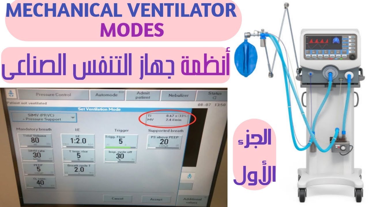
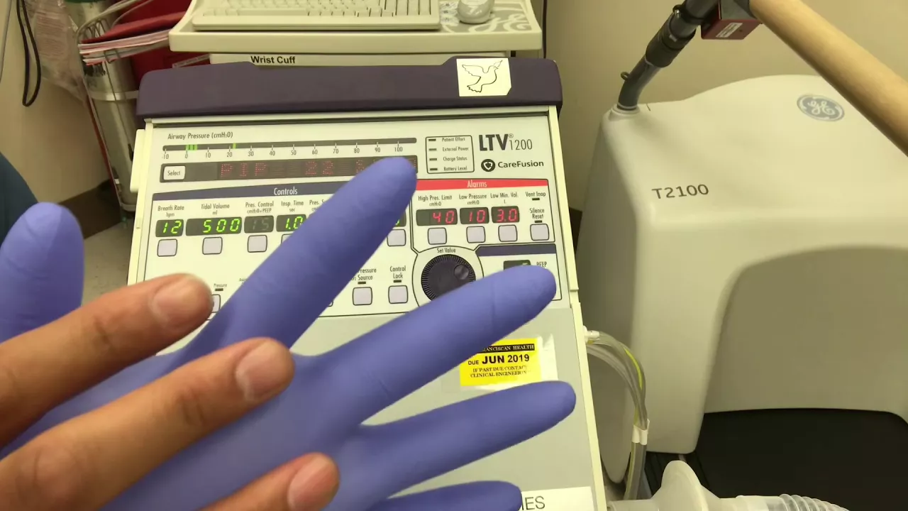

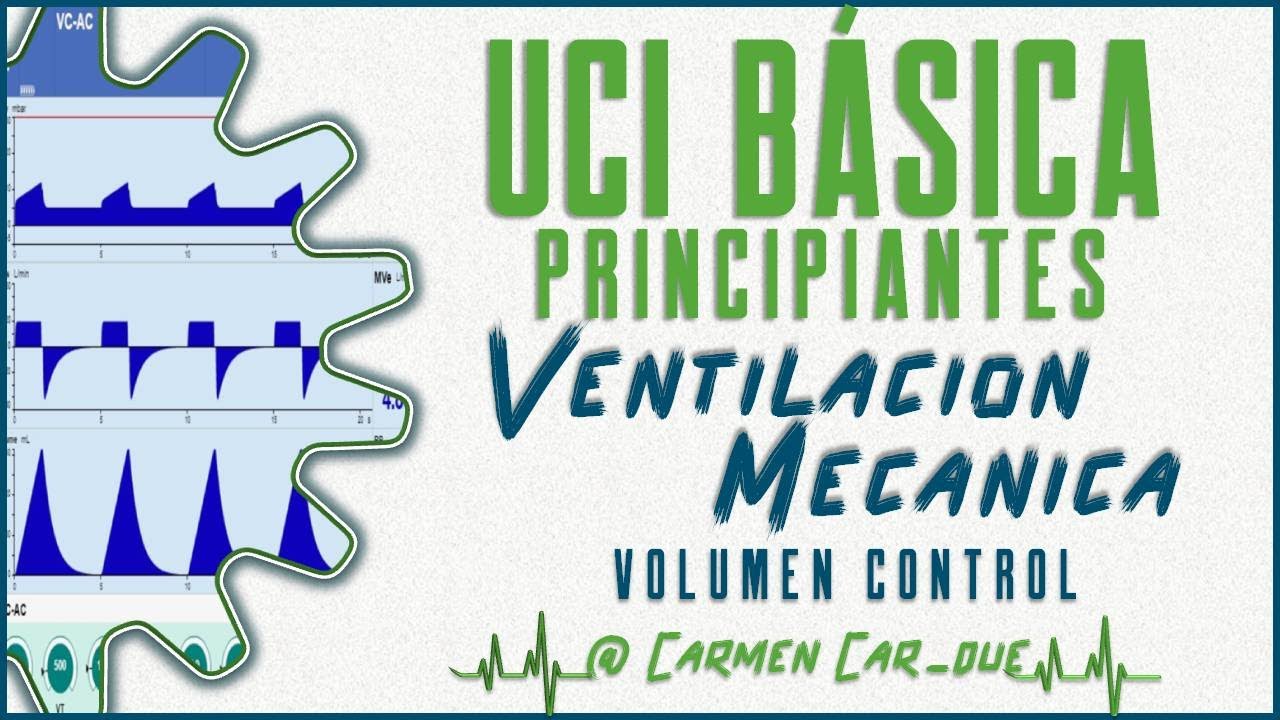




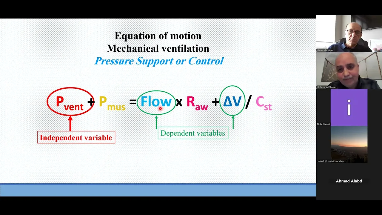








Ventilator scalars are waveforms that plot a single parameter (pressure, flow, or volume) against time, providing critical visual information for mechanical ventilation management. Pressure-time scalars show square waveforms in volume control mode and varying patterns in pressure control mode, helping assess peak pressure, plateau pressure, auto-PEEP, bronchodilator response, and patient-ventilator synchrony. Flow-time scalars display constant flow in volume control (square waveform) versus decelerating flow in pressure control, revealing airway resistance, secretion accumulation, and expiratory flow dynamics. Volume-time scalars track tidal volume delivery and can detect leaks and air trapping. Fixed scale settings (pressure 35-40 cm H2O, flow 120-150 L/min, volume 500 mL) are essential for early detection of clinical deterioration, especially in resource-limited settings where auto-scaling should be disabled.
Ventilator Waveforms: Scalar Basics in Critical Care
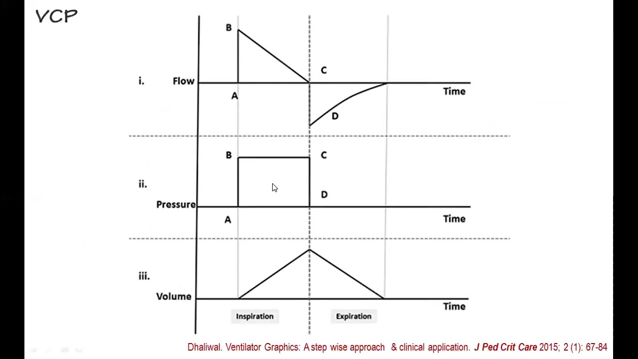
Added:[Music] today we'll be talking about the scalars and basically the waveforms are seen on the ventilator screen last time we had covered about the basics part of mechanical ventilation and today we'll talk about scalars and the wave forms which are seen on the ventilators so if you look at this ventilator screen this is a standard ventilator screen in any of the ventilator irrespective of the make of the company what I wanted to emphasize here is if you look at uh the screen the 75% of the screen is almost covered by the graphics and say around 20 25% is covered for the rest of the parameters the numbers and everything and if the company people or if the clinicians and the scientists who have given a thought into bringing this thing so they must have thought a lot of thing and probably that emphasizes the value of this all different Graphics which are shown on mechanical ventilator unfortunately what happens is most of the times maybe because of the way it has been we focus more on the numbers and we tend to ignore the screen or the wave forms which are seen on the ventilator but understanding the ventilator wave forms becomes very important and it gives a very quick and easy diagnosis for most of our problem so last time when we were talking about ventilator we talked about how the three important parameters that is pressure flow and volume are there and how they make the basics of mechanical ventilation just to re summarize that thing there's a pressure generated by the ventilator it creates a flow of gas and then ultimately it results in a change of volume in the lung and that is basically tidal volume so these three things are represented in the waveform in terms of pressure flow and volume time scalar now before I go for this thing what do I mean by scalar we all know there's a x-axis and y axis and when we talk about the scalar this means that the pressure the flow and the volume they are represented against the axis of time that is it's a continuous beat to beat variation when I say a loop when I say a loop this is basically a pressure and flow Loop so there's not a time there's only one bit which will be seeing but the two parameters across the X and Y axis will be pressure and volume or flow and volume so if there are two parameters we call them as Loop and if there's a single par meter which is plotted against time that is something we call as scalar and there are pressure time scalar flow time scalar and volume time scalar now you'll be seeing that in most of the newer anesthesia workstations the advanced work stations and even the basics ones which are coming right now which have an integrated ventilator into them at least there will be one waveform which will be scalar and most of the them there will be two waveforms if you look at the advanced ventilators they will almost have pressure volume and flow scalars as well as have loops present into embedded into their screens so this is the basic difference between scalar and Loops now before we talk about individuals like what are the things which we look into all these three scalers today I'm going to talk only about the scalars and I'll skip the loop part today but the most important thing to before I go for the scalar description is we need to understand that we need to say no to Autos scaling now what do we mean by autoscaling if you look in most of the ventilators in most of the ICU setups many of times you will see that they are on Autos skill mode now what do I mean by Auto scale mode what happens is suppose this is a imagine this is a pressure scalar and you have 0 to 60 cm of water kept as your scale it's like a graph isn't it it's like a graph and we have parameters kept here now what will happen is if you are doing it on a auto scaling what will happen is suppose for some reasons this pressures initially were 30 and now the PR have gone to 50 so what will happen is with the time the initially this this scale was probably 0 to 30 now your pressures for some reason has increased now the pressures reading are around say 45 and now what will the machine will do is it will increase the scale back to 60 so that all your waveforms comes looks very nicely in the center of that scale it is adjusting the scale according to pressures now people might say that okay this is a very good thing I'm able to continuously see the scale very nice nicely but this is a big problem and this is a big problem especially in resource limited setting we all agree that in an IU there should be one is to one ratio for nursing at least and doctors also should be in sufficient amount especially for mechanically ventilated patients but more often than not what we see is dir of shortage in most of the public setups even at times in private setups and you'll have three or four nurses managing at times a single patient now what will happen is if you put a somebody on the auto skill you are not able to actually go to the patient every time you're seeing from far and from far you are not able to always make out the numbers but you are looking at the bigger part of the screen that is covered by the waveforms now if it starts Auto scaling on its own you won't realize the problem at times till it is too late the moment you keep your scales fixed what happens is if your pressures are going up you'll see a change in wave form that my pressures are missing if you see a change in your flows you will see your flows are missing if you see that volume has become suddenly very increased or it has changed because of the change in lung condition whatever be the reason you will suddenly notice that there's a change in the height of the your graph the scalar graph and that will alert you immediately so the most important thing the first thing when you are setting the ventilator you set the basic parameters and then you go to the ventilator setup where you sort of say no to the autoscaling and you fix your sces for pressure flow and volume and this is something which should be uni form for the whole ICU basically it should not be like one consultant is doing as per his own this thing or the other consultant is doing for that whole unit the your scale should be fixed generally what we do at our placees we fix the pressure at around 35 CM or 40 cm then flow around 150 or 120 generally depending upon which unit we're looking and the volumes we keeping at 500 mL so now what will happen is for example a patient who was nice sleeping ventilated for some reason he develops a barot trauma his pressures have increased and his volumes have dropped even if I'm far away from the pressure you'll see the alarms are definitely going on but at the same time you'll see what is happening is that your graphs they are all being like it is not Autos scill so you'll see that there is not a complete graph your pressures will be going very high and in the same thing you'll see that the height of the volume graph has come down and you will be able to detect problems very early second thing is alarms we set in the very sometimes it's not a good way to say alarm but too many alarms at times becomes a problem so what everybody will do they'll keep the thresholds very high they'll say oh the patient is breathing very high let me keep the respiratory rate alarm very high let me keep the peak pressure alarm very high let me keep the volume alarm very high otherwise the volume alarms will keep on increasing so rather what will happen is the alarms will also become lit so this visual thing I'm emphasizing more on this part is the Autos scaling should generally be not be encouraged at least in a setup where there are resource constraint this becomes very very important before we go for the next part now we talk about types of waveform in pressure and volume modes now if you see we had last time also discussed in a volume mode you will have a fixed volume a fixed pattern of flow and depending upon your lung compliance whether your lung is a very nice lung or whether it's a ARS lung with a pure compliance your pressures will keep up and down so a parameter which is fixed will always be shown by a square wave form so you can see here that this is the flow is waveform is square pattern and the pressure waveform here is a square pattern in pressure mode so even if I'm covering the volume mode and pressure mode and I'm only shown this diagram I'll be still able to say that because the pressure mode is a square wave form this is a pressure control ventilation and here because the flow is a square wave form this is a volume control ventilation so now let's see these three scalars in detail so first thing is we'll talk about pressure time scalar or simply pressure scalar now we have seen already this part in a volume targeted Moree depending upon the lung it will change its pattern so it will be either an accelerating deating depending upon whatever we are setting and the flow in a the pressure scalar in a pressure targeted mode will always be a square wave form if it was other way around in a volume control ventilation the flow would have been a square wave form and the in a volume control ventilation you'll have the pressure which is varying now what are the uses of pressure waveform at least these are the major uses which we can see in day-to-day use in our I and operation theater we can talk about Auto peiping we can see it very nicely on pressure waveform we can talk about Bronco dilator response we can talk about active exhalation bread type that is pressure versus volume we can talk about Peak inspiratory pressure p plat pressure we talk about asynchrony and Trigger effort now let's see all of this in slightly different now imagine this is a patient and time control what is happening is here there is no patient effort so you can see if I have set a PE of F this is a pressure mode and this is a time so here there was no effort from the patient so the machine decided that okay there is no effort from the patient and I am giving the breath now look at this this is not a square wave form so what will be the mode of ventilation this one this is going to be probably a volume control ventilation now here what has happened is the patient has tried to trigger a breath the moment he's triggering a breath last time we talked about flow triggering and flow triggering you'll see that there is a dip in the pressure wave form so you can detect that the patient is also trying to breathe the in particular things so you can differentiate from a pressure scalar whether it's a patient generated breath or whether it's a machine generated breath now this is something which all of us should know very nicely I'll slightly take time on this thing this is basically a pressure time scalar how it looks like in a volume controlled ventilation now this is your starting phase from here you'll have the initial part the initial pressure is required to overcome the ventilator resist we have put a HMA the tra and all those things and then from here in your tra to broncus and all these things will go so it is basically reaching the peak pressure from here you'll have a slight change and this is where your alular pressures or plate pressures will be represented so what I want to emphasize again on this thing is if you look at the pressure scalar you have an inspiratory time and after this plative pressure is coming down you have an expiratory time like a normal breath now another important concept is a peak pressure and a plate pressure Peak pressure combines the pressure of all things including plate also and that will include your pressure to overcome the resistance of the your Airway conducting tubes if any hme filter is there if you have put a small tube or all those things whereas plat pressure is something which will be representing your end alular pressure and that is what our targets are generally I'll talk about this in more detail also if you can see the starting point is something what we have said if I not set a peep probably My Graph will start from zero if I set a peep of five or six whatever depending upon my setting my flows I mean my pressure will start from above that peep so this is very important inspiration time expiration time peak airway pressure and plate airway pressure now if you remember last time we talked about compliance isn't it so we'll look into this next SL what do you mean by Static compliance and what do you mean by Dynamic compliance now imagine this dotted line shows you a normal graph okay now you see for some reason you are ventilating a patient he was suppose an ards patient or you have operation theater a patient and you see the peak airway pressure has gone up on the ventilator and then when you see and when you check your PL pressure press remains more or less same for some reason in this patient so what you will understand is that your Peak pressure has been going up but your plate pressure remain the same probably the problem most likely is not in the alvillar level but problem is somewhere in your conducting Pathways now when I say conducting Pathways it can be not necessarily only a bronos spasm it can be right from your ventilator and machine and to write your till your terminal bronol are there it can be a problem anywhere it can be a simple Broncos spasm it can be a lingos spasm there might be some blood clot of your hme filter might be blocked or something so it depends so troubleshooting is a different part right now what we are looking at is identifying the problem so the peak pressure has gone up but the plate pressure has remained normal probably it is because of the airway resistance now look at this another graph again the dotted line is showing a normal graph and now if you look at the peak pressure graph the peak pressure will go up but you look at the plate pressure the plate pressure also has changed from a baseline of say around 15 to a 40 so that means probably the problem is in the aluli it can be in a broncus or something but because the difference in Peak and plate pressures is less or more or less normal what was here here also same thing isn't it so the problem most likely is either in the Alvi or the interstitium because your Peak the plate pressures has gone up this implies that there is a dict inrease in compliance of the lung now again this is identification why your compliance can go down and there can be a number of reasons your ala are flooded with fluids ards pulmonary edema there is interstitial fibrosis whatever be the reason isn't it so you need to identify the difference between a static that is Peak pressure and the dynamic part that is the compliance now when we talk about the ards ventilation strategy we say that we will allow a p plate pressures of around 30 cm in OB at 35 CM people casually tend to say that we allow a pressures of 30 and 35 those pressures actually imply a plate pressure and not a airway pressure so this is something we need to be very clear about so this is the important difference between Peak pressure and plat pressure and it is very easy to do if you are on a volume control ventilation especially if your patient is like sedated paralyzed you can put a end expiratory hold and you'll be able to see what is happening to this particular patient to see the pressur I'm sorry and inspiratory hold now the next thing is autop PIP we all talk about Auto pip like what is happening is Auto auto peip is very simple there is a pressure which is generated in addition to whatever peep we are giving that is end respiratory pressure most commonly it is because there is already some amount of air which is getting trapped in and when you are forcing an additional air that basic air which what was trapped in or the your pressure which was there the pressure is getting added to it the most common reason is seen in generally in patients where you are not giving enough amount of expiratory time for the exhalation so what is suppose COPD patient asema patient you have kept a respiratory rate of say 14 15 and you have kept I time of say 1 is to two patient is having activ vising so what will happen is suppose if I'm giving 400 mL of tidal volume maybe 300 ml is coming in two seconds but now it is taking another one or 2 seconds time because we know expiration takes time but if you're not giving that time that air will start trapping in it will start sort of build a pressure and you will be able to see a peep so this five pip is something which I have said and now when you can start seeing the next bre that this Auto pip that is somewhere around nine so the total pip which is going is almost around 14 so this is something you'll see on the numerical scale and you'll see on the graphical scale also so Auto peeping becomes very important if you I want to emphasize too much on this point but these are the graphs which you see many times what we call as the flow starvation graph so what is happening is like probably your patient is hyperventilating initial septic patient who was intubated recently isn't it he's having a high minute volume why is he having a high minute volume this is a common mistake what we do at times we are having a patient who is breathing at a respiratory rate of 25 30 having a high minute volume because his body is generating lot of acid and that acid will come out by compensatory hyper metabolism what we do is the moment we put a tube we keep our normal setting for this patient something like what we do in a normal a one patient will keep a respiratory rate of 14 a tital volume of 400 now this is not enough for that patient to breathe and especially if we have C and paralyze the patient is not able to generate the bread this becomes a problem again so flow station is something which we need to be very aware about even in routine breath if there is a patient is requiring flow starvation that means you are not giving enough amount of pressure enough amount of flow and you need to increase the flow and you or you need to increase your delivery time of that flow so depending upon your ventilator setting this is something which can be done again we are not going for troubleshooting as to how to do it but we are identifying currently on the flow star now rise time again not very important but in some cases yes it makes a difference so it is amount of time it rest is to take the desired airway pressure or Peak fluid now if you see something graph like this this is too straight a graph so what will happen is suddenly all of the air is going inside so this is probably too fast and if you take see a long sling SL that is probably that the patient is here you'll have a probably a curve and you'll say that the pressure is my ventilator is delivering the flow very slow so you need to sort of optimize this thing most of the machine will do it on auto mode most of the machines will have a preset value and generally most of the cases it will work very well but we need to be aware about the graph like whether it is too fast or too slow now coming to the second scalar that is flow time scalar we talked about pressure time scalar we'll talk about flow time scalar now again because it's a volume control ventilation you'll have a constant flow so you will see a square wave of pattern in a flow time scalar mode and in a pressure mode what we will see that the flows will keep on changing I'm reemphasizing this thing in a volume control ventilation your volume is fixed your flow is sort of predetermined so you will have a square wave pattern in your flow but because your lungs are always in the changing position your pressures will keep on adjusting up and down dep depending upon how your lung is in a pressure control mode your pressures are fixed isn't it so in a pressure control mode your Square waveform will be seen by a pressure scalar so this is something which we need to be very clear about now in a flow time scalar you'll have a positive axis and you'll also have a negative axis generally which is not seen in other things that in pressure If you see it's only mostly a positive wave if you see in a volume it's a positive wave but here in a flow scalar you will have an inspiration which is conveniently represented by a positive axis and you will have an expiration which is shown by a negative AIS so only volume targeted ventilation offers a choice in slow waveform pattern as to how do you want isn't it in a pressure targeted ventilation to maintain the constancy of pressure only the decelerating waveform is used so this is something a decelerating waveform which is there so we need to be very clear about various types of flow waveforms which are seen in this patient now what are the uses of flow time scalar again it can be used to see auto peiping Airway obstruction Bronco dilator response again as we discussed which is the mode of ventilator how do you see an inspiratory flow asynchrony and triggering effort so if you see many of these are overlapping with the pressure time scalar so we sort of combine all the waveform interpretation together to come to a conclusion as to what is happening to my patient again the same thing autoing will be much better appreciated on a flow scalar rather than on a pressure scalar so the same thing which I was explaining some time back you have an inspiration and now this dotted line shows a normal expiration now you see this dotted line is touching your base line and you still have some amount of time before your inspiration is starting that means we have significant time for expiration all the air which was pushed in the patient's lung is being taken out before the next breath is started now for some reason imagine that this patient has a COPD and he's having a long expiration time but we didn't adjust the ventilator so what will happen is inspiration is there now the patient is taking some amount of time and it has not touched the Baseline there is still some amount of air this air trapping will generate an auto peiping and what you will see that whenever your flow scalars are not touching the Baseline and your new inspiration has started this is something what will generate an auto peip which will be seen in the pressure scalar as well as your number and most importantly very nicely appreciated on a flow scalar so as you can all agree and appreciate here now this same patient imagine that he was having a COPD and everything and we are you can see there's a long inspiration time which is required for this patient luckily we adjusted our ventilator nicely we gave enough expiration time so we could decrease the autop but we still see that my ventilatory I'm sorry for the quality of the slide there's a long period of time which is required so probably there is some amount of COPD or a bronos spasm or something I gave a Bronco diil to this patient and what you can seees my Peak expiratory flow rate that is this is the representation of peak expiratory flow rate has increased and because this has increased my expiratory time has become shorter and that is something helping me isn't it my lungs are expanding nicely and rapidly so you can see to a response of Bronco in a flow scalar how it is working out now we all set eyes to e ratio and everything but sometimes the patient has a tendency to breathe either slowly or too fastly so we'll have to see whether we are setting it in a nice way or not now if you see a pressure control mode what will happen is you will see that you will have enough amount of inspiratory end and then you'll be able to see an expiration I'll come to this particular graph again pressure support or volume support as to how we set it because they are like spontaneous mode but here you can see that I am having a good amount of inspiratory time and and then I'm starting expiration so I'm not hurrying in my inspiration then if you see I'm allowing enough amount of expiration time probably too long and if my patient needs a rapid breath I still have the chance the scope to increase my respiratory rate without creating a worry about whether I will have a auto pipping or not so this is something we need to understand now coming to pressure support and volume support mode all these modes are control mode control mode in the sense that the machine is delivering at a set rate if the patient is not breathing pressure support mode or volume support mode is something which is spontaneous breathing all I'm breathing is spontaneous like CPAP BiPAP and my pressure my machine is supporting the ventilator so how does the machine understand that when I need to cycle from inspiration to expiration here it is pretty clear I set a fixed in pressure I set a fixed amount of time and after that time is there my machine will change but what happens in pressure support So now let's see what happens in a pressure support or a volume support mode you can see here I have a trigger flow and there is something called as inspiratory cycle of now we'll see slightly more in detail you can see here you have a peak pressure you have a respiratory rate and of course the F2 and everything and you can see the patient is breathing continuously here now let's see what do you mean by inspiratory cycle of this is very important for all of us to appreciate when we talk about inspiratory cycle of what I mean to say is 100% of patients Peak inspiratory flow has reached here now at it will start coming down at what point of coming down I want to stop my inspiration and convert it into expression if you see this button is something we'll be adjusting in your pressure support mode the PS mode in bigger ventilators isn't it and generally it is kept at around 20 25 as a standard but we need to understand the logic behind it if you keep at 100% that means the moment the cycle is the patient has started reached his b he will start expiration this will make the breath very small more and if you keep it say at say late 10% that means the patient wants to breathe out but the machine is going to provide inspiratory flow and this is again going to create a sort of an active exhalation the work of breathing will increase so probably we need to decide based on the patient's condition where we want to keep the patient flow and somewhere around 25 to 30% is most of the time accepted in these cases so when the Funda is again this inspiratory cycle of at what percentage of peak inspiratory flow so this is something which all of us can be remembering now again I talked about short eye time and long eye time here you can see my inspiration was not completed but before that only my inspiration started so I'll need to increase my inspiration time now here you can see that I have a lot of inspirat time so and probably if I needed I can probably cut down my inspiration time so again this is something which is very nicely being deped by a flow time scalar short inspiratory time long inspiratory time now this is something which is commonly seen most probably the most of the time the reason is it is because of Airway secretion you'll have multiple Airway secretion and you can see a sort of an oscillatory pattern in your flow scalar so we need to see especially if we are using not using a hme filter or if we are using a heated plate the condenser will come come and will accumulated in the circuit we need to clear and drain it off sometimes this might be seen with cardiac oscillations in a thin build patient who is having tuy cardia now coming to the last part of my presentation that is volume time scal so in a volume time scaler again you'll see it's a positive waveform and either you can have a mountain peak or you can have something like a plate plague the reason is how much time of inspiration you are giving if you have given a good amount of inspiration time and there is some amount of inspiratory pause you will see that there is a plateau in the volume time scalar as well now the scalar volume time scalar uses are air trapping leaks tidal volume and asyn now let's see what again so this is basically inspiration and then this is basically expiration then again you'll have a new breast starting in so whenever it is starting to coming down that is the beginning of expiration now this is something we commonly see in our ventilator see what is happening is the title volume has gone in Inspiration has was there there was a probably pause and then if you see at expiration suddenly you will see there's a loss now at the same time if you look into your machine you'll have a two numbers vti VTE that is tidal volume inspiratory and tidal volume expiratory here what is happening is for some reason the title volume insir say it was 400 ml and if you see look at the VT it was 300 ml probably there's a curve fak or something that initially 200 300 ml is coming through the ventilator which is being measured but then there's a loss of 100 ml from around the space and what we need to Simply do the most common reason is your cuff is leak and you'll need to the curve properly to a desired pressure and this should be done ideally in every shift to see that it is there is no loss of volume the bigger problem is if you have a loss of volume if you have a curve Fleek in ards patient especially you'll not be able to reach a good oxygenation Target because your Airway pressures will not be generated because there is a leak in the system now air trapping again very important we talked about Auto peep in COPD patient isn't it in acute asticus patient young patient suppose every time I am talking about 400 ml title volume 300 C 100 remaining Next Breath Again I I have not corrected it so air is keeping on accumulating so what will have is your lung volumes you see will start increasing so this is actually a medical emergency if you see your lung volume going so high it's almost creating a condition like tension neumotorax rather than in plura your lungs are expanding so much that it is creating a very positive intrathoracic pressure it will decrease your cardiac output it might cause bloody cardia and if not corrected it will cause a this happens very commonly in aticus patient acute status aticus patient who are intubated and whose ventilator setting has not been done so what you need to do is you need to change your inspirator expiratory time but before that you simply need to disconnect the ventilator let the air which was collected inside that auto peip needs to be released and then connect the ventilator with a proper setting this can be a lifethreatening emergency if not corrected in time [Applause] [Music]
Up Next

NIHSS Item 6: Visual Field Testing & Scoring Guide
@BácsĩNộitrúAdmin
63.4K views•2013-11-20

Graphic Medicine: Comics for Collaborative Healthcare Communication
@nationalpatientadvocate
189 views•2023-12-04

Ventilator Loops Basics: PV & Flow-Volume Waveforms
@OzoneAnaesthesiaGroup
182 views•2024-08-01

Stages of Labor and Vaginal Birth | Childbirth Animation
@nucleusmedicalmedia
52.1M views•2017-08-18
Related Study Plans & Knowledge Roadmaps
Structured learning paths in Medicine