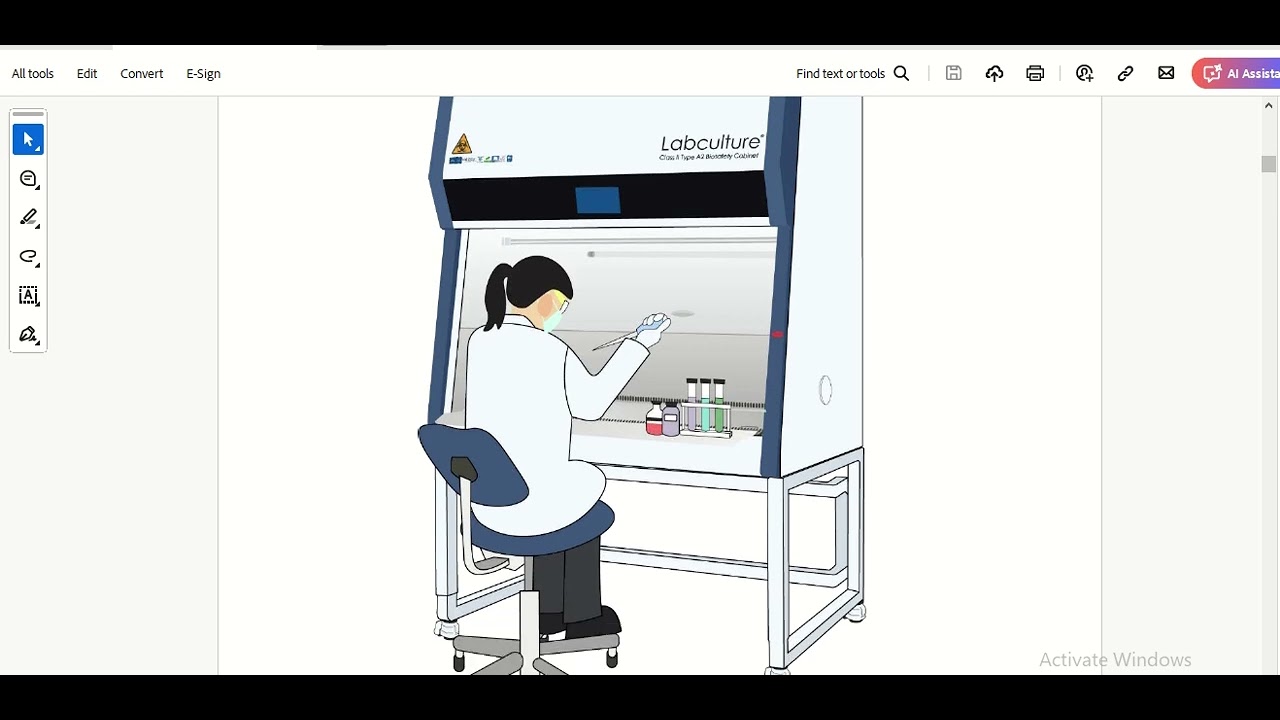


















Mammalian cell culture involves growing cells in sterile conditions within a Class II safety cabinet, using tissue culture flasks (T75 or T25) with cells attached to the plastic bottom surface under liquid growth medium; the complete medium consists of a base solution (like DMEM) supplemented with 10% fetal calf serum, and cells are passaged by washing with PBS, detaching with trypsin, counting using a hemocytometer, and replating at desired densities for experiments.
Mammalian Cell Culture Protocol: Counting & Passaging
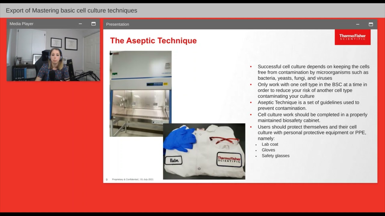
Added:Fundamentals of aseptic technique and maintaining sterile environments, including the proper use of biosafety cabinets.
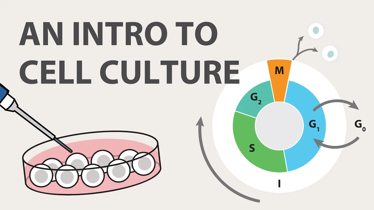
Basic eukaryotic cell biology, specifically mammalian cell growth cycles, cell morphology, and the difference between adherent and suspension cells.
The functions of key cell culture reagents, such as trypsin-EDTA for cell detachment, phosphate-buffered saline (PBS) for washing, and complete growth media containing serum.
Basic light microscopy principles needed to visualize cells and focus on a grid system.
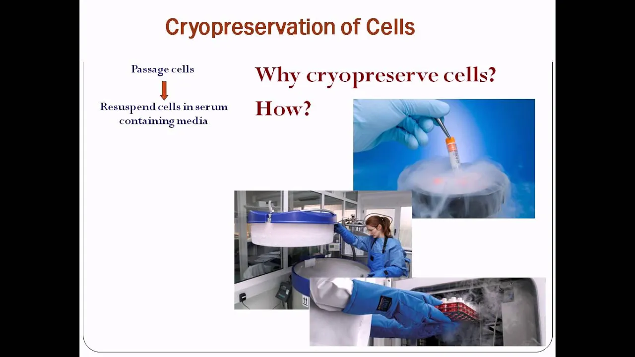
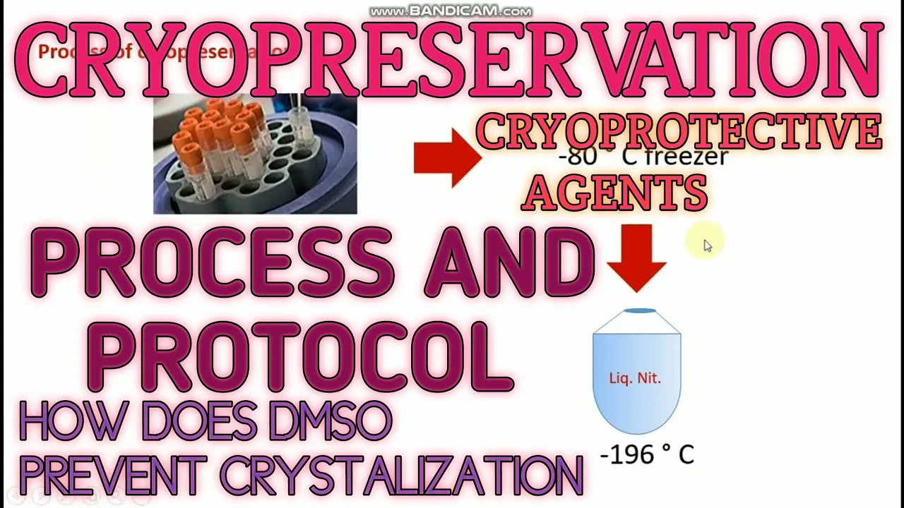
Cryopreservation and thawing protocols, including the use of cryoprotective agents like DMSO for long-term cell storage.
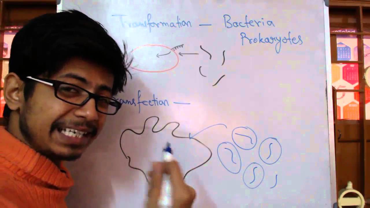
Transfection and transduction techniques to introduce foreign DNA, RNA, or viral vectors into mammalian cells.
Downstream functional assays utilizing seeded cells, such as cell viability assays (MTT/CCK-8), flow cytometry, or immunofluorescence imaging.
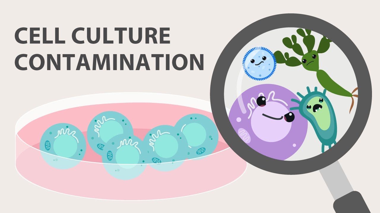
Troubleshooting contamination issues, specifically detecting and treating bacterial, fungal, and mycoplasma contaminations.
Advanced physiological models, such as transitioning from 2D monolayer cultures to 3D cell cultures (spheroids and organoids).
126.9K views3.1Klikes23:37Original Release: 2020-09-28
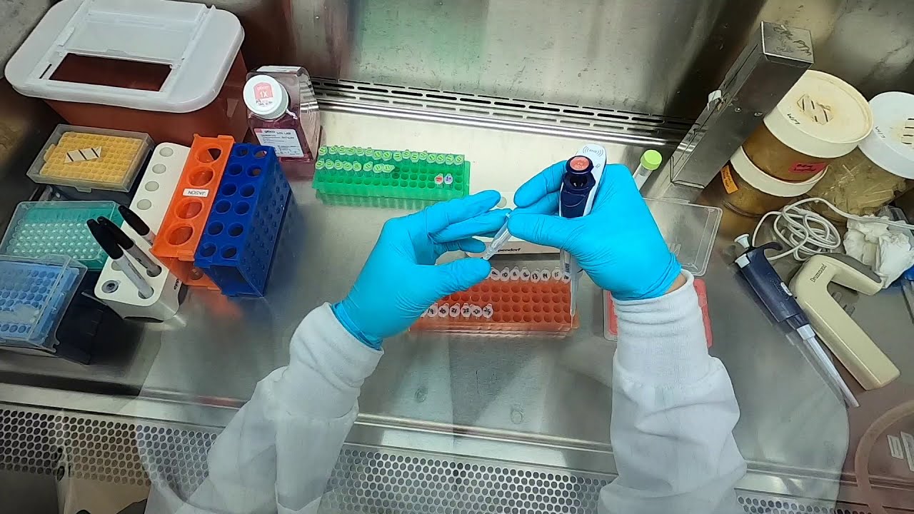
okay this is uh dr neil cross and i'm going to take you through a short video on how to do mammalian cell culture so mammalian cell culture goes on in laboratories like this and this what we're looking at here is a class two uh safety cabinet which is um a cabinet that has recirculated filtered sterile air to keep the cells safe from our own pathogens now we work within these cabinets and we have to put our hands inside this cabinet this sterile air here there's dirty air out here and we try to avoid working on this little grid here now we grow our cells in these tissue culture flasks this is called a t75 flask because it's 75 centimeters squared and the cells are growing on the bottom surface under a liquid layer now if you look really carefully you might just be able to see that the bottom of that layer is opaque and those are all the cells attached to the bottom layer of the plastic with liquid medium on top of them this is a smaller t25 flask 25 centimeter square flask so we use both of these routinely cell culture we'll refer to them as either t75s or t25s it's just part of the language that we use so to grow these cells [Music] we grow them overlaid with this liquid medium and we call this growth medium and growth medium is a solution of salts amino acids buffers that just keep the cells isotonic and happy but provide amino acids for growth glucose for growth and you can see from the bottle of medium this is growing in dmem which is dulbecco's modified eagle medium that's just a one of the growth mediums that we use and we add into that 10 fetal carb serum which is the acellular part of blood from a neonatal cow fetal calf serum and that is added to make complete medium so that's what is in that bottle there and that's what is in the flask there so complete medium has everything that the cells require to grow and keep them happy the other solution that we're going to be using today is pbs phosphate buffered saline and we use that to wash all of the medium off of the cells now what we're going to do is pour this medium into a waste pot wash the cells with phosphate buffered saline to get rid of that medium and then we're going to enzymatically remove the cells from the plastic using trypsin trypsin is an enzyme that will remove the cells from the plastic i'm going to go and show you all of these steps once we've done all of that we can centrifuge the cells count them resuspend them back into the growth medium and add them back to the flask at whatever density we want them to be at density in terms of cells per milliliter okay now we're going to look at the cells down the microscope so we're on a microscope here those are the cells in the flask on the microscope and this is what the cells look like down the microscope and each of these here are individual cells growing on that plastic surface so we can see that about maybe 50 percent of that plastic is covered with cells and we call that 50 confluent when it's 100 covered it's 100 confluent and we can have a look at those under a higher magnification and there we can see all of those individual cells these are breast cancer cells and they grow very very rapidly these cells will double every single day okay so i'll just take you through how we handle the cells in the lab so those are the cells they've got their liquid medium we can simply pour that off into a waste pot take our phosphate buffered stay line watch i'm holding the lid use my little finger hold it like that take off the lid and pour some liquid in you could put it in i tend to pour things and then give those cells a really good wash with the phosphate buffered saline now the reason we're doing that is to get all of the fetal calf serum that's in the growth medium off of the cells because what we now want to do is add some trips into that flask to strip the cells off the plastic okay so in cell culture we use these sterile pipettes and usually use power pipettors and i'm going to measure this out i want to add about a milliliter and a half of trips in to the flask so just simply add it to the flask and swirl it around so that all of those cells are covered with trypsin if you don't wash the fetal cast serum off the cells then there's lots of protein stuck to the cells the trypsin is added and all it does is chews up on the fetal calf serum and the cells remain stuck to the plastic and then you have to repeat the process again and again to get them off so that's why you wash your cells with pbs before adding your trips in these cells are now going to be incubated at 37 degrees in a cell culture incubator for two to three minutes or until the cells come off the plastic okay so this is a cell culture incubator 37 degrees with 5 co2 in the atmosphere that's standard for cell culture and the cells can sit on there for five minutes maybe three minutes and then we'll have a look at them down the microscope okay these cells have had a couple of minutes in the incubator and what you can see is the cells are now rounded up and they're detaching from the plastic okay that's that's a sign that the trypsin has eaten away all the proteins that are sticking the cell to the plastic and those cells are now detaching from that plastic now i've just given the flask a very slight know tap a very little light tap like that and all of those cells have now detached from the plastic now it's a really important point to add some growth medium to the cells to stop that trypsin from eating away the cells if we don't do that the cells will be dead within a few minutes so i'm now going to add some growth medium to them and that will inactivate that trypsin okay so here's some growth medium i've just put into a 50 ml falcon tube i'm just going to add that to the cells to inactivate that trypsin okay so now the trypsin has been stopped from eating away at the cells what i now need to do is centrifuge these cells because i want to get rid of the trypsin i want to get rid of the medium that's there so i'm going to centrifuge these cells at five 500 to a thousand revs per minute probably about about about 400 g and we're going to centrifuge those now so prior to centrifugation we're going to just put the cells into a centrifuge tube and just pour them in there and then we're going to centrifuge that uh and we'll require a balanced tube that's got exactly the same volume of medium as in that tube okay so this is the benchtop centrifuge those are our cells that is a balanced tube you see they're indirectly opposite each other in the rotor set it to 5 000 1000 rpm which is about 400 g for five minutes okay these cells have now been centrifuged and you may not be able to see that there is a pellet of cells at the bottom of that tube it will be much more obvious once i've pulled off the medium so now i've poured off that medium you should be able to see that there are some cells in the bottom of that tube it's sort of a grayish color of cells you might have to trust me but they are there now what i've got to do now is just resuspend that pellet to break it up a little bit so give it a little flick maybe a little bit of a grate on the grill and then we can add some medium to those cells prior to counting to finding out how many cells are in that tube there's probably in the region of a couple of million cells in there but we need to count them accurately so the first thing i'm going to do i'm going to add three mils of medium to the tube okay so we've got three million we've got three milliliters of cells we just don't know how many cells we have got and to do that to find out how many cells we have got we're going to use a hemocytometer now this is a hemocytometer it's a thick glass slide and you may just be able to see there's like a cross etched onto this shiny silver surface now this is a as little microscopic grid on there for counting cells and we use it in conjunction with a cover slip so what we have to do to use this hemocytometer is first breathe on it which is difficult with a face mask on but i will try so i'm going to breathe on it and you see this condensation on there now that condensation is now going to evaporate so i'm going to breathe on it and then quickly put a cover slip on it okay so i've put the coverslip on there and the cover slip has stuck and what we can now do is load the cells onto the hemocytometer okay so those are the cells i'm going to give them a shape because they settle out really quickly so just using a sterile tip what we now do is load the cells either side of the coverslip onto the shiny surface so that the liquid draws underneath the coverslip so that should take about 50 microliters of cells to load that coverslip now we're going to look at it down the microscope so there's a slide the hemocytometer on the microscope this is what it looks like and this is that grid that you could just about see and as i move it around there are basically nine large squares and each of those large squares is made up of either 16 or is look at the central one lots of tiny little squares and what we're going to do is count the number of cells in one large square there and one large square at the opposite corner take an average of those cells within those squares and multiply that by 10 000 and that will give us the number of cells per milliliter of medium so if there's a hundred in there and let's say a hundred in there the average is a hundred multiply it by ten thousand that equals one million so i'm now going to count those okay so i've counted those cells in one large square there was 118 and then the other one there's 122 so there is an average 120.
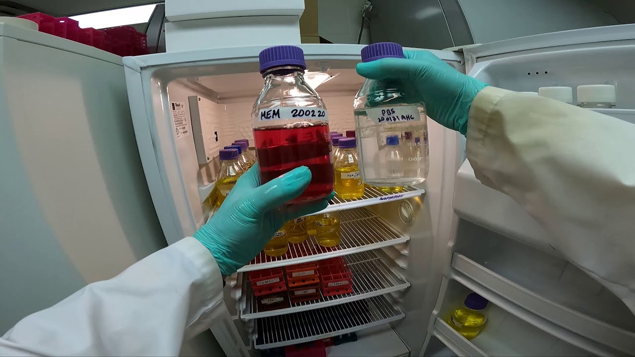
if we multiply that by 10 000 10 to the power 4 that gives us 1.2 million cells per milliliter of growth medium if you remember i added three milliliters of growth medium and we have 1.2 million cells per ml that gives us 3.6 million cells from that flask so we know exactly how many cells we've got and we know exactly what their density is okay so for this particular experiment i want to use some of these cells but i want to keep some of them growing for my experiment next week so i'm going to do i'm going to split these cells 50 50. so i'm going to put half of the cells back into the original flask okay so those are the cells that was just some medium okay and all i need to do now is top up this flask with medium um to keep those cells happy for the next five days or so okay this is just a tube of growth medium from this bottle i don't like putting my pipette into the growth media bottle for risk of contamination so i always pour a little bit into a falcon tube and then transfer it into my flask and this flask will require about 20 to 25 milliliters of growth medium so these cells can go back into the incubator and i'll use those next week these cells are the ones i'm going to use for my experiment and i'm going to plate them out a very specific density and the tutorial that i've put online the cell counting tutorial tells you how to do that if you work your way through that cell counting tutorial remember i got 120 cells in my large square on my hemocytometer i've got 1.2 million cells per ml if you use those numbers i want you to work your way through that tutorial to calculate how to play cells out into a 96 well plate however i will try to demonstrate that to you now okay so here's the calculation i counted 120 cells in a large square multiplied it by 10 to the power 4 that equals 1.2 times 10 to the power of 6 cells per ml that is 1.2 million cells per ml i want for this particular experiment 50 000 cells per milliliter and i need about 15 milliliters of cells in total for the experiment which i'm going to be putting these cells into a 96 wall plate you're going to see that in a minute this is a calculation that i do i take 15 milliliters which is what my final volume is multiply it by the density of cells that i want which is 50 000 cells per mil 15 mils times 50 000 cells per ml the units cancel out and the answer is i need 750 000 cells now i've got 1.2 million cells per ml so if i divide 750 000 cells by 120 let's say 750 000 cells divided by 1.2 million cells per mill the units cancel out that mill ends up on the top line of the calculation and that results in a volume of 0.625 milliliters of cells is what i need i'm going to take 0.625 milliliters of cells and make it up to a final volume of 15 milliliters that 15 milliliters will have a density of 50 000 cells per mil okay now the tutorial that i've supplied for you to work through is exactly the same as this only it's more step-by-step and numbers are different but i'd like you to work through that tutorial and see if you understand and follow what i'm doing okay so i've just finished adding 0.625 milliliters of cells to a tube containing uh 14 points uh one so 14.35 milliliters of medium so there's now 15 milliliters in there 0.625 of that are the cells so that should now be 15 milliliters what we now need to do is transfer these cells into a 96 well plate that is a plate with 96 wells and we're going to put 200 microliters in each of those wells okay for this we use a multi-channel pipette so our cells can go into the trough this is the sterile trough we then use our multi-channel pipette and i will put six tips onto there okay we've got six tips on there there's a good reason for that when you fill in a 96 well plate and as you can see as i release the plunger i can lift six lots of 200 microliters and i can just then just fill the plate so that each well is getting 200 microliters and we can fill a plate in a matter of seconds now the reason i'm using six tips is because when you're putting cells into a 96 well plate you only you don't use the outer edge for cells you put cells in the middle um 6 by 10 rectangle and then we're going to put pbs phosphate buffered saline around the outside edge okay so this is just some phosphate buffered stair line six tips again and i'll do the ends this is just how i do it do the ends first and then we can do the rest of the outer edge so we've now got a plate that is full of cells apart from the outer edge which has got pbs and that pbs prevents the cells from dehydrating if you had cells in that corner or that corner they would dehydrate and the results wouldn't be representative across the whole plate now once you've plated your cells out it's always a good idea to look at them on the microscope just to make sure you've actually got some cells in your wells and that is a well you can see some cells have already settled out as we focus up and down through the liquid medium you can see there are lots of cells floating around just by experience i'm fairly happy that that's probably 50 000 cells per mil we don't count them at this point we just accept that we have got cells in our wells and if you go to any other well across the plate you should see that the density looks fairly similar okay so that's just a little last minute check that i do then these cells are going to go into the incubator for a few days and i'm going to treat them with some drugs okay so those cells are going to go in the incubator all we need to do now is do the tidy up that is essential to cell culture we always wipe down our hemocytometers with a bit of ethanol and we make sure we never throw these away so they go back into their little boxes because they're quite expensive the cover slips go into a glass waste bin which is that one the cells go straight into the incubator so they're gonna be there for next week i'll do something with them and then we just need to clear up all of the mess so with cell culture it doesn't come across as being quite wasteful but everything that we use in cell culture just goes into the yellow bins all the plastic wear anything that we've used all the plastic tubes i'm afraid they all go into these yellow bins for incineration um the waste medium that's in this flask this will be treated with so it's in a uh a beaker that'll be treated with bleach after 24 hours it'll be thrown down the sink so that's just how we dispose of things and then all of our medium make sure the lids are all sealed up again and they will go and be still stored in the fridge for long-term storage okay that summarizes as much as you really need to know about mammalian cell culture one final thing to add is that when you split cells the flasks all have what cell line they are so these are mda mb231s they were last split on the 25th of the ninth and they were one in two split and uh that was um passage so that says passage two sorry so they were split on the 25th of the ninth passage two it's today it is the 28th of the ninth and then now up to passage three so what i have just done is passage those cells i've split them i've written on the flask what date it was who did it what passage they're up to now they were at passage two they now at passage three and i put a note on there to let them know i've done a one in two split so basically i've used half the cells i've thrown half the cells either away or into an experiment so you always label your flasks in this way so that when you come back to them you know how often you know when you last split the cells now these cells need probably a one in four split twice a week they grow that fast that is literally taking 75 of the cells out of the flask 25 back into the flask and top it up with medium twice a week so keeping these cells happy is you know it's a part-time job just keeping your cells alive and happy okay that's it that's the end of the video
Up Next

10:09
Hybridoma Technology: Monoclonal Antibody Production Explained
@FrankLectures
214.2K views•2017-10-12
14:12
Quantitative Real-Time PCR (qPCR): Principle, Method & Data Analysis
@animatedbiologywitharpan
129.2K views•2023-09-06
23:33
Microbial Degradation of Plastics: Biodegradation Pathways & Sustainability
@majeedhammad
2.9K views•2021-04-11

16:04
CRISPR and Genetic Engineering: How Gene Editing Works and Why It Matters
@kurzgesagt
30.5M views•2016-08-10
Related Study Plans & Knowledge Roadmaps
Structured learning paths in Biotechnology
biotechnology
AAV Production: Transfection, Prep & qPCR
01Introduction to Gene Therapy and AAV Biology
02Mammalian Cell Culture: Suspension HEK293 Cells
03Upstream Bioprocessing: Triple-Transfection Optimization
+ 2 more learning modules
18h5 modules
Explore Roadmap
biotechnology
Building Gut-on-a-Chip: PDMS, Cells & TEER
01Foundations of Microfluidics and Gut Anatomy
02Cleanroom Fabrication & PDMS Soft Lithography
03Epithelial Cell Culture and On-Chip Seeding
+ 2 more learning modules
26h5 modules
Explore Roadmap
biotechnology
Optimizing CFPS: E. coli Extract & Yield
01Fundamentals of Molecular Biology and Translation
02Introduction to Cell-Free Protein Synthesis (CFPS)
03Preparing E. coli Crude Lysate (The Extract)
+ 2 more learning modules
19h5 modules
Explore Roadmap
biotechnology
Haematococcus PBR: Design, Stress & Astaxanthin
01Biology and Life Cycle of Haematococcus pluvialis
02Bench-Scale Photobioreactor (PBR) Design and Engineering
03Culture Media Formulation and Inoculation
+ 2 more learning modules
15h5 modules
Explore Roadmap