
















The brain is organized into four major lobes with distinct functions: the frontal lobe controls voluntary movement, planning, and executive functions including speech production (Broca's area); the parietal lobe processes somatic sensation; the occipital lobe handles visual information processing; and the temporal lobe manages auditory processing, language comprehension, and memory formation via the hippocampus. The brainstem (medulla, pons, midbrain) connects to the spinal cord, while the corpus callosum enables communication between hemispheres. Gray matter (cell bodies) forms the outer cortex, while white matter (axons) provides internal connectivity. Understanding this anatomical organization is essential for neurological localization—identifying where disease processes occur in patients.
Neuroanatomy Brain Dissections: Lobes, Gyri & Localization
Added:Welcome to the University of Utah Neuroan Anatomy Laboratory. We're going to go inside and do some dissections of the brain and look at some of the important structures that are useful for understanding neurological principles and localization.
The purpose of neuro anatomy and all of its tortuous tracks is to allow you to do neurological localization. That is to say, decide where in the brain of your patient the disease process is occurring. That's the total reason for most of you to want to understand the connectivity of the brain.
Now let's look at the surface of the brain. We've removed the coverings the leptomening the pia and the arachnoid.
And I want to just show you the different loes of the brain as an introduction.
In addition on this brain you can see the brain stem. The brain stem extends from this region where my probe is a little bit difficult to see down all the way here to where the spinal cord was attached. In addition, you can see the cerebellum uh involved with coordinated motor control as well as the surface of the cerebral cortex. This is the vententral surface of the brain. And the most prominent feature here is the temporal lobe which I am outlining.
Here is the temporal lobe on the other side. And in front of it is the under surface of the frontal lobe. Cranial nerves are attached such as a visual uh optic nerve and the sensory nerve from the face the trigeminal nerve and blood vessels such as this large basler artery are running on the surface or you can even see the large internal corateed artery. We will come back and look at the nerves and the arteries and the other connections in greater detail.
But I just want to introduce you to the region of the medela, the pawns and the midbrain. And together they make up the brain stem.
Let's look at the surface now of the brain and look at the different loes.
You already saw the inferior surface of the temporal lobe coming back to here.
And we just kind of arbitrarily draw a line and say that this is approximately the end of the temporal lobe posteriorly and the tip of the temporal lobe anteriorly.
And then we look for a sulcus on the surface of the brain. Sulsi are these grooves and the surface structures that are coiled are the gyri. So we have sulsi and we have gyri.
The gyri have names but we're not going to learn most of them. However, this major sulcus, the central sulcus because it runs from the midline all the way down to the lateral fissure separating it from the temporal lobe.
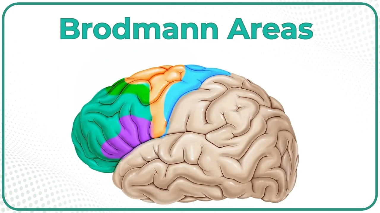
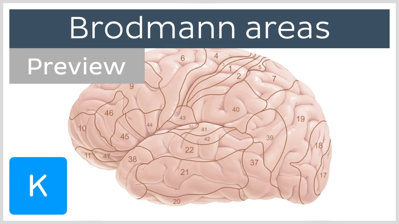
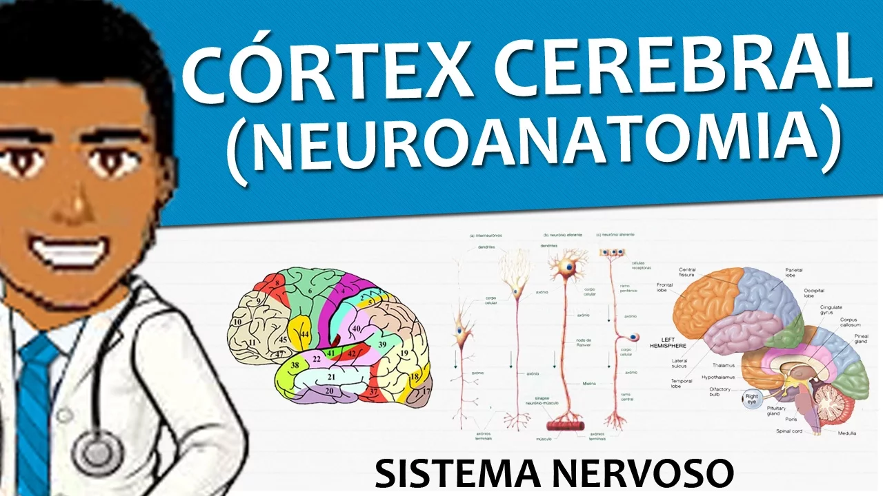
This central sulcus separates frontal lobe in front of it from parietal lobe behind it. And very interestingly, the cortex involved in voluntary movement is in front of the central sulcus. And the cortex involved with primary somatic sensation from your body is the gyrus behind the central sulcus. So this is frontal lobe, this is parietal lobe, this is motor cortex in general and this is sensory cortex in general. So we're just getting our bearings. The frontal lobe is further sort of divided and it's hard to see the distinction, but this band along here is the superior frontal gyrus. Then we have the middle frontal gyrus and the inferior frontal gyus.
The motor cortex in front of this pre-entral gyrus as we call it because it's in front of the central sulcus is an area involved in the left hemisphere which we're looking at with the production of speech and you may have heard of it before. It's called brocus area.
The rest of the frontal lobe is either motor function, planning motor function or executive function such as judgment and planning.
Let's look at the parietal lobe. Now the parietal lobe is separated from the occipital lobe by an arbitrary line that we draw between the pridal and the occipital regions.
these little notches here. So if I were to display here, I would say that approximately this is the occipital lobe. The occipital lobe is very important in humans because this lobe is involved with visual information processing.
It's a wonderful brain that we have and we are very specialized as humans uh for visual computation or an orientation and analysis. So we have our three loes.
Occipital lobe involved with vision, parietal lobe involved with sensation and integration of multiple modes of sensory input and a frontal lobe with motor and higher order function. And this temporal lobe that is down here is very much involved with auditory information which is coming into this fissure here that I'm pulling down. This is the lateral fissure and auditory information sound comes to both hemispheres and is processed here and this area behind it the superior temporal gyrus this back part here is involved in the comprehension of language.
So you've got two language areas, an interpretive language area and a production language area. And when we study the consequences of cerebral stroke, we'll see that some strokes can knock out one area and you'll have problems producing a uh intelligible speech, while other areas leave you hearing perfectly fine, but not understanding what you're hearing. and other lesions or damaging strokes that involve all of this area can leave you with a large deficit which we call global aphasia.
Aphasia is a language disturbance. The temporal lobe in addition to having an auditory function has an area underneath this gyrus here which is required in one hemisphere for forming new memories.
It's called the hippocampus but it's not visible here. So temporal lobe, auditory and new memories, occipital lobe, visual information processing and recognition.
Parietal lobe, somatic sensation from the face and the body.
frontal lobe motor execution voluntarily planning and execution of of uh appropriate uh judgment and actions. The two hemispheres are not identical and later we'll talk about special localizations between the hemispheres but right now I just want to point to the fact that the two hemispheres are held together by a large band of fibers called the corpus colosum.
What I have here is a coronal section through the brain. What we've done is we've taken our brain and we've sliced it like through this way and so this is called a coronal or frontal section and I'm going to rest it here so that you can see the internal structure of the brain.
We'll deal with these in more detail, but right now you can see that those gyri that we were looking at and the sulsi, the dips in between can be easily distinguished. And the gyri are covered with a nice layer beige in color. And that is your gray matter, as we say, of the cerebral cortex. Maybe we should have called it beige matter. And beneath it is a lighter area which we call the white matter. In general, gray matter consists of cell bodies, both neurons and GA. And white matter are axons. Axones are the processes of neurons either coming to the cerebral cortex or leaving the cerebral cortex and descending to lower brain structures.
An example of white matter here is this band of fibers called the corpus colosum. It was that band that held the two hemispheres together. When we were looking down through that inner hemispheric fissure, at the base of that was this band of white matter connecting similar areas or homotopic areas of one hemisphere with the other. And this is a way that the right and the left hemispheres can communicate.
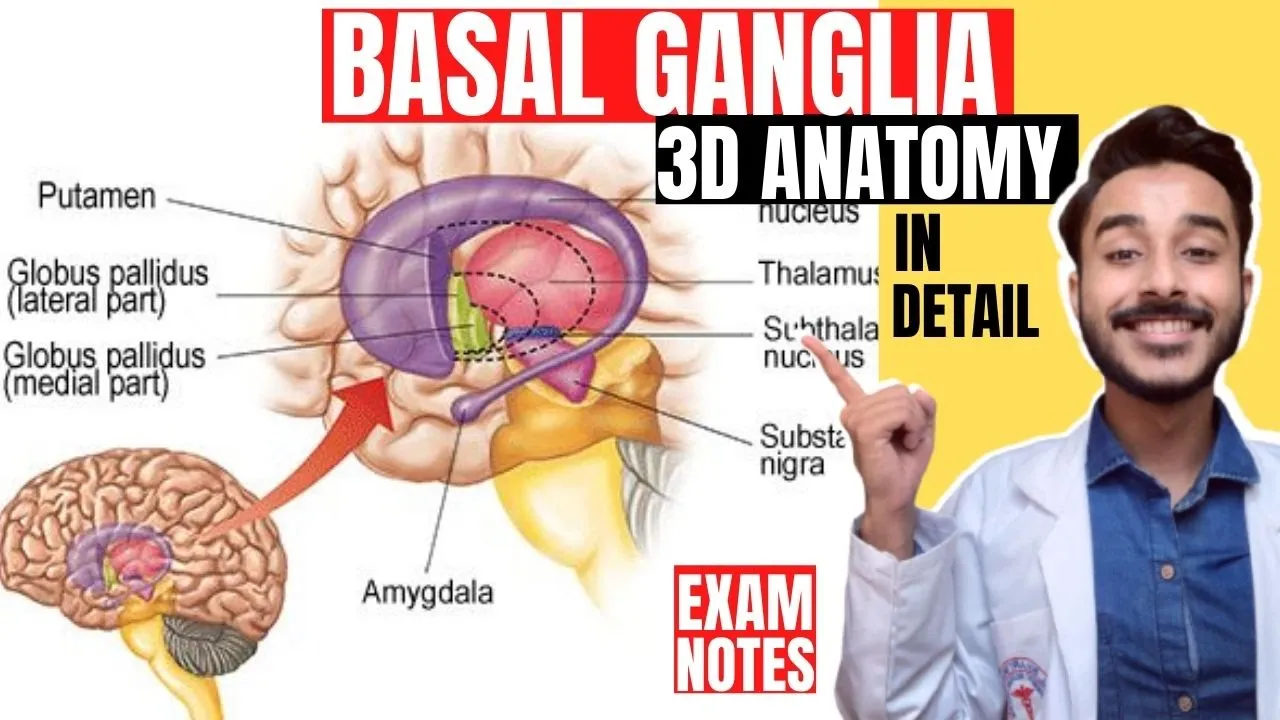
In addition, they can communicate through structures like this deep area.
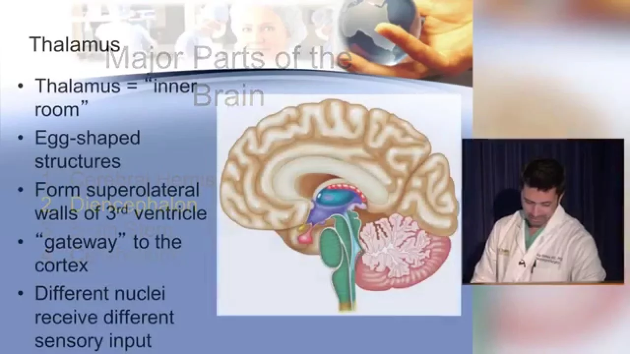
You notice it's beige, so it's mainly cell bodies. A deep structure called the phalamus. And here we can see the temporal lobe that we were looking at on the surface. Notice here if I prop this up and pull it down a little bit, you can see that large lateral fissure that we saw on the lateral surface. That means that this is the temporal lobe.
You can even see a blood vessel in there. This bit of cortex buried underneath the hemispheric temporal and parietal loes is the insula. We're not going to to deal with that, but it does have a name. It's where the cortex got buried as it became more and more convoluted.
This is parietal or it could be motor cortex. I can't tell with an isolated section exactly if we're in the frontal lobe or the parietal lobe. It doesn't really matter. And remember I told you that one of the functions of the temporal lobe in addition to the auditory region was an area involved in memory. That area is called the hippocampus and it's curled up here right in this medial part of the temporal lobe.
The other major structure you see in in a cross-section like this or a coronal section are these holes in the brain.
One here, one here, one here, a little bitty one here and here. These form part of the ventricle system which we will explore further but right now just know that they're filled with cerebral spinal fluid and the leptomening or the pia and the arachnide on the surface. If we can zoom in a little bit we can see them delicately covering the surface of the brain. The main thing I want you to see on this brain is the brain stem. So this is the cerebellum that we saw before up to the side and the brain stem recall was in the middle.
Here we have the brain stem on a midsagittal. This is cut in the middle.
So this is a midsagittal section. Here's a bit of the medela. Here is the pawns with a big basler artery creeping up over it. And here is the midbrain. Often hard to see. And finally then we have deep structures such as the phalamus and we can see many of the vessels like the anterior cerebral artery here uh as well as spaces such as the ventricular system which we'll look at in more detail.
Yeah.
Up Next
Neuroeconomics: How Orbitofrontal Cortex Neurons Drive Choices
@NeuroscIQ
1.9K views•2020-11-07

Deciphering Neural Circuits Controlling Anorexia in Mice | Richard Palmiter
@AllenInstitute
2.3K views•2011-10-10

Vagus Nerve (CN X): Anatomy, Nuclei & Functions Explained
@Alilamedicalmedia
305.2K views•2022-10-31

How Exercise Benefits Your Brain: Science Explained
@TED
11.4M views•2018-03-21
Related Study Plans & Knowledge Roadmaps
Structured learning paths in Neuroscience