











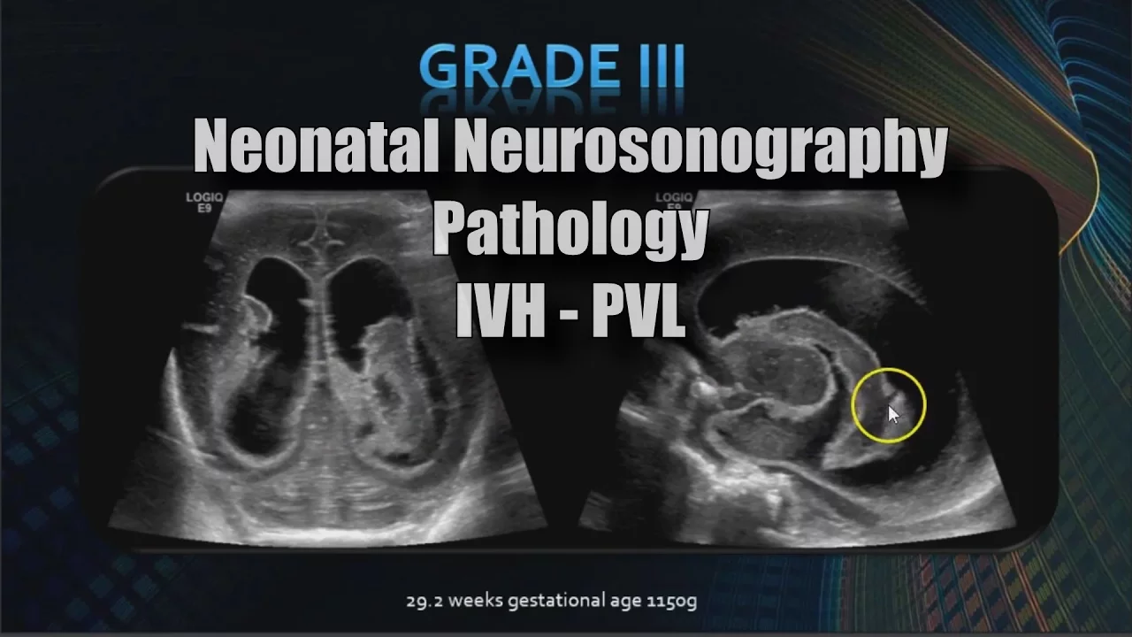






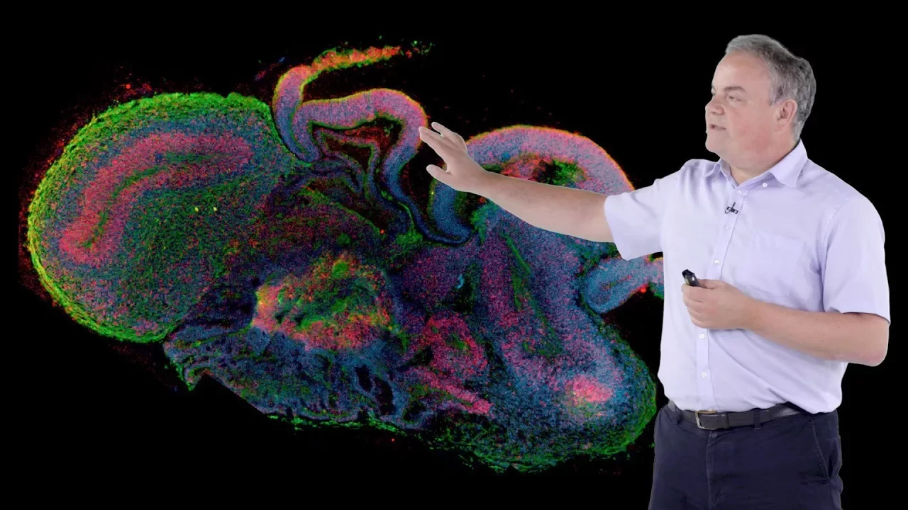

In the prenatal human brain, angiogenesis and neurogenesis are tightly coordinated through dynamic interactions between vascular and neural cells, with vascular cells serving as signaling hubs that influence neural stem cell behavior; the ganglionic eminence (GE) serves as a critical angiogenic niche during the second trimester, characterized by abundant endothelial filopodia contacting dividing neural progenitors, and this region's vascular immaturity, combined with potential hypoxia-reperfusion injury, contributes to the high susceptibility of preterm infants to germinal matrix hemorrhage.
Angiogenesis & Neurogenesis in the Prenatal Human Brain
Added:Fundamentals of embryology, particularly the early stages of human central nervous system development and neurulation.
Basic cell biology of stem cells, including the concepts of multipotency and the differentiation pathways of neural progenitor cells.
The physiological process of angiogenesis, specifically how endothelial cells migrate and proliferate to form new blood vessels.
An understanding of neurogenesis—the biological process by which neurons are generated from neural stem cells.
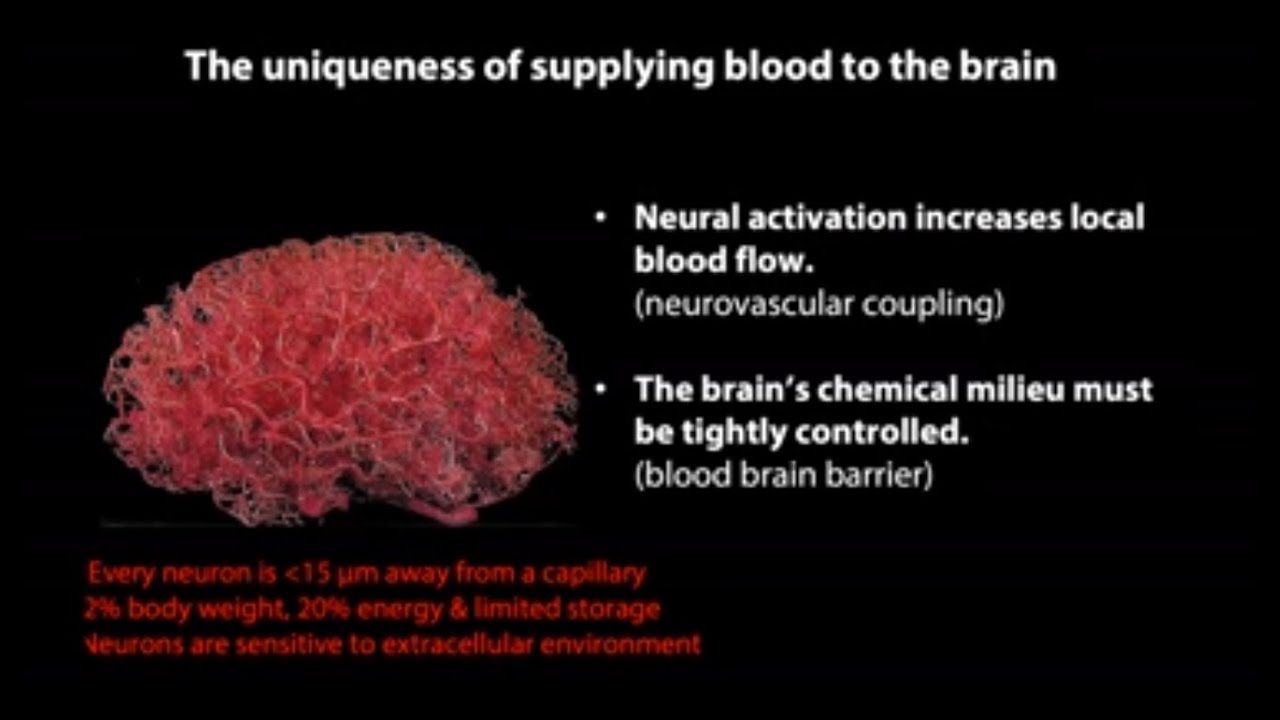
The formation and function of the Blood-Brain Barrier (BBB) and how neurovascular coupling regulates cerebral blood flow in the mature brain.
Pathologies related to disrupted prenatal neurovascular development, such as congenital brain malformations, cerebral palsy, and hypoxic-ischemic encephalopathy.
Applications of neurovascular interaction research in regenerative medicine, specifically in developing therapies for stroke and traumatic brain injury (TBI) repair.
The use of advanced research models, such as 3D brain organoids and microfluidic 'brain-on-a-chip' systems, to study human-specific neurodevelopment in vitro.
325 views4likes53:10Original Release: 2023-06-15
exactly I think the intro okay we're recording and I can't see the chat but I can see myself and some people so I'll monitor the chat sounds good thanks Eve so hi everybody we're going to get started and uh let me just ask before we start did anybody um offer to Roberta Keller to introduce Betsy Crouch is there anybody on who got that assignment if nobody's volunteering then we're gonna ask Betsy to introduce herself because she will she's best uh positioned to tell you all about her background Betsy all right well thank you and thanks to Roberta who is in Paris we all realized um so good for her but she um you know kindly gave me the invitation to speak today uh I am going to for those of you who've seen me speak before um I've been you know lucky to be really well supported here at UCSF and I'm very grateful to continue to work here uh I'm going to put in some very fresh data so you know please excuse me for that but I also you know wanted to stimulate some discussion for those of you who don't know me my name is Betsy Crouch and I obtained my MD PhD from Columbia after I graduated from there I had a a really fortunate conversation with David roach who at times the time was the neonatology division Chief here at UCSF and he told me that if I wanted to be a physician scientist in neonatology I had to come to UCSF he after that left us to go be the department chair of pediatrics at Cambridge but I've always been very grateful to him for that piece of advice because he was spot on I did my residency and I fast-tracked into Fellowship I'm in neonatology here at UCSF I did the PSS psdp program the national K-12 award that's now run by Sally permar That was just really instrumental in how I you know positioned and thought about myself as a physician scientist and then I was very fortunate to be awarded the UCSF pssp in 2020 and started my own small group there in the stem cell building in the broad stem cell center and that's where my lab is located now so today the topic of my talk is angiogenesis and neurogenesis and the prenatal human brain and as I understand the convention is for this seminar I'll ask you to please hold your questions until the end and I'll try to leave you know a good five to ten minutes for a discussion so in the Crouch lab we study brain vasculature and much more than passive conduits for oxygen and nutrient delivery vascular cells are emerging as a signaling Hub of the neurovascular unit our perspective is similar to the 17th century Dutch anatomist Frederick Royce who said that blood vessels are ubiquitous and tissues are only blood vessels that are variously arranged if you're unconvinced by the Dutch anatomist the blood vessels are much more than passive conduits look at the recent literature on this topic so on the this is and this is also kind of an area of strength through UCSF so I'll point out a lot of my esteemed colleagues in this area so this is Ethan Winkler's paper here and let me get out my pointer there we go this is Ethan Winkler's paper here with Dan Lin who's also a neurosurgeon along with Ethan and Tom nowakowski who studied or who published a single cell atlas of the normal and malformed adult brain human brain vasculature this was published last year in addition here's Andrew Yang's paper who himself is a Sandler fellow who also has a lab here at UCSF and he when he was a grad student in Tony week's Corey's Lab at Stanford published this paper also published last year called a human brain vascular Atlas reveals diverse menus of Alzheimer's risk performing single cell RNA sequencing on adult human brain vasculature and Alzheimer's cases um and we recently added to this topic describing the prenatal human brain vasculature and data that I'll discuss today so in part my interest in this topic began as a stem cell biologist interested in how neurovascular interactions affect stem cell Behavior so there's a lot that has been published in neurovascular interactions for a long time but we arrived on this topic recently in a collaboration with fully up to Alfonso's lab in Germany and like many good stories this one started with an observation and these are these beautiful filipodia which are colored in Gray from the vasculature which are contacting dividing neural stem cells which their nuclei are colored in in fuchsia in this diagram now this image is taken from the mouse ganglionic evidence it's a area of the brain which you'll hear much more about today but in short what we Quantified here was the phillipodial contacts with the dividing neural progenitors and found that in fact it was statistically significant and that this interaction increased over time now in trying to discern why we had these contacts we then moved to the human brain where now we've switched up the markers but the vasculature in these pictures are are in white and you can see we also saw abundant philippodia in the vasculature in the human um same region the ganglionic Eminence and we Quantified this interaction and also saw that there was a statistically significant relationship between the philippodia and the dividing neural stem cells so in some we created this model at the end of this paper where the radial glia the stem cells in this region they secrete a molecule called fedjaph which makes the philippodia from the vasculature extend and in Mouse models we were able to manipulate the number of philippodia and we found that when there were too many philipodia that this caused the neural stem cells to prematurely exit the cell cycle and generate neurons at the expense overall of sulfernuel so there are many other examples but this is just one from our work to highlight how tightly coordinated angiogenesis and neurogenesis are in the developing brain now in addition to a stem cell biologist and my work as a neonatologist I take care of premature babies and unfortunately um one of the sequelae from their prematurity that happens relatively frequently is something called intraventricular or general Matrix hemorrhage now this unfortunately again occurs to 20 percent of our youngest babies so elbw stands for extremely low birth weight we generally think of this in the babies who are born less than 30 gestational weeks it's very stereotyped to occur under the cortex next to the ventricles this is the region called the GE which I was just showing you in the previous images and this region will become over time the caudate and the thalamus this um condition carries a high risk of mortality and morbidity and is associated with intellectual disability and cerebral palsy we have no disease disease modifying treatments as many of you are probably aware now the outcomes after severe gmh are poor and just to again review briefly for those of you who aren't familiar with this condition um a grade 4 Hemorrhage has now been renamed a pvhi which is periventricular hemorrhagic infarction when the babies have especially a bilateral grade 4 or pdhi Hemorrhage this carries a 40 mortality rate and also has a very high cerebral palsy rate practically this means many of these children with a severe Hemorrhage will likely never walk and may never talk and have intellectual disability so why do we have no treatment for the Hemorrhage well this is a difficult area to study so these are gross pathological specimens when in specimens without Hemorrhage collected from my postdoctoral mentor Dr Eric Wong and these were images were published in a beautiful paper by Mercedes Perez at all that I'll reference a couple times in this talk that was published in science last year so in these gross pathological specimens we've highlighted in the coronal sections beneath the area that constitutes the ganglionic Eminence also called The germinal Matrix the area which is prone to Hemorrhage and I hope you can appreciate in these images how the GE takes up proportionally more surface area at the younger gestational ages and then over time by 33 gestational weeks there's only a Remnant left and perhaps this developmental time frame underlies some of the vulnerability of this region to hemorrhage so we're starting to study these structures in humans Mercedes Paredes paper was one of the first but we also do know more about these structures from Mouse so these are this is um a schema of the developing Mouse brain and here are coronal sections where the lateral ganglionic Eminence LGE medial ganglionic Eminence and caudal ganglionic evidence has have been highlighted and in general these structures will produce the majority of gaba-containing interneurons which are born in this region and then migrate up and integrate into the cortical circuitry but let's take a step back where do brain vascular cells come from and how do they develop so these images are from Tom Arnold's paper where he labeled blood vessel cells with cd31 in green and showed that this is about halfway through Mouse gestation that the blood vessels start outside of the brain and what's called a perineural vascular plexus they dive in and then elaborate up to the um most dorsal and then further the most medial aspects of the developing brain and then they elaborate out with the growing neural tissue but when I started in this work during my fellowship now six years ago there was very little known about human brain muscular development and that's where we decided to focus our efforts so here's our outline for today and first we're going to discuss angiogenesis and the human GE this is also a Shameless plug for a review that actually just came out last week online in Trends in neuroscience and Trends in neurosciences where we highlighted all of the recent work using single cell RNA sequencing in the human brain and I encourage you to dive into it if this piques your interest now to study vascular cells in the developing human brain my lab focuses on two main cell types endothelial cells which can compose the Lumina blood vessels throughout the body in addition these endothelial cells in the brain are connected by tight junctions which are important for the blood-brain barrier neural cells are an umbrella term that I use to include parasites smooth muscle cells and fibroblasts although the lateral inclusion is controversal in general these are heterogeneous relatively undefined support cell population we know that they're required for the formation of the blood-brain barrier and on the opposite end of the life Spectrum they've been implicated in neurodegeneration although many of these Studies have been performed in Mass so to investigate blood vessel cells during human development we first started performing immunostaining in sections from the early second trimester ganglionic eminence this is that region again prone to Hemorrhage so I'll abbreviate ganglionic Eminence GE and here's a schema of a hemichronal section of the developing human brain and these pictures are taken by the in the area highlighted in the red box so first we use cd31 which is uh you know a well-known cell surface marker for endothelial cells we used ng2 to label mural cells it works well for them I'll say before 30 gestational weeks after that it starts to label what we think are oligodendrocytes and so after that it gets complicated Now pdgfr Beta is the canonical mural cell marker but in the human brain it's complicated because it also labels radialglia which are the neural stem cells so just focusing on the ng2 something that was very apparent when we first looked at this data was that there's a prominence of vascular cells mural cells specifically located right next to the ventricular surface and in addition it's less dramatic but the endothelial cells also seem to be preferentially enriched in this region now looking over gestational time frames so at 22 gestational weeks you can see similar patterns where there's a predominance of cd31 and ng2 localized next to the ventricle but by 39 gestational weeks things are very different so now we think ng2 and pdgfr beta are labeling glial cells right located right next to the ventricle but cd31 is still very good to label endothelial cells to quantify this data we divided up the GE into three regions based on different neural progenitors that can be found in this region Zone one is also called the ventricular Zone this is filled with densely packed radioglial neural stem cells zone two was the subject of Mercedes Paredes paper these are areas which look like they have holes in the tissue but in fact these are not holes the regions which are filled which are just negative for these immunostains and filled with those densely packed newly borne neurons and zone three are the area's lateral to zones one and two so as you can appreciate by eye we also saw when we Quantified cd31 surface area by percentage that there's an enrichment in the ventricular Zone Zone one at the early and late part of the second trimester but that this trend disappears by the time that babies might be born at turn and similarly as you saw by eye there's an enrichment of the neural cells also in the ventricular Zone at the early part of the second trimester so these are high powered confocal images emphasizing the vascular gradient and then we did an Ameris rendering on the right I'm showing the ng2 expression in a gradient of expression in a gradient of colors with red denoting High expression right at the ventricular surface and the cooler colors a little bit further back from The ventricle um I've shown shown beneath in this region at this magnification you may also be able to see more detailed features of the vasculature such as the endothelial filipodia that are poking out right next to the ventricular surface to focus on these Phillip podia which of course I talked about at the beginning of the um of the seminar as well here's another video this the top of the ventricular surface is here and now I have the endothelial philippodia in green and the overlying mural cells in blue and this is just zooming into different regions to help you appreciate how many more philippodia there are the ventricular surface so when we Quantified this data we saw that again as you can see by that there are more phillipodia in the ventricular Zone compared to zones two and three about the early and the late part of the second trimester and this allowed us to start having a hypothesis about what was going on in this region so we know that the ventricular zone is the canonical neurogenic Niche with the build with abundant radial glia but this data allowed us to start to generate the hypothesis that this was also an angiogenic niche an angiogenic vasculature should also be have more Branch points so we Quantified that parameter and saw also there was increased branching so in addition to increased in addition to increased Philip podia and branching angiogenic vasculature should also be dividing or proliferating so to test this parameter we performed immunostaining with c31 and pdgfr beta as well as ks67 many cells including the neural cells are dividing in these regions these are immunohistochemistry using a confocal microscope and here is an Ameris rendering showing uh this area of this vasculature highlighting that you can see a dividing endothelial cell right captured right here and when we Quantified this there are very few dividing vascular cells in general and especially you know vascular cells are only 10 to 15 percent of the surface area but we were able to find a statistically significant enrichment for them in the ventricular Zone as well as for the mural cells and we did an Ameris rendering of a dividing mural cell here captured from this vasculature region in II now we are also fortunate in these experiments to be able to collaborate with a Spanish group uh Garcia Manuel Verdugo lab based in Spain as well as Arturo Alvarez buya and the postdoc who was doing the analysis was a wonderful scientist called aransha Sia and here I'm showing uh transmission electron microscopy from 17 gestational weeks and 23 gestational weeks and something I thought was really striking about these images is that here she's highlighted the blood vessels in fuchsia and and what was really noticeable is that there are very few vascular lumen in this region and of course angiogenic vasculature is also not patent it's not having blood flow through it yet because that would be devastating obviously um so what we're able to see is just that there are very few lumens in the ventricular Zone and even in the inner subventricular Zone at 17 gestational weeks but by 23 gestational weeks now we're starting to see some Peyton lumen in this area so again just just able to capture the angiogenesis process and different snapshots so to summarize this part of the talk I'm going to show this schema that was nicely made for us by a woman called Sarah Pyle where she is capturing the vasculature in terms of tiles which are making up this region and here I've highlighted with arrows the different angiogenic subtypes of vascular cells which are found enriched in the ventricular Zone at least during the second trimester so these initial histological observations made us curious to use orthogonal techniques to study the vasculature including the now popular facts flow cytometry and single cell RNA sequencing now this is a Disney thought from a single cell RNA sequencing data from a paper published now six years ago from the creekstein lab at UCSF to interrogate individual cells in the developing human brain from six gestational weeks to full term but I've highlighted here with this arrow that there were very few vascular cells captured in this entire data set and up until last year there were very few vascular cells in any data set because as you could see from the previous data they just compose a small part of the overall cellular composition of the brain so we needed to develop a way to enrich for them um you know here I'm skipping a lot of data but in short we decided to use flow cytometry which of course is a is a way to use self-surface markers that are present on the cells to be able to use fluidics and capture them and then study them in more detail so I'm not showing the flow cytometry plots here that include the degree dead cells and doublets but what we decided to do was use cd45 conjugated to a fluorophore called PE size 7 which is what the machine uses to to separate out these populations um this allows us to separate out microglia as well as perivascular macrophages as well as some small population of infiltrating immune cells in the in the divide in the developing human brain and I give these to other people who are interested in studying those cell types I'm interested in the cd45 negative cells which then I can use and pep ABC on the y-axis and cd31 conjugated to a fluorophora called Alexa 48 on the x-axis to pull out distinct populations of mural and endothelial cells so these are umaps summarizing the Single Cell RNA sequencing data of approximately 150 000 endothelial and mural cells so each dot in these plots represents an individual cell and we were first very pleased to see that in this data set which is using seven cases all from the second trimester that endothelial and mural cells um they know who who they are and they're able to separate into their cell type identities in these plots which is a way to reduce all of the dimensionality of single cell RNA sequencing data you have cells which are similar to one another in the RNA molecules that they're expressing their transcriptomes are localized right next to each other now interestingly we set up these experiments to understand the difference between the GE the vasculature which is prone to Hemorrhage and the cortex which is a control region but we really didn't find anything that is that significant yet and I'm happy to talk about that in further detail similarly we we found some sub subtle differences by age that we're now going into more detail by but I'm not going to talk about it today I did want to offer that we put this data up on a publicly available web browser and so you know everybody can look up their favorite Gene of interest and sort of correlate with subtype might be expressing it in the second trimester of human brain if you don't know about the cells.ucsc.edu resource it's a phenomenal resource with many different cell browsers and so I was talking to somebody studying glioblastoma the other day and interested in the role of the vasculature in that regard and we were able to look up a glioblastoma single cell RNA sequencing database as well so first let's focus on the different subtypes that we found in the second trimester of human brain again here are some umaps showing all the different data and when we looked at this our first question was we know that there are arteries and veins and capillaries throughout the body but will we see these distinct subtypes as early as the second trimester and in fact we could find them already this early so here's our group of cells which are predominantly arterial endothelial cells here are capillary serovanes and then we pulled out some subtypes which are specific to development such as mitotic endothelial cells and the tip cells are the ones with the Abundant philopodia so we were grateful to see that they actually appeared in our database as well if anyone is curious I have feature fonts showing the specific genes that we used um here and on this slide and I'll note that um you know this is development so no gene expression here is strictly on and off so there's a gradient of expression in many of these genes so then we needed to use RNA scope which is just a fancy and situ hybridization to be able to localize these cells to validate the bioinformatics so here's a schema of a hemichronal section from the second trimester human brain specifically 17 to 27 15 to 17 gestational weeks and you know in this area that I'm capturing one which is a little bit um back from the ventricular Zone in the ganglionic Eminence you can see that here are some beautiful individual puncta um which is uh the RNA adgrg6 which in our data set we found is a venous capillary marker next when I was looking for tip cells we used the RNA molecules adrenal Medellin and angiopoietin 2 to label these cells and we were happy to see that indeed they were localized right in these blood vessels which are right next to the ventricular surface as they should be based on our histology and finally when we looked for arterial endothelial cells we found them outlining individual the cytoplasm of individual cells in a beautiful way in this bigger blood vessel which is a little bit back from the ventricular surface so then very little is known actually about the way that these vascular cells mature I think about it a lot as a neuroscientist that we know the canonical developmental trajectory of neural cells that you have radioglia which are the stem cells that then go through an intermediate State and give rise to neurons as well as you know other other cell types over time but the the maturation trajectory of the vascular cells is still not well um clarified so as a first pass to un to get at this question we use something called RNA velocity which is an algorithm bioinformatically that looks at the pre-splice to splice mRNA ratios to understand who's a more immature and who's a more mature cell so we looked at two of our cases separately the 15 and 23 gestational weight cases and in both of our data sets we saw that there are mitotic or venous cells which are more stem cell-like which then give rise to a capillary intermediate and that arterial endothelial cells are the most differentiated at least in our data set and this was gratifying because in previous morphological observations um in different model systems like zebrafish people have have shown that that this is the way that that it seems to go morphologically so now we have some single cell RNA sequencing data that is in line with those observations as well moving on to the mural cells I wanted to provide a little bit of a primer on these cells because they're less talked about so here um crystals is a scientist based in in Sweden and he's really the The Godfather of all of the parasites and so these are um from some of his papers on the left is a review where his focus is really on the pericite which is this mural cell found in the capillary microvasculature but as you go back to the bigger um blood vessels the arteries these are certainly smooth muscle cells the cell type that's this intermediate between the smooth muscle cells and the parasites I'll just say is very controversial as well as the the mural cells which are found on the postcard capillary of annuals and the venules these some people call them venous smooth muscle cells but they aren't very well defined at all in addition in a single cell RNA sequencing paper that looked at the adult Mouse vasculature they were able to capture this new fibroblast-like cell type which is found in the bigger blood vessels the arteries and the veins again in the adult mouth spring so using the marker genes from this data set we then looked back at our data set to see what type of mural cells we had in the second trimester and we were able to identify the smooth muscle cells the classic parasites as well as a small population of fibroblasts and in addition capture a larger group of mitotic mural cells which again was consistent with our histology so in the interest of time I'm not going to show you all of the RNA scope for this but you can find it in the paper if you're um if you're so inclined but I will show this movie that I really like where we did RNA scope for atp1a2 and kcmj8 which are classic parasite markers which should be found in these smaller blood vessels especially I put in an arrow at the one which was found at the branch points and you know we saw that they localized as we expected them to which was great in addition of course this marker is not exclusive to the vasculature we saw atp1a2 expressing cells in other areas of the brain which which we think are likely new neurons now the stages of mural cell development in the brain had not been shown in any animal model to date at the time that we published this paper and so we also wanted to use RNA velocity to try to understand the maturation trajectory of these cells and again looking at the 15 and the 23 gestational week case what we saw was there's a mitonic group of mural cells which then seems to give rise to smooth muscle cells and then there's a small group of classic pericites which are present at 15 gestational weeks and by 23 gestational weeks now you can see that there's um you know a nice flow from the smooth muscle cells to the classic parasites this is something that we're working on currently because I'll just say that in the mouse heart this is not the way the trajectory goes Christie redhorse is a brilliant scientist who did her PhD here at UCSF and now is at Stanford in a hhmi-funded and she showed that in the mouse heart it goes the opposite way the classic parasites are the progenitors for smooth muscle cells um so you know I think this is very exciting but now we're trying to do some functional as well as histological assays to understand if if this is just it could be a bioinformatic kind of Quirk but but we don't think so for a couple of reasons that I can get into now on the horizon one of the ways that we're starting to validate this is using this new tech called hypex RNA scope I had shown you the the RNA scope previously for where we could do two probes at the same time along with some immunohistochemistry but I have a wonderful Master's student in the lab now whose name is Edward Valenzuela and he's starting to do this High Flex RNA scope um experiments where we can do 12 probes simultaneously so here's a picture of the developing human brain here's dorsal and ventral and medial and lateral this is an image that he's taken using these high Plex RNA scope probes and he's capturing the blood vessels in different regions and here of course is the ganglionic Eminence so here's a zoomed in image of one of these blood vessels that he's capturing here where you can appreciate the um the different RNA scope probes in this bigger blood vessel which is kind of thought um caught uh cut tangentially and then another one here which is more parallel to the tissue section so here are some of the individual RNA scope probes we're doing a number of different pan mural cell markers here pdgifr beta we've all been found in new marker rgs5 which seems to work really well at the RNA scope level but there isn't an antibody so this has been really helpful and then using some specific markers for subtypes and trying to parse out where are the different subtypes present and how do they appear over time in the human brain specifically I'll I'll say that sdc2 is a new smooth muscle cell marker that we found as well as acta2 smooth muscle actin which is kind of the canonical one and we were very pleased to see that in this bigger blood vessel which is likely an artery or an arterial we have abundant sdc2 and smooth muscle actin um RNA puncta showing up but here's a smaller blood vessel down here which has pdgifr beta and rgs5 and but is absent for the smooth muscle cell marker so that's good and this was encouraging so we're using this technique to map out all of the different mural cells and that's work that's ongoing so in addition we wanted to use this data to think about the ways that endothelial and mural cells might communicate with one another to create the vasculature and maybe particularly the blood-brain barrier in the second trimester so to do this this was a work from Claire Howard who at the time was a Pediatrics resident at UCSF and she was in the molecular medicine program and she had a month to do a little bit of lab research time and so she figured out bioinformatically how to run this algorithm and created this beautiful plot so what we did was using again our RNA sequencing data this plot is mapping the number of significant interactions and what was really interesting was that we when we looked at this to understand the interactions between endothelial and mural cells the collagen and lamin signaling pathways are the ones that came up as the most enriched and this was also very gratifying because as many of you likely know that mutations in call 4A can be associated with severe uh intracranial hemorrhages that can present at birth and so this molecular data now casts some light on why that is collagen is so important it's the number one signaling pathway in the second trimester human brain so mutations in one of these signaling subunits would be expected then to be very detrimental so in addition trying to understand some of the blood-brain barrier we did find some of these adherence Junction proteins like the jams pecam1 is also it's an endothelial marker but it's also an adherence Junction proteins as well as some of the coherence these are further down um in terms of our number of significant interactions but they're still present I think this was starting to hint that the blood-brain barrier must be barely immature during this time and then finally the tight Junction proteins the occludans and the clottens are present but again they're they're much less significant or much less abundant so I think this is all hinting that the blood-brain barrier is very immature at this time Point as has been suggested by others for a long time but now we have molecular data for this so in addition we went back to our trans transmission electron microscopy to corroborate these data um and this is just showing a blood vessel from the 17 gestational weak human brain and here are some of the different adherence Junctions which are present but I'm not showing an example from the the adult human brain but the hearings Junctions are supposed to be surrounding the entire vascular cells so they're present but they're again like much smaller and less um than uh than expected for a mature blood-brain barrier in addition we know that another way molecularly that the blood-brain barrier is present is that the mural cell talks to the endothelial cell and really shuts down transcytosis which is the way that the endothelial cell is bringing things in from the vascular Lumen but at this 17 gestational weak case we see pretty abundant evidence of transcytosis that's actively occurring so again this is other evidence just to to promote the fact that the blood-brain barrier seems to be very immature so finally we wanted to start to do some functional experiments with these vascular cells and we decided to do a transplant into cortical organoids so to perform these experiments we took our facts Paradigm pulled out the endothelial the mural cells we labeled them with a virus which is non-specific and just makes all of the cells Express gfp and then put the vascular cells on top of organoids to understand how they develop and and in one way model angiogenesis so the growing vascular cells in a known neurogenic model the cortical organoid we let these cells go two weeks in culture at first we are very pleased to see that the cells they integrated they seem to be happy in these co-cultures and then we went on to analyze them after two weeks first I'll show the data from the endothelial cells so we knew based on the Single Cell RNA sequencing that we would start with five different subtypes of endothelial cells arterial venous capillary the tip cells and the mitotic ones but what was really interesting was when we transplanted them into this model after two weeks the predominant subtype was entirely tip cell so here I'm showing the RNA scope probes for adrenal Magellan and angiopoietin II and here's a rough quantification on the right of the abundance of these probes but this was really curious and and then we went back and read some of the organoid literature where we know that the organides are predominantly radialglia so again this is really just recapitulating that ventricular Zone where all the endothelial cells become a tip cell and the neural cells are predominantly radioglia and we're hoping to use this in the future to study those interactions now with the mural cells again we knew based on the Single Cell RNA sequencing that we started with four different subtypes but again when we transplanted them into this very radioglial heavy cortical organoid we saw that they also had a um a favorite phenotype so in the mural cell instance it's the mol9 and transgeline expressing smooth muscle cells and then I Quantified this on the right truthfully we don't know a lot about the smooth muscle cells in this region that's one of the reasons why we're doing that Plex RNA scope to try to map out where exactly they are and so hopefully I'll have a good explanation for you as to this phenotype in the future so coming back to the schema I've showed you using single cell RNA sequencing that there's a variety of different subtypes of vascular cells which tile the prenatal human brain and they do have some micro Regional specificity as shown in this nice schema by Sarah okay so I think we're going to move on now from the published data into some more emerging experiments and again with apologies for the fact that it's not as polished but I'm very excited about this data so one of the um techniques that I've gotten really excited about SS everyone is spatial transcriptomics um and to make a long story short I investigate a number of different ways to do this but currently we're using um a panel from nanostrain and the reason is because Nano string is a pathology company and again like I'm not getting any money from them in fact I've given them a lot of money to do these experiments um but but nanostring is a pathology company and so we can use all of our archive samples my postdoc Mentor Eric Wong is a neuropathologist and he's been banking samples from babysus unfortunately passed away from our NICU for many years but it's all fixed in PFA and the technology to be able to do transcriptomics on PFA tissue has taken some time but but Nano string I think has really perfected it so now what I've started to do is we were able to dissociate the cells and then get the Single Cell RNA sequencing but then mapping them back to their micro regions has been more challenging so this is a way that I can capture three different regions of interest in us in a similar brain region and the three different regions is for statistics but now I can capture specifically the vascular transcriptome um in this ventricular Zone compared to the subventricular Zone compared to these are bigger blood vessels which are further out from the ventricular surface and I can do this in the GE which is prone to Hemorrhage of course compared to the cortex in the same section here's some preliminary data I think the other brilliant thing about this technology is that it allows us to do the segmentation just of the of the blood vessel cells compared to the surrounding regions so here for those of you who who may be familiar this is similar conceptually to laser capture micro dissection where we can pull out the vascular cells stained with cd31 and as well as some other mural cell markers and compared to the regions here's just proof of concept where I'm showing you if you focus here on the pecam1 this is the RNA which was captured under what we're calling the vessel positive regions and the specificity is just wonderful where you can see there are highly high amounts of the pecam1 RNA captured under the cd31 positive immunostained blood vessels in all of our different regions of Interest so the question I really wanted to use this technique to um query is that here again is just showing you the different types of blood vessel morphologies this is a section from uh the 17 gestational weak human brain where the blood vessels right here remember the ones that are really angiogenic they have all these phillipodia but the ones here are much more mature looking and again these areas which are look like the big holes in the tissue so the areas which are filled with densely packed newly borne neurons in the GE which will go on to migrate up to the cortex and become inhibitory neurons so this is showing again some preliminary data from doing the spatial transcriptomics comparing the ganglionic Eminence svz so this region to the ventricular Zone VZ here and what's really exciting is now we can start to understand the genetic programs which are underlying these different vascular phenotypes so in the GE we see things like degradation of the extracellular Matrix and a lot of extracellular Matrix organization you know associated with probably the very active phillipodial State and the migration the migratory capacity of these cells as well as some aspects of um of cell proliferation as expected in contrast the ones that are in the svz are are expressing genes that are associated with transmembrane transport again these are probably more um mature blood-brain barrier related genes but also there's something really exciting that we just found recently so this is a heat map comparing our three different regions of interest that are in the ventricular Zone compared to the subventricular zone and there was this group of genes which are associated with Gaba synthesis release reuptake and degradation so specifically here's a feature plot of one of these genes SLC 6a12 and this is showing a gene that again is associated with Gaba and I was really excited to see that in our different subtypes of vascular cells these classic parasites and fibroblasts are ones that are found a little bit back from the ventricular surface so that's good this is all consistent and I want to remind you that again this is the area that's filled with these densely packed newly born neurons this is the schema from Mercedes paper so I think this is suggesting that before the astrocytes come into this region because astrocytes haven't been made yet in the 15 gestational week developing human brain that potentially the mural cells are actually functioning in an astrocytic role so helping to you know modulate the neurotransmission of these cells and I think this is really exciting so finally I'll also end with some preliminary data talking about germinal Matrix on rash now to date there has been some very nice studies to understand the cellular mechanisms of germinal Matrix hemorranged but we haven't really been able to build off of it so I think I'm trying to work on this as well as some other awesome scientists especially here at UCSF like Mark Peterson um so thus far Praveen balaba had done these studies um here's one um that he published in journal Neuroscience in 2007 where looking at the second trimester human brain here are sections from 17 21 and 25 gestational weeks he performed immunostaining for cd34 for the endothelial cells and ng2 for the mural cells and it's quite apparent by eye although there's no quantification here that the blood vessels in the white matter so the area not prone to Hemorrhage have much more mural cell investment compared to the ones in the GE at the same gestational age and in addition they did a stain for fibronectin in another publication which is an extracellular Matrix protein and showed that there's less extracellular Matrix present in The germinal Matrix compared to the cortex and now I think we can start to have this make sense based on our cellular trajectory so I think that the pericites are the more differentiated subtype and this is all saying that for a long time we thought that the ganglionic Eminence one of the regions why it's prone to Hemorrhage is that the blood vessel cells in this region are immature but now potentially I think we can start to identify which cell types are mature or more immature and hopefully move towards way to start protecting or accelerating the maturation of the less mature cells in addition we have some data using the same facts to single cell RNA sequencing Paradigm of two cases that were generously donated from families after their baby and unfortunately passed away with a severe germal Matrix Hemorrhage where we collected the endothelium and mural cells as I described previously this is a umap laying out the different cells and here I'm just showing data integrating these cases with our 23 gestational weak control cases which is our closest age match control and we were happy to see that the vascular cells they can be studied after this terrible Hemorrhage but that you know we can still pull them out and they they have good data but as we looked more carefully at the different subtypes which are present we saw that there seems to be a notable absence or certainly a decrease in the number of mitotic endothel and mural cells this potentially makes sense because of course I just showed you that the mitotic cells are the ones more present in the ventricular Zone but there might also might be some underlying metabolic vulnerabilities here and that's something we're trying to understand with our single cell range sequencing data also in addition so for a long time based on clinical data it's been hypothesized that that hypoxia or potentially a hypoxia reperfusion injury is also contributing to General Matrix Hemorrhage and when we did a differential expression analysis looking at the difference between our control and our germinal Matrix Hemorrhage cases shown here in a volcanopot we saw that there was this increase in oxygen oxygen levels and some hypoxia or hip one signaling that's upregulated in the germinal Matrix Hemorrhage cases so in addition I apologize for this very busy slide but now we're looking at comparing our germinal Matrix damages to all our control cases and we're starting to see things um you know again oxidative phosphorylation my mitochondrial membrane organization and some reactive oxygen species substantiating the hypoxia idea or some dysregulation in the oxygen tension as well as hinting at metabolic vulnerabilities that we're trying to follow up now um and here are some feature plots just showing um some of these genes that we're interested in specifically like hip 1A um hip2 is also called E-Pass one um the E here stands for endothelial so it's known to be a vascular specific if isoform that might be underlying some of this vulnerability and here's ship 3A and I'll um also give some appreciation to M and meltepe who's been helping us kind of figure out next steps for this data with his expertise and hypoxia um and one final note is that um for a long time people also thought that maybe there was some dysregulation of the blood-brain barrier in terminal Matrix fem range but at least so far we don't see that as playing a role using this data set so here's um some of the adherence Junction proteins that I'm showing in these violin plots looking at the different genes specifically in our cases and controls and then in our all of our different vascular subtypes on the x-axis and here are some of the occlusions to represent the tight Junction proteins so in summary we're building toward an updated two hit model for germinal Matrix Hemorrhage where we think that there's an underlying vascular cellular immaturity in this region and on top of that that there's some cellular damage yeah hypoxia or oxidative stress and I welcome your comments on this model um so that's all the data that I have today I'll just give some appreciation um to my lab whose members as of last summer are shown here which is a really you know hard-working and inspiring group of young people I can I'm listing some of my mentors on the right including my postdoc Mentor Eric Wong Fernando of course who runs a molecular medicine program which got me started on this Tippy McKenzie who's now the stem cell Institute director and really a lot of appreciation to the NICU parents and families um I'm very active on Twitter and so you can talk with me there where you know I learn about I love science Twitter I find it very productive and also you know I'm very grateful to some of my funding agencies to all coin funding agencies some of which I mentioned at the beginning of the talk and I'll just highlight the tsdp as well as the pre-term birth initiative which was also very instrumental the serum Alpha Stem Cell clinic funded me as well as during my fellowship and that was really helpful and of course the UCSF position scientist Scholars Program and with that I'll leave you on a mesmerizing GIF of some of our vascular cells and I can take questions thank you Betsy that was that was marvelous uh let's see if people have questions for Betsy mark hey Betsy that was that was terrific um um and congratulations on your career trajectory which looks very very positive I wanted to ask a hematology uh question which has to do with the globe and switching which as you know in in in the in the age uh gestational age infants that you're talking about the the fetal to adult globe and switch is already pretty far along and I'm just curious if if the fetal hemoglobin if you could extend that or keep it induced which has a higher affinity for oxygen so it wouldn't deliver as much oxygen to the tissues would that be would that be helpful or is it actually the opposite that you would want to do which is to to accelerate Global switching and get the adult hemoglobin yeah you know thank you for thinking about this you know it like sort of in a in a different uh with a different perspective now that we can start to understand like the vascular vulnerabilities one of the things that I'm a little stuck on the moment is like exactly how to use this data to think about Therapeutics so yeah I appreciate that I'll say that some of the things that we're trying to figure out is you know is this really is it hypoxia or is it like an oxygen-free rattles free radical toxicity so the fortunate thing is that we can take these vascular cells and grow them in culture so we're starting you know along with um with emin's advice to um to just do some culture experiments and hypoxia or you know actually normal oxygen tension which would be hyperoxic for these babies to to understand which direction we need to go so I think once we figure that out then I'll be able to answer your question about whether fetal hemoglobin would be helpful but yeah I mean you know I think we are very interested um and and I'll you know give appreciation to you for you know for for the alpha Stem Cell clinic Fellowship because I think the way that you know blood cell people are starting to mobilize Therapeutics we just haven't gotten there in Neuroscience yet something I'm really excited about is the idea of using like therapeutic blood-derived vascular cells because in I don't know if we've talked about this but in adults who have received bone marrow transplantation or you know different hematopoietic malignancies there's a current clinical trial by a company called angiocrime biosciences where they're infusing endothelial cells circulating endothelial cells and they find that they migrate to the areas that are injured specifically in the gut is the most helpful and they really decrease their rates of diarrhea and mucositis so we're wondering if we could Infuse therapeutic endothelial cells in babies after Hemorrhage and um you know for this they have a huge Advantage because we can actually get them from their own umbilical cord so that's something that I've been talking to you know Liz Rogers and others about potentially getting that going after we do some proof of concept with animal studies thanks hey Jim so I've asked this question of others I don't want to keep asking the same but whenever I see transcriptomic data like this I'm intrigued by connectivity map drug repurposing have you considered any of that as a um possible therapeutic Avenue for some of these data sets yeah I'd love to talk about this more I I think we I'm not sure that we have a good functional assay you know I showed you the organoid transplants that was I think a good first pass but we're working on let's say a better more representative functional assay you know to represent developing vascular cells um maybe we can talk a little bit offline on whether you think I have a good essay these are very very nascent ideas so I may in um an enthusiastic um promoter of it um we've um we've not tried it yet with some of our um placental and preeclampsia stuff but we're we're on the verge of it so there is a maven here as well um great yeah let's talk more okay great well as Dr Hirsch was saying I did just talk at the fellows college so I think there aren't any further questions I'm happy to you know you can hit me up via email or on Twitter thanks Betsy both talks were fabulous uh so uh thank you for presenting again on such uh so quickly after last week but they were both really exciting talks and you're doing great work really appreciate uh hearing about it and uh we'll wish everybody a good week yeah we'll give everyone five minutes back so thanks again for your attention thanks
Up Next

14:44
Kinship and Chosen Families: Anthropology of Same-Sex Relationships
@erikaalpert3759
721 views•2022-03-14

50:12
Bacterial Communication: Quorum Sensing and Biofilm Formation Explained
@JHUAAP
12.6K views•2011-06-28

5:35
Enteric Nervous System Explained: The Gut's Brain | Neurobiology Lecture
@alumniu6029
438 views•2018-09-12

7:08
Bacteriophages: Earth's Deadliest Killers and Future Antibiotics
@kurzgesagt
34.6M views•2018-05-13
Related Study Plans & Knowledge Roadmaps
Structured learning paths in Biology
biology
RNAi: Biogenesis, RISC & Gene Silencing
01Molecular Biology Foundations: DNA to Protein
02Introduction to RNA Interference (RNAi)
03Molecular Biogenesis of miRNA and siRNA
+ 2 more learning modules
10h5 modules
Explore Roadmap
biology
Mycorrhizae: Signaling, Biophysics & Ecology
01Foundations of Mycorrhizal Symbiosis
02Molecular Signaling and Root Colonization
03Biophysics and Bioenergetics of Nutrient Exchange
+ 1 more learning modules
11h4 modules
Explore Roadmap
biology
Circadian Clocks: TTFL, SCN & Periphery
01Foundations of Molecular Biology & Gene Expression
02Introduction to Chronobiology and Actograms
03The Core TTFL: Molecular Architecture of the Clock
+ 3 more learning modules
14h6 modules
Explore Roadmap
biology
Population Genetics: Math, Drift & Selection
01Genetic & Probability Foundations
02The Hardy-Weinberg Principle and Baseline Models
03Mathematical Modeling of Natural Selection
+ 3 more learning modules
18h6 modules
Explore Roadmap