














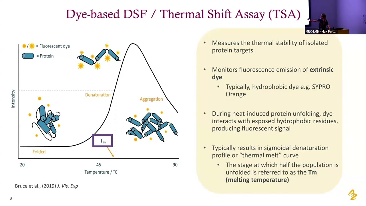








Differential Scanning Fluorimetry (DSF), also called Fluorescence Thermal Shift or Fast Thermal Melt, is a technique that measures protein thermal stability by detecting when proteins unfold using a fluorescent dye (such as SYPRO Orange) that binds to exposed hydrophobic regions upon unfolding; the melting temperature (Tm) is determined by identifying the peak of the derivative of the fluorescence versus temperature curve, with higher Tm values indicating more stable proteins that require more heat to unfold.
Differential Scanning Fluorimetry: A Guide to Protein Stability
Added:Fundamental protein biochemistry, including levels of protein structure (primary to quaternary) and the non-covalent forces that govern protein folding and denaturation.
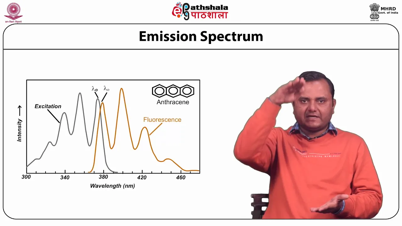
The principles of fluorescence spectroscopy, specifically how fluorophores absorb and emit light, and how environmental changes (like exposure to hydrophobic regions) affect their signal.
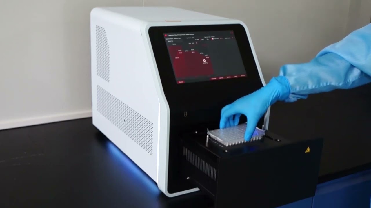
The basic mechanics of quantitative PCR (qPCR) instruments, focusing on their dual capability of precise thermal ramping and real-time optical fluorescence detection.
Introduction to chemical thermodynamics, particularly the concepts of thermal stability, Gibbs free energy, and the definition of a protein's melting temperature (Tm).
Application of DSF in high-throughput drug screening, specifically using thermal shift assays (TSAs) to detect small-molecule ligand binding to target proteins.
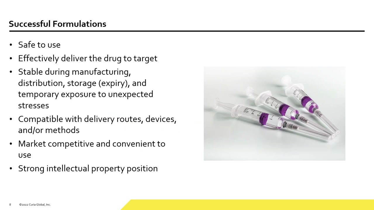
Biopharmaceutical formulation optimization, learning how DSF is used to screen buffers, pH, salts, and excipients to maximize the shelf-life of therapeutic proteins.
Comparative analysis of alternative biophysical techniques for studying protein stability, such as Differential Scanning Calorimetry (DSC) and Circular Dichroism (CD) spectroscopy.
Data analysis techniques for complex DSF profiles, including how to fit raw fluorescence curves to thermodynamic models to calculate enthalpy and entropy of unfolding.
516 views32likes9:05Original Release: 2026-01-27
Differential scanning flareometry, DSF, sometimes called a fluorescent thermal shift, is a quick and easy way that you can screen proteins happiness under different conditions. And so when I'm talking about happiness, I'm talking about kind of the sturdiness of the protein, how much heat it takes to unfold the protein or denature the protein.
And the way that it works is that you use a qPCR machine to heat up the protein sample. And as the protein sample unfolds, then you have a dye that is going to bind to the unfolded protein. And the way that it does this is because when your protein unfolds, it exposes the hydrophobic regions, the water excluded regions of the protein that are normally in the center of the protein. You heat them up, then they get exposed, the dye binds, and it flueses.
And this machine, not only does it heat it up, but it also then takes a picture of the fluoresence. And that is going to then allow you to see at what temperature your protein unfolded. And if your protein was sturdier, it was happier under certain conditions, then it's going to have a higher TMA, so an apparent melting temperature. Whereas if your protein was unhappy, then you would have a low TMA, so a low melting temperature. And this is a quick and easy screen that you can do in order to test a variety of different conditions.
Things like different salts, different phes, the presence of a lian. So a binding partner might help your protein kind of certify and then increase the TMA. And because higher TMA means what?
>> Uh higher uh stronger bonds in between the protein.
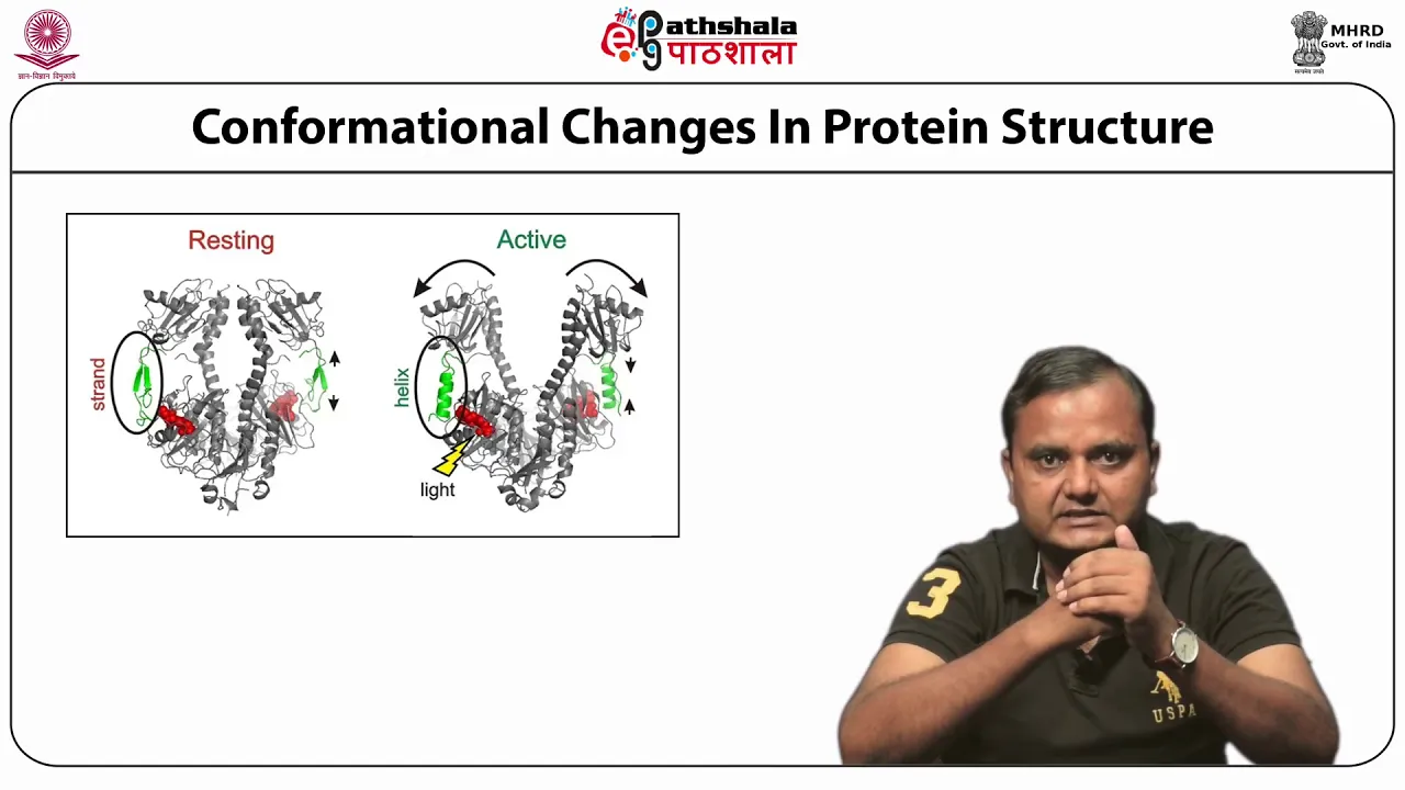
>> Yeah. When you heat up proteins, you allow them to wiggle. And when they're wiggling, they're able to break free from those non-coovvealent interactions, those intermolecular forces. And so the more of those forces there are holding the protein together, the sturdier the protein is, the more heat it takes for them to go and unfold. And we call unfolding of proteins. We say they denature.
And so the sturdier the protein is, the more heat it's going to take to unfold.
And then we just need a way in which we can detect that the protein has unfolded.
And so there are different ways that you can do this but a lot of them require fancy equipment such as circular dicroism.
The beauty of this FTS or this DSF is that all you need is a qPCR machine. And so these are typically used to measure the concentration of things like cDNAs.
So to measure expression of a gene, you make the DNA copy of the messenger RNA.
So that's the cDNA. And then you measure the copies get made over time. And we are using it however for this fluorescent thermal shift. And so this PCR machine can get hot and cold and hot and cold and hot and cold. But we're just going to go have it go from being cold or like coldish to being really hot. And we're going to have it do it in this gra gradient ramp. So it's going to slowly increase the temperature.
And as it increases the temperature, every minute it's going to take a picture along the ramp. And as it's taking the pictures, what's happening is you're seeing fluoresence. And the fluoresence is the indicator that the protein has unfolded. And you can see that you get kind of this curve. So RFU stands for relative fluorescent units and that's basically the amount of fluoresence and as you increase you kind of you start at the baseline you keep going you're you're increasing the temperature but nothing's happening because your protein is still sturdy but then you reach the temperature at which your protein starts to unfold and then you get kind of this sigmoidal shape where the protein unfolds the die binds you see fluoresence is that you'll see that the curve dips down and the curve dips down after it reaches the peak because what happens is it's thought that like the protein then clumps and when it clumps it's kind of hiding the binding sites for the die and you can see that under different conditions the protein might have a higher apparent melting temperature. So the TMA is the temperature at which half of the fluoresence is reached. So basically the TM the melting temperature would be the amount at which half the protein has unfolded but what we're seeing is an indirect indicator of that we're seeing this die fluoresence and so then we we get is the TMA the apparent TM and that is what we can measure based on this and so it would be the amount the heat at which you get like half maximum fluoresence if you're doing it um this way there also like a curve fitting model that we use and basic but the same basic idea applies.
And so what you do is then you can see if a protein has changed its TM under different conditions such as different pH, different salt or the presence of a lian. So a binding partner. You can actually use this as a technique to see if molecules are binding and how strongly they're binding by doing like different concentrations of different compounds. So people often use it as a screening technique to see what conditions their protein might be happy or unhappy at, as well as to see whether they're binding to certain things. And you can also use it in order to see whether if you make mutations to a protein that actually changes the stability of a protein. So there are a lot of really great things that you can use it for as a screening tool. And so if you're only testing a few different things, then you can do it in like PCR strips. But if you want to screen a bunch of conditions, then people often use um like a 96 well plate or even going up to like 384 various things. And a lot of those things then you're dealing with robots when you get really high up.
But with all these, you kind of want to use a white tube. And the white tube, this opaque tube is going to make it so that the fluoresence doesn't bleed between the different plate, between the different wells or whatever. And but you want to make sure that the camera can actually access and to see the fluorescents. And so then you're and shine through and stuff. And so then you're going to need an optically clear lid or they have like optically clear seals that you put on your plate because what's going to happen is that up here there's going to be a camera that's going to actually take pictures of things. And so this is going to heat it up and this is going to take its picture as you're heating it up. And as you're heating it up, the protein's going to unfold. When the protein unfolds, the dye binds. When the dye binds, you see fluorescents.
The dye that we use is like cypro orange. And it was like not even a dye that's actually like designed for this, but people found that it's really useful to do this. And so this dye is sold as like a protein stain, like a gel stain, but it's really useful for this. You can export the data. And if you export the RFU, the relative fluoresence units, you get this data that then you can upload into a tool called DSF World. And DSF World is a really great website that's going to then allow you to do kind of custom curve fitting to the curves to get a better idea about the actual apparent TM.
But at the end of the day, you can hopefully see whether the conditions that you tested have changed the protein's sturdiness or not. And so the higher the TM, the or TMA, the sturdier the protein is basically the tightly more tightly it's held together. Or it can also often when you bind to a lian, it kind of certifies things up a little bit. And so, but you could also have some like sometimes you might get a opposite effect type of thing, but then you can at least see a change if it bind compared to if it didn't bind. And so, if you see an increase, the TMA is kind of indicating that things are getting tighter and held together more tightly.
It's taking more heat to unfold them.
Whereas, if the TMA goes down, then you're destabilizing the protein. And so if you make a mutation to the protein and you see that the TMA goes down, then it might be an indicator that your protein is not liking that mutation that you made to it. Sometimes people use this as a screening tool for then doing some sort of structural biology work.
And so maybe they're trying to find an optimal buffer for your extra crystalallography or for your cryom or various things like this. Um, screening can be a quick way to kind of optimize your conditions for those other experiments you want to do.
And that is the basics of this technique.
Up Next

18:56
DeepXDE Tutorial: Solving 1D Poisson Equation with PINNs
@elastropy
5.4K views•2024-10-24

178:38
Whole Brain Architecture: First International Workshop 2024
@thewholebrainarchitecturei9784
640 views•2024-07-22

9:49
FastAPI vs Flask vs Django: Choosing the Right Python Web Framework
@TechWithTim
302.5K views•2024-05-26

1:42
Game of Thrones Opening Credits: A Cinematic Analysis
@gameofthrones
46.3M views•2011-04-18
Related Study Plans & Knowledge Roadmaps
Structured learning paths in General & Interdisciplinary Studies
general-studies
Artificial Light: History, Tech & Human Impact
01The Pre-Electric Era: Fire, Oil, and Gas Lighting
02The Incandescent Revolution and Grid Electrification
03Gas Discharge and Fluorescent Illumination
+ 3 more learning modules
14h6 modules
Explore Roadmap
general-studies
Elevator Engineering: History, Physics & Cities
01From Hoists to Safety Brakes: The Dawn of Vertical Lift
02Hydraulic vs. Traction: The Engineering Physics of Elevators
03Modern Speed, Safety, and Algorithms
+ 2 more learning modules
12h5 modules
Explore Roadmap
general-studies
Rubber: Material Science, History & Geopolitics
01Origins of Natural Rubber & Mesoamerican History
02The Chemistry of Natural Rubber & Vulcanization
03The Geopolitics of the Rubber Boom & Colonization
+ 2 more learning modules
12h5 modules
Explore Roadmap
general-studies
Containerization: Evolution, Logistics & Trade
01Ancient Trade & Bulk Cargo: Amphorae to Barrels
02The Industrial Revolution & Cardboard Packaging
03The Containerization Revolution: Malcom McLean
+ 2 more learning modules
12h5 modules
Explore Roadmap