This video demonstrates a simplified, non-enzymatic cell harvesting protocol for HEK293 cells grown in VitroGel HEK293 hydrogel, enabling efficient recovery of 3D spheroids without the use of trypsin or other proteolytic enzymes, with a complete process achievable within 5 minutes and suitable for scaling in flasks or bioreactors.
HEK293 3D Suspension Cell Harvesting Protocol | Enzyme-Free
Added:Deep Dive
Basic biology and common research applications of the HEK293 (Human Embryonic Kidney) cell line.

HEK 293 cells: types and uses
8K
HEK 293 cells are an immortalized human embryonic kidney cell line developed in 1973 by Dutch scientist Alex Van der Eb and further adapted by postdoc Frank Graham through adenovirus transformation; these cells became immortal when approximately 4.5 kilobases of adenovirus E1A and E1B genes integrated into chromosome 19, disrupting cellular quality control systems like cell cycle regulation and apoptosis, allowing indefinite growth; they are widely used in laboratories for recombinant protein production, gene therapy vector development, and drug testing due to their high transfection efficiency and ability to perform authentic human post-translational modifications, though researchers must work under Biosafety Level 2 conditions and use early-passage cells to minimize mutation accumulation and maintain experimental consistency.
Click to watch →
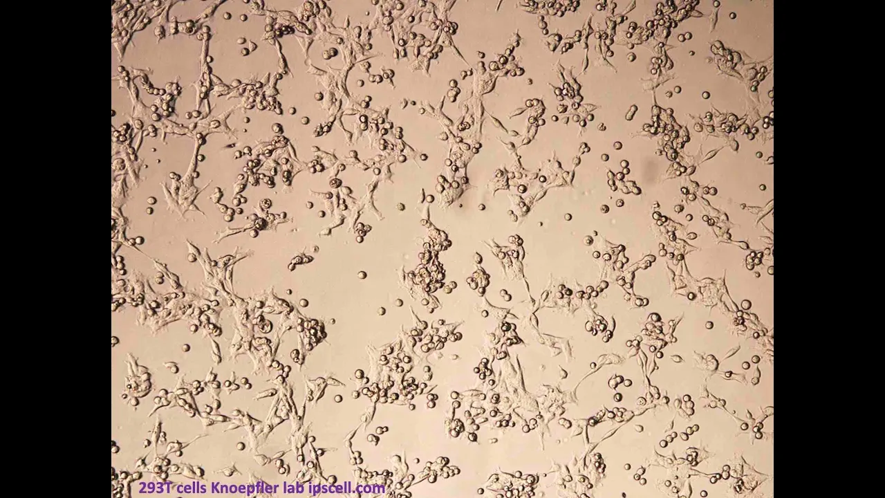
What are HEK 293 cells & their roles in research including on COVID-19?
4.3K
HEK 293 cells are human embryonic kidney cells that have been used in scientific research for nearly 50 years; they are immortal cell lines derived from fetal kidney tissue and are widely utilized as protein production factories and virus manufacturing platforms, playing a crucial role in vaccine development including the production of some COVID-19 vaccines, though it is important to note that no actual cells are present in the final vaccines and these cells are not stem cells but rather epithelial kidney cells.
Click to watch →

What the HEK? A Beginner’s Guide to HEK293 Cells
4.9K
HEK293 cells are human embryonic kidney cells originally isolated by Alex Fonder Eb in the early 1970s and transfected with adenovirus DNA by Frank Graham, who named them after his 293rd experiment; these cells are highly versatile for recombinant protein production due to their rapid doubling time (~36 hours), ease of transfection, and ability to grow in suspension or monolayer, making them the second most widely used cell line after HeLa cells in applications ranging from vaccine development to cancer research, though they require careful handling as extended culture beyond 20 passages degrades cell health and increases contamination risks.
Click to watch →

Октябрь. Коты - Сосуды Красоты!
38.6K
HEK 293 cells (Human Embryonic Kidney 293 cells) are a widely used cell line in biotechnology and vaccine development. These cells, originally derived from human embryonic kidney tissue, have been modified to efficiently produce viral proteins and vectors. They are used in the production of various vaccines, including those for coronavirus, and in the development of viral vectors for gene therapy. The cells' ability to support high-level protein expression makes them invaluable for biopharmaceutical manufacturing.
Click to watch →

أسرار صناعة الغذاء: هل نتناول طعاماً معدلاً بخلايا بشرية؟
490
HEK 293 (Human Embryonic Kidney 293) is a cell line created in 1973 by researcher Frank Graham at McMaster University. The cells were derived from embryonic kidney tissue obtained from a pregnancy termination. Scientists genetically modified these cells using adenovirus 5 to prevent natural cell death, transforming them into immortal cells that can grow and replicate indefinitely in laboratory conditions. This cell line has become invaluable for medical research and vaccine development, including vaccines for measles, mumps, rubella, and hepatitis B. The cells are used because viruses require a human-like cellular environment to replicate, and HEK 293 cells provide a sterile, suitable environment for this purpose.
Click to watch →
Deep Dive
The structural and physiological differences between traditional 2D monolayer cell cultures and 3D spheroid cultures.

3D Cell Culture | 3D Culture Techniques
12.1K
This section explains why 3D cell culture is superior to traditional 2D monolayer cultures. Standard 2D cultures inadequately represent the in vivo environment, making them unreliable predictors of drug efficacy and toxicity. In 3D cultures, cells grow in all directions with natural cell-cell and cell-matrix interactions occurring. 3D systems develop natural gradients for nutrients, oxygen, pH, and other factors, whereas 2D systems lack these gradients entirely. 3D tumor spheroids replicate in vivo tumor architecture with peripheral proliferative cells, intermediate layers, and necrotic cores, mimicking oxygen and nutrient diffusion patterns. Living tissues contain stromal cells (fibroblasts, adipocytes, glial cells, endothelial cells, immune cells) that interact with epithelial and neuronal tissues, regulating disease progression and therapeutic responses. These complex 3D environments enable more accurate modeling of cellular behavior and responses to treatments.
Click to watch →

Considerations & Testing: Using Primary Hepatocytes in 2D and 3D Cell Culture | Dr. Rafal P. Witek
327
3D spheroid cultures offer significant advantages over 2D systems including: (1) Higher physiological function - spheroids achieve nearly double albumin secretion and substantially increased phase I/II enzyme activity compared to 2D; (2) Extended longevity - enabling 10-30 day cultures versus 5-7 days for 2D, allowing long-term studies like fibrosis modeling, inflammation studies, and disease progression research; (3) Applications for implantable tissues - spheroids can be encapsulated in alginate or printed for therapeutic applications; (4) Better prediction of idiosyncratic toxicity - 3D systems better model complex cellular interactions involving Kupffer cells and stellate cells that contribute to unpredictable drug reactions.
Click to watch →

El lado resistente del cáncer mecanismos que desafían la eficacia de la terapia - Dra. Noemí Salinas
209
Drug effects on cancer cells differ significantly between 2D monolayer and 3D spheroid culture systems. In 2D conditions, 5-fluorouracil reduced complement-dependent cytotoxicity induced by cetuximab. However, in 3D spheroid conditions, the same treatment increased cytotoxicity. This difference may be explained by tumor microenvironment structure affecting drug penetration and cell-cell interactions. Spheroid culture conditions themselves can induce phenotypic plasticity, causing non-stem cells to acquire stem cell characteristics. Cell proliferation analysis revealed that drug effects on proliferation versus sphere formation are concentration-dependent, with higher concentrations inhibiting proliferation while lower concentrations maintain proliferative capacity.
Click to watch →

Dr Dimitris Bouzianas on 2D and 3D cell cultures. SCAFFY Project Workshop March 8 2016
183
Cell cultures are classified by tissue origin (epithelial, osteosarcoma, etc.) and growth format (adherent or suspension). Traditional 2D monolayer cultures are easier to use and process but lack physiological accuracy. 3D culture systems better mimic in vivo conditions by incorporating extracellular matrix components. Three major 3D methods exist: scaffold-based cultures, aggregate cultures, and microcarrier systems. Microgravity conditions further enhance physiological relevance by reducing mechanical stress between cells and their environment.
Click to watch →

Cell culture technology 3D and scaffold based
5K
Cell culture technology has evolved from 2D monolayer to 3D spheroid systems. In 2D culture, cells grow as flat layers on plastic substrates (Petri dishes or flasks), unable to mimic in vivo microenvironment or cell-to-cell interactions. Cells may lose natural morphology. 2D culture is economical, easy to perform, and has high throughput capacity, making it ideal for large-scale drug screening. In contrast, 3D culture uses natural extracellular matrix, creating spherical aggregates that resemble original tissue. The 3D spatial arrangement affects cell surface receptor organization, proliferation, differentiation, signaling, and gene expression. While 3D culture is expensive and laborious with low throughput, it is essential for tissue engineering and regenerative medicine where physiological relevance is critical.
Click to watch →
Deep Dive
The role of extracellular matrices (ECM) and hydrogels, such as VitroGel, in supporting and encapsulating 3D cell growth.
What is VitroGel®? A xeno-free hydrogel replacement to animal-based ECMs for 3D cell culture.
312
VitroGel® is a xeno-free, synthetic hydrogel designed for 3D cell culture that offers advantages over traditional animal-based extracellular matrix materials, including room temperature stability, batch-to-batch consistency, and injectable properties with shear-thinning behavior that enables easy handling and application in various research settings from bench to bedside.
Click to watch →

Using Animal-Free VitroGel Hydrogel in Comparison to Animal-based ECM for Xenograft - WEBINAR
302
Synthetic hydrogel systems like VitroGel offer a well-defined, animal-free alternative to traditional animal-based extracellular matrix (ECM) for xenograft PDX and CDX models, providing consistent mechanical properties, biocompatibility, and the ability to control growth factors and supplements, while maintaining similar tumor growth characteristics compared to animal-based ECM across various cancer cell lines.
Click to watch →

Dr Claudia Loebel: Metabolic Labeling in 3D Cell-Hydrogel Studies (11th June 2020)
498
The extracellular matrix (ECM) is a three-dimensional scaffold surrounding cells, providing biophysical and biochemical support. Cells constantly remodel the ECM through building, shaping, and reassembling it, and disruptions in these mechanisms cause diseases like fibrosis. Hydrogels—water-swelling polymer networks—have evolved to mimic ECM properties for studying cell behavior. Early breakthroughs showed cells respond to mechanical signals like elasticity, leading to more sophisticated designs incorporating viscoelasticity. However, a fundamental challenge emerged: cells rapidly secrete their own ECM proteins upon encapsulation, forming a nascent matrix that displaces the engineered hydrogel, limiting long-term control over cellular behavior.
Click to watch →

VitroGel Hydrogel Helps in Novel Pre-Clinical Model of the Normal Human Breast -Dr. Hannah Harrison
658
A novel preclinical model of normal human breast tissue using VitroGel hydrogel maintains epithelial proliferation, hormone receptor expression (estrogen and progesterone), and immune cell residency (T cells and macrophages) over 7 days without the hyper-proliferation, loss of hormone signaling, or short treatment windows that limit other organotypic tissue culture systems, enabling more accurate testing of breast cancer preventative agents.
Click to watch →
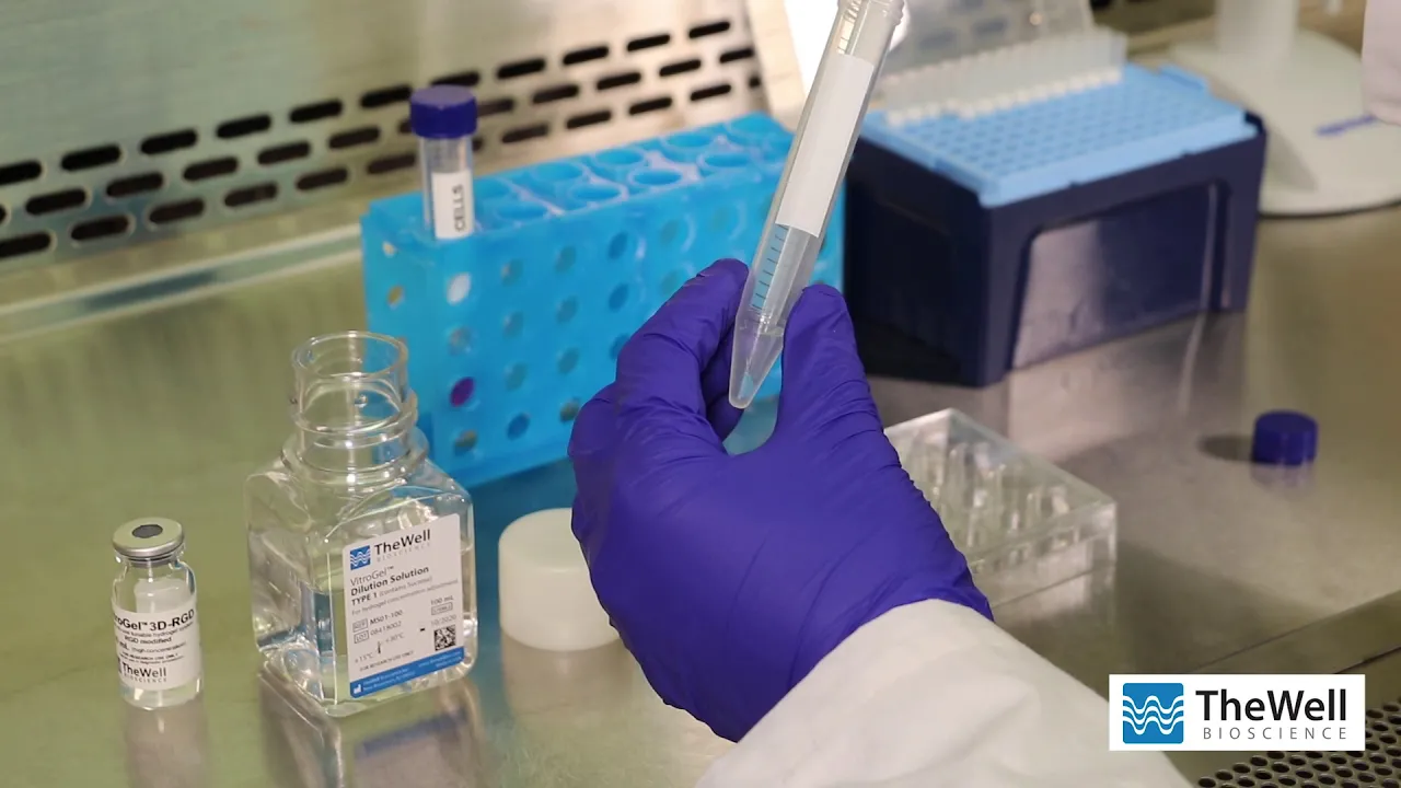
2D Cell Coating with VitroGel® Hydrogel System - Method 2 : TheWell Bioscience
651
The VitroGel® hydrogel system enables researchers to coat cell culture surfaces with varying stiffness and texture, which influences cell behavior including migration and invasion patterns; this 2D coating method supports long-term cell culture as media can penetrate the hydrogel matrix to provide nutrients while allowing easy microscopic observation of cell growth.
Click to watch →
Deep Dive
Standard cell-harvesting concepts, specifically the mechanism and limitations of traditional enzymatic dissociation (e.g., trypsinization).

I Genetically Engineered *MYSELF* to Fix Lactose Intolerance
618.5K
Trypsin is an enzyme that breaks down proteins holding cells to culture surfaces. When added to cells, trypsin causes them to detach and become suspended in liquid. The solution becomes cloudy as cells release from the dish. After trypsinization, cells are collected by centrifugation, which pellets them at the bottom of tubes for further processing or analysis.
Click to watch →

Peptide Digestion for Protein Sequencing: Trypsin, Chymotrypsin & More! | CSIR NET Life Science
4.5K
Trypsin is an endopeptidase that cleaves peptide bonds at the carboxyl side of arginine and lysine. Key limitations include: cannot cleave at terminal positions (first or last amino acid), cannot cleave when proline follows the target residue (due to cis-peptide bond recognition), and cannot cleave at N-terminal positions where trypsin would need to act. These restrictions are essential for understanding why certain sequences remain intact during digestion.
Click to watch →

Disaggregation of cells - Animal Biotechnology Lecture 5 @leelamani4091
603
Enzymatic disaggregation uses enzymes to dissolve collagen between cells gently. Trypsin is used for moderate cells, while collagenase is preferred for sensitive cells that would be damaged by trypsin. The trypsinization process is classified as cold (ice-cold trypsin, 1-6 hours soaking) or warm (37°C trypsin, 3-4 hours with continuous magnetic stirring). After disaggregation, centrifugation separates live cells (which settle) from dead cells and debris (which float). The live cell pellet is collected for inoculation into appropriate culture media.
Click to watch →

Trypsinization Process (Animal Cell Culture )
237
Trypsinization is the enzymatic process used to detach adherent cells from culture flasks for passaging. Cells are either adherent (attach to flask surface) or suspension (grow freely). Trypsin, a serine protease, breaks down proteins anchoring cells to the surface. The process requires PBS, trypsin-EDTA solution, complete medium, sterile pipettes, CO2 incubator, and inverted microscope. All materials must be pre-warmed to 37°C to ensure optimal enzyme activity and prevent thermal shock. The old culture medium is removed under biosafety cabinet conditions to prevent contamination.
Click to watch →

Cell detachment with trypsin/EDTA: the biochemistry going on when you take cells off!
6.2K
Adherent cells must be passaged before reaching 80% confluence to maintain optimal growth. Trypsin/EDTA solution is used to detach cells by breaking protein-protein connections. Trypsin is a serine protease that cuts various proteins, while EDTA acts as a chelator that removes calcium ions, weakening calcium-dependent adhesion proteins like cadherins. This combination effectively releases cells from the culture surface and from each other.
Click to watch →
Prerequisite Knowledge
- Concept 01Basic biology and common research applications of the HEK293 (Human Embryonic Kidney) cell line.
- Concept 02The structural and physiological differences between traditional 2D monolayer cell cultures and 3D spheroid cultures.
- Concept 03The role of extracellular matrices (ECM) and hydrogels, such as VitroGel, in supporting and encapsulating 3D cell growth.
- Concept 04Standard cell-harvesting concepts, specifically the mechanism and limitations of traditional enzymatic dissociation (e.g., trypsinization).
Subsequent Learning
- Step 01Downstream characterization techniques for intact spheroids, including immunofluorescence staining, flow cytometry, and Western blotting.
- Step 02Methods for evaluating cell viability, recovery yield, and functional integrity post-harvest.
- Step 03Scaling up 3D suspension HEK293 cultures for bioprocessing applications, such as viral vector (AAV/lentivirus) or recombinant protein production.
- Step 04Exploring how chemical or physical modifications to hydrogel matrices affect spheroid growth kinetics and ease of harvest.
Opening
Tension
Focus Start
Key Theme
Detail Work
Application
Synthesis
Close
Opening
0:04Playing Section
- 1
Video begins with musical introduction.
- 2
No spoken content or visual topics presented yet.
- 3
Atmosphere sets tone for upcoming material.
Enzymatic Digestion and the Limitations of Non-Enzymatic Harvesting
While non-enzymatic harvesting protocols avoid the potential risks of protease-induced cell damage, they face significant criticism regarding efficiency and cell viability. Researchers often favor enzymatic digestion (using collagenase, trypsin, or specific hydrogel-depolymerizing enzymes) for several reasons. First, non-enzymatic methods, which often rely on mechanical disruption or temperature shifts, can cause high shear stress, leading to mechanical cell damage and reduced viability. Second, they frequently fail to completely dissolve robust extracellular matrices or synthetic hydrogels, resulting in incomplete spheroid recovery and low cell yields. For quantitative downstream applications like flow cytometry, western blotting, or single-cell RNA sequencing, enzymatic dissociation remains the gold standard because it ensures complete matrix degradation and generates highly viable, uniform single-cell suspensions without the selective loss of cells trapped in undigested hydrogel remnants.
Deep Dive
Downstream characterization techniques for intact spheroids, including immunofluorescence staining, flow cytometry, and Western blotting.

Tumor Specific 3D Cell Culture Models for Accurate Testing of Anticancer Agents
181
Multiple downstream analyses can be performed on 3D spheroids including: (1) Flow cytometry for cell characterization; (2) Histology with embedding for analysis of cell type distribution; (3) Immunohistochemistry for marker detection; (4) Any marker analysis based on research objectives. The spheroids can be treated as miniaturized versions of tumors, enabling comprehensive analysis of cellular composition and response.
Click to watch →

Disease model generation: 5 steps to a 3D cancer spheroid model
3.1K
Immunofluorescent experiments can be performed on 3D spheroids, including markers for DNA damage. DNA damage kits are available that allow simultaneous study of DNA damage and cytotoxicity. These kits have been created or optimized using 2D models but have also been adapted for use in 3D culture systems.
Click to watch →
Learning Lab LIVE! Q&A with IF Expert Virginia Bain, PhD
455
Flow cytometry and immunofluorescence have different fluorophore requirements. Flow cytometry uses single-cell suspensions with brief light exposure, so fluorophores don't need high brightness or photostability. Immunofluorescence requires photostable, brighter fluorophores due to extended light exposure on tissue. Some flow dyes like PE are not photostable and should be avoided. Different detection techniques can be combined in multiplex panels: conjugates with indirect experiments (unique hosts), conjugates of same host as primary (stain before blocking), and amplification added to any channel.
Click to watch →
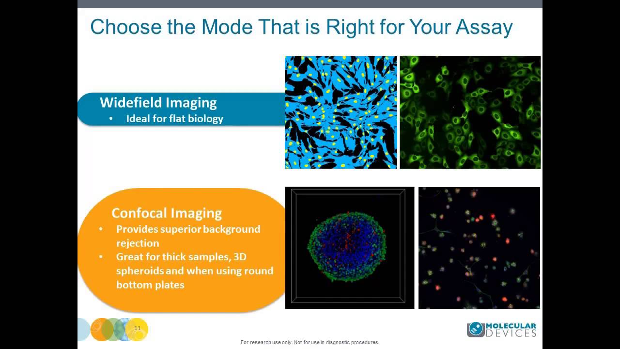
Jayne Hesley - Grischa Chandy - Multidimensional High Throughput Imaging with the New ImageXpress Mi
580
Spheroids form via hanging drop, scaffold, or 3D gel matrix methods. Ultralow attachment plates enable easier media changes. Staining challenges include dye penetration through tightly packed cells and wash steps disrupting spheroids. Optimization requires 2-3 hour incubation at 37°C and 2-4 times higher dye concentrations than 2D assays. Z-stack imaging from bottom to halfway through provides consistent samples. Confocal mode provides better image quality for thick samples where background rejection is needed. Custom analysis modules identify specific parameters including morphological changes, total cell count, and viability markers. Data can be collected on spheroid diameter, area, and live cell intensity.
Click to watch →
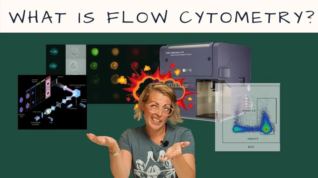
What is FLOW CYTOMETRY (Intro to Flow Cytometry - Episode 1)
13.2K
Unlike western blotting which pools all cells together and provides only an average expression level, flow cytometry keeps individual cells intact and analyzes each cell separately. This distinction is critical because two different scenarios (few cells expressing high amounts vs many cells expressing low amounts) would appear identical in western blot data but can be distinguished using flow cytometry by examining shifts between negative and positive populations.
Click to watch →
Deep Dive
Methods for evaluating cell viability, recovery yield, and functional integrity post-harvest.

Bioestimulación en el cultivo del Aguacate
23.8K
Post-harvest recovery assessment uses SPAD chlorophyll meters, with readings of 60-70 indicating healthy recovery capacity. Field evaluation of bioestimulants involves measuring chlorophyll content, enzyme activities (superoxide dismutase, catalase, peroxidase), and antioxidant status. Amino acids enhance antioxidant defense through increased enzymatic and non-enzymatic antioxidant production. Root absorption of amino acids occurs through hydroponic studies, with rapid absorption of glutamine, glycine, and arginine, while proline and lysine absorb more slowly.
Click to watch →

Exploring the five pillars of cell sorting: Viability, recovery, efficiency, purity & proliferation
514
Four techniques for measuring recovery: (1) Volumetric analyzer - weighs tubes before and after sorting, runs viability dye, and calculates total cell count from gate statistics, (2) Hemocytometer or cell counter - suitable for smaller cell numbers, (3) Counting beads - added to sorted sample to determine cell-to-bead ratio, and (4) Microscopy - visualizes plated cells in wells to verify correct cell numbers.
Click to watch →
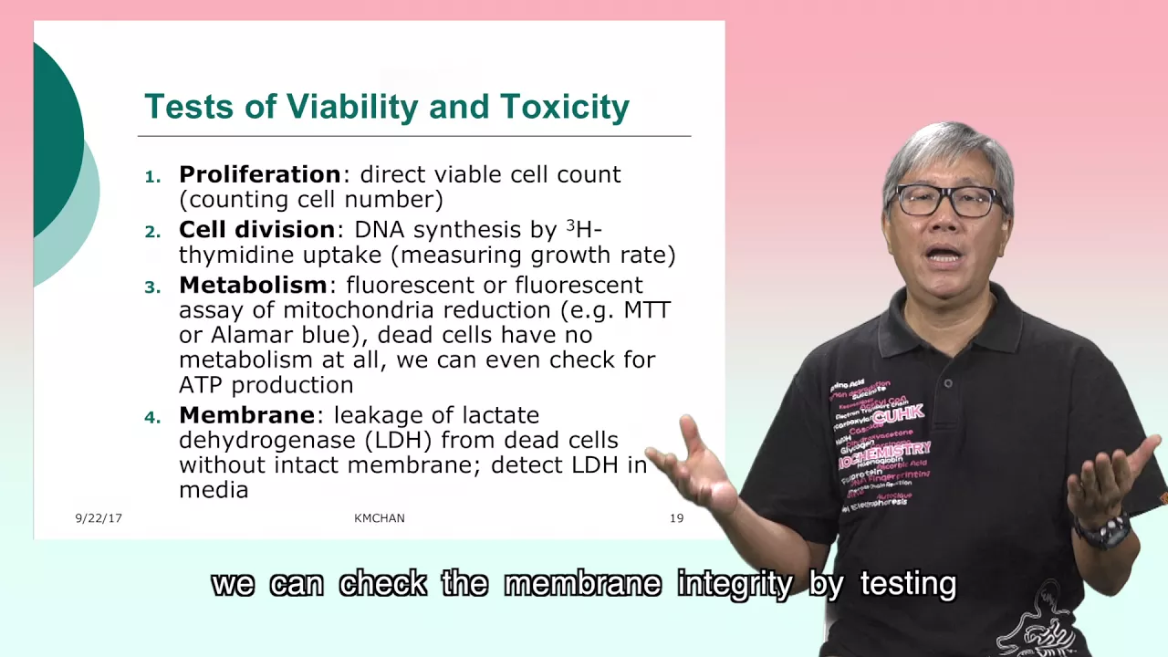
Cytotoxicity Assays (2): How to perform cytotoxicity assays
23.3K
Five common methods assess cell viability and toxicity: (1) Cell counting under microscope using hemocytometer; (2) Trypan blue exclusion assay where dead cells stain blue; (3) Crystal violet staining of viable cells with absorbance read at 595 nm; (4) Neutral red staining of lysosomes read at 540 nm; (5) Metabolic assays measuring mitochondrial function. Additional approaches include tritium thymidine uptake to measure DNA synthesis and ATP production to assess metabolic activity. Membrane integrity can be checked by testing lactate dehydrogenase (LDH) levels in media from dead cells.
Click to watch →

DBT BET 2025 | Operation BET Series | Animal cell Culture Explained by Aashita Ma’am
1.1K
Three main methods are used to assess cell viability: (1) Trypan Blue test - live cells exclude the dye and remain unstained, while dead cells with compromised membranes appear blue, (2) LDH (Lactate Dehydrogenase) test - detects cell death by measuring enzyme release from damaged cells, and (3) MTT assay - live cells convert yellow MTT dye to purple formazan crystals through NADP-dependent oxidoreductase enzymes. These tests are essential for determining which cells are viable before subculturing and for quantifying cell proliferation and cytotoxicity effects.
Click to watch →

CSI: Produits stimulants de l’immunité des plantes en agriculture biologique
1.7K
Cell viability is assessed using: (1) XTT assay (metabolic activity); (2) Trypan blue exclusion (dead cells take up dye); (3) Propidium iodide staining (enters only dead cells with compromised membranes). These methods allow quantification of cell death and assessment of product toxicity.
Click to watch →
Deep Dive
Scaling up 3D suspension HEK293 cultures for bioprocessing applications, such as viral vector (AAV/lentivirus) or recombinant protein production.

BalanCD HEK293 Viral Feed: Accelerating AAV and LV Production for Gene and Cell Therapy
191
Chemically defined feed media, when added 24 hours post-transfection in fed-batch suspension cultures, can significantly enhance viral vector production yields in HEK293 cells for both adeno-associated virus (AAV) and lentivirus (LV) production, with improvements ranging from 2-8 fold for AAV across different serotypes and 3-fold for LV genome and functional titers, while also demonstrating compatibility with various transfection reagents, growth media, and culture formats.
Click to watch →

Platform Based Scalable Suspension Process for LVV Production
142
IDT Biologika has developed a platform-based scalable suspension process for lentiviral vector production using HEK293 suspension cells with transient transfection, which enables efficient optimization of critical parameters such as DNA load, plasmid ratios, and complexation time to achieve approximately two-log increases in productivity, with typical crude bulk harvest titers reaching 1-2 × 10^10 physical titers and 1-2 × 10^8 transduction units per milliliter; this approach addresses the industry-wide shortage of viral vectors by enabling early process optimization and robust scale-up from shake flasks to bioreactors.
Click to watch →

Optimizing HEK293 Cell Culture for Gene Therapy Webinar
389
Gene therapy utilizes viral vectors (adenoviruses, lentiviruses) to deliver therapeutic genes in vivo or ex vivo. As of 2021, adenoviruses dominated with 575 clinical trials. HEK 293 cells serve as ideal host cells due to their human derivation and ability to produce human-infectable viruses. However, adherent HEK 293 cultures face scalability challenges requiring two-dimensional expansion, high labor, and significant cleanroom space. Transitioning to suspension culture offers better scalability through volume-based expansion. Multi-cabinet incubators with independent control of temperature, CO2, agitation, humidity, and orbit diameter enable parallel optimization of these critical parameters, addressing the complexity of developing scalable viral vector production platforms.
Click to watch →
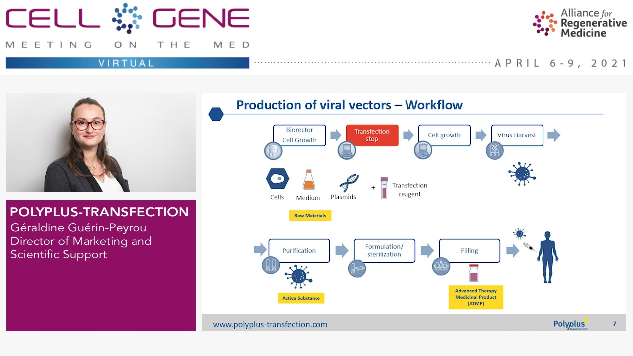
Polyplus transfection
104
Viral vectors (AAV and lentivirus) are the preferred delivery method for nucleic acid therapies, carrying genes into target cells via direct injection (gene therapy) or transducing patient cells (cell therapy), manufactured in HEK 293 cells through transient plasmid transfection. By 2020, over 1100 clinical trials were ongoing globally, primarily in oncology. Viral vector requirements vary dramatically by indication—ophthalmologic treatments need 1×10^12 particles/dose while hemophilia requires 1×10^15 particles/dose (1000× difference). Meeting growing demands requires improving productivity through suspension culture adoption and process optimization. Three main manufacturing challenges exist: achieving consistent reproducible yields despite parameter variations, scaling from adherent to suspension cultures (time-consuming), and addressing increasingly stringent regulatory requirements for raw materials early in clinical development.
Click to watch →

BalanCD HEK293 Viral Feed: Accelerating AAV and LV Production for Gene and Cell Therapy
156
A chemically defined feed medium (BalanCD HEK293 Viral Feed) added 24 hours post-transfection in fed-batch suspension or adherent HEK293 cell cultures can significantly increase viral vector yields for gene and cell therapy production. Studies demonstrate that this feed improves adeno-associated virus (AAV) titer by 3-10 fold across multiple serotypes (AAV2, AAV5, AAV9) and increases lentivirus genome and functional titer by approximately 3 fold, while maintaining compatibility with various transfection reagents, growth media, and culture formats including shake flasks, deep well plates, and bioreactors.
Click to watch →
Deep Dive
Exploring how chemical or physical modifications to hydrogel matrices affect spheroid growth kinetics and ease of harvest.

The Next Step in Evolution of 3D Culture
3.8K
Fully formed spheroids can be harvested intact from low-adhesion plates since they are free-floating rather than attached to the substrate. Spheroids are typically fixed first, then pulled from the plate and embedded in paraffin for histological sectioning. This allows downstream analysis including immunohistochemistry and morphometric measurements.
Click to watch →
Dr. Ece Öztürk I Tümör Mikroçevresini Modellemede Doku Mühendisliği ve Yeni Nesil Organoidler
134
Organ hydrogel derivation creates tissue-mimicking scaffolds by removing cells from organs, enzymatically digesting tissue, and thermally gelating the remaining extracellular matrix. This preserves native ECM components like collagen and sulfated GAGs while enabling controlled experimental manipulation. Different preparation methods (freeze-thaw, Triton X-100, SDS) yield hydrogels with varying mechanical properties and cell viability. Freeze-thaw and Triton methods produce hydrogels supporting better cell survival. Two hydrogel models were created: healthy tissue-mimicking (non-sulfated hyaluronic acid) and tumor-mimicking (sulfated hyaluronic acid). Cancer cells showed dramatically different behaviors: in healthy-mimicking matrices, cells remained quiescent, while in sulfated tumor-mimicking matrices, they exhibited remarkable proliferation and formed tumor-like nodules. Sulfated matrices also induced epithelial-mesenchymal transition (EMT) markers including vimentin, fibronectin, and transcription factors. Inhibiting PI3K completely reversed the growth effects, identifying it as the key downstream mediator.
Click to watch →

Defensa de tesis de Simioni, Yamila Roxana
310
Nanoparticles were incorporated into carbopol hydrogels for topical application, as direct application causes particle runoff. Hydrogel preparation involved mixing carbopol with nanoparticles, vortexing, and centrifugation to ensure homogeneity. The hydrogel showed pH 5-6 (optimal for skin), maintained nanoparticle size and PDI over 5 months, and demonstrated appropriate rheological properties (yield stress, consistency, pseudoplastic behavior). Vitamin D3 release was measured at pH 7.4 and 5.2 (psoriatic skin pH), showing higher retention in the hydrogel. 3D spheroid models were developed as intermediate models between 2D cultures and animal studies, consisting of fibroblast cores surrounded by keratinocyte rings. These models allow communication between cell types impossible in 2D cultures. Studies showed hydrogel with archaeal lipid nanoparticles maintained size and PDI, showed appropriate rheological properties, and demonstrated anti-inflammatory effects in 3D spheroid models.
Click to watch →
Мучаю шарики ОРБИЗ в мясорубке и блендере! Alex Boyko
6.4M
This segment demonstrates how different mechanical processing methods transform hydrogel materials. When Orbeez are processed through a meat grinder, they transform into a gelatinous, jelly-like substance. When blended in a blender, they become a smooth, puree-like consistency much faster. The choice of processing method significantly affects the final texture and consistency of the processed material.
Click to watch →

Artificial Vascular Systems – ArtiVasc 3D on the Way to Bio-Artificial Tissue
1.9K
To provide a nature-like environment for cells, researchers use biopolymers from the natural tissue matrix. These biopolymers must first be chemically modified so they can react with each other to form stable hydrogel matrices. By adjusting the degree of chemical modification, scientists can control the viscosity of biopolymer solutions and customize the softness and stability of the gel matrix.
Click to watch →
Opening
Tension
Focus Start
Key Theme
Detail Work
Application
Synthesis
Close
Opening
0:04- 1
Video begins with musical introduction.
- 2
No spoken content or visual topics presented yet.
- 3
Atmosphere sets tone for upcoming material.
Enzymatic Digestion and the Limitations of Non-Enzymatic Harvesting
While non-enzymatic harvesting protocols avoid the potential risks of protease-induced cell damage, they face significant criticism regarding efficiency and cell viability. Researchers often favor enzymatic digestion (using collagenase, trypsin, or specific hydrogel-depolymerizing enzymes) for several reasons. First, non-enzymatic methods, which often rely on mechanical disruption or temperature shifts, can cause high shear stress, leading to mechanical cell damage and reduced viability. Second, they frequently fail to completely dissolve robust extracellular matrices or synthetic hydrogels, resulting in incomplete spheroid recovery and low cell yields. For quantitative downstream applications like flow cytometry, western blotting, or single-cell RNA sequencing, enzymatic dissociation remains the gold standard because it ensures complete matrix degradation and generates highly viable, uniform single-cell suspensions without the selective loss of cells trapped in undigested hydrogel remnants.
Basic biology and common research applications of the HEK293 (Human Embryonic Kidney) cell line.
The structural and physiological differences between traditional 2D monolayer cell cultures and 3D spheroid cultures.
The role of extracellular matrices (ECM) and hydrogels, such as VitroGel, in supporting and encapsulating 3D cell growth.
Standard cell-harvesting concepts, specifically the mechanism and limitations of traditional enzymatic dissociation (e.g., trypsinization).
Downstream characterization techniques for intact spheroids, including immunofluorescence staining, flow cytometry, and Western blotting.
Methods for evaluating cell viability, recovery yield, and functional integrity post-harvest.
Scaling up 3D suspension HEK293 cultures for bioprocessing applications, such as viral vector (AAV/lentivirus) or recombinant protein production.
Exploring how chemical or physical modifications to hydrogel matrices affect spheroid growth kinetics and ease of harvest.
9.8K views0likes2:34@ThewellbioOriginal Release: 2021-01-20
[Music] [Music] [Music] [Music] [Music] [Music] [Music] you
Up Next

15:22
mRNA Purification: Globin & rRNA Depletion Protocol for cDNA
@adwoabiotech
169 views•2025-04-16

12:54
Triumph of Orthodoxy Icon: Byzantine Art & History Explained
@BenCallan
2.1K views•2024-08-06

9:49
FastAPI vs Flask vs Django: Choosing the Right Python Web Framework
@TechWithTim
302.5K views•2024-05-26

1:42
Game of Thrones Opening Credits: A Cinematic Analysis
@gameofthrones
46.3M views•2011-04-18
Related Study Plans & Knowledge Roadmaps
Structured learning paths in General & Interdisciplinary Studies
general-studies
Elevator Engineering: History, Physics & Cities
01From Hoists to Safety Brakes: The Dawn of Vertical Lift
02Hydraulic vs. Traction: The Engineering Physics of Elevators
03Modern Speed, Safety, and Algorithms
+ 2 more learning modules
12h5 modules
Explore Roadmap
general-studies
Rubber: Material Science, History & Geopolitics
01Origins of Natural Rubber & Mesoamerican History
02The Chemistry of Natural Rubber & Vulcanization
03The Geopolitics of the Rubber Boom & Colonization
+ 2 more learning modules
12h5 modules
Explore Roadmap
general-studies
Containerization: Evolution, Logistics & Trade
01Ancient Trade & Bulk Cargo: Amphorae to Barrels
02The Industrial Revolution & Cardboard Packaging
03The Containerization Revolution: Malcom McLean
+ 2 more learning modules
12h5 modules
Explore Roadmap
general-studies
Textile Tech: Evolution, Labor & Global Impact
01Pre-Industrial Textiles: Handloom & Cottage Industry
02Mechanization: The Spinning Jenny to the Power Loom
03The Factory System & Global Labor Transformation
+ 2 more learning modules
10h5 modules
Explore Roadmap